© Borgis - Postępy Nauk Medycznych 8/2014, s. 536-541
Karolina Wawiernia, *Barbara Bukowicka, Wiesław Tarnowski
Ostre zapalenie wyrostka robaczkowego w ciąży – przegląd piśmiennictwa i doświadczenia własne
Acute appendicitis in pregnant – review of the literature and our own experience
Department of General, Oncological and Gastrointestinal Surgery, Medical Centre of Postgraduate Education, Professor Witold Orłowski Independent Public Clinical Hospital, Warszawa
Head of Department: prof. Wiesław Tarnowski, MD, PhD
Streszczenie
Wstęp. Ostre zapalenie wyrostka robaczkowego (OZWR) u kobiet w ciąży stanowi istotny problem kliniczny. Odmienności fizjologiczne związane z ciążą mogą być przyczyną opóźnienia w rozpoznaniu i prawidłowym leczeniu.
Materiał i metody. W latach 2004-2013 operowano 35 kobiet w ciąży z podejrzeniem OZWR. Śródoperacyjnie potwierdzono zapalenie wyrostka robaczkowego w 29 przypadkach. W pozostałych 6 przypadkach przyczyny dolegliwości były związane z inną patologią wewnątrzbrzuszną.
Wyniki. W grupie 29 pacjentek z OZWR średni wiek wynosił 28,4 roku. OZWR najczęściej występowało w II trymestrze ciąży. We wszystkich przypadkach stwierdzano ból w prawym dolnym kwadrancie brzucha, w 78% nudności i wymioty, wzrost leukocytozy powyżej 11,0 K/uL w 75% przypadkach 89.7% pacjentek było operowanych w pierwszej dobie od chwili przyjęcia do szpitala.
Wnioski. Leczenie chirurgiczne OZWR w ciąży powinno być wykonane w ciągu pierwszych 24 godzin od przyjęcia do szpitala.
Summary
Introduction. Acute appendicitis in pregnant women is essential clinical problem. Physiologic differencies of pregnancy can cause problems in diagnosis and proper treatment.
Material and methods. In period 2004-2013 35 pregnant women were operated on with clinical diagnosis of acute appendicitis. The diagnosis was proven at the time of operation in 29 cases. Causes of remain 6 cases were related to other intra-abdominal pathology.
Results. In group of 29 patients with acute appendicitis mean age was 28.4 years. Appendicitis occurred most often in second trimester of pregnancy. In all cases occurred pain in the lower right quadrant of the abdomen, in 78% nausea and vomiting were observed and in 75% leukocytosis was higher than 11.0 K/uL. 89.7% patients were operated on in first 24 hours after admission to the hospital.
Conclusions. Surgical treatment of acute appendicitis in pregnant women should be taken in first 24 hours after admission to the hospital.

Introduction
Acute appendicitis (appendicitis) is the most common surgical disease in pregnant and is a risk factor for a healthy pregnancy (1). Moreover it causes an important diagnostic problem. The incidence is estimated at between 1 in 1400 to 1 in 1600 pregnancies (2-5). According to the statistics it occurs slightly more often in the second trimester of pregnancy (2-5) and is most common in patients between 20 and 30 years of age (6).
Diagnosis of appendicitis in pregnancy is associated with many difficulties at the stage of history taking and physical examination. Physiological pregnancy as well as complicated one are associated with many symptoms which are common with appendicitis, such as nausea, vomiting or eating disorders.
Other diagnostic difficulty may be caused by the location of pain. Patognomical location of pain in appendicitis in the right lower quadrant of the abdomen in typical cases during the pregnancy may not be present, because of the uterus enlargement and in turn intestine displacement especially after the fifth month of pregnancy (7-9). However, the pain in the right lower quadrant of the abdomen is reported to be a constant symptom of appendicitis (3, 10). Pain located in the lumbar region and laterally may be associated with appendicitis located behind ceacum, but also with urolithiasis or inflammation of the urinary tract. Anatomically, the right side is particularly predisposed to purulent urinary tract infections because the pressure on the right ureter caused by right sided flexure of the uterus and hormone-dependent decrease in motility of the ureters (11). These two phenomena contribute to urine retention and bacteriuria found in the urine analysis (12).
Many patients have no evidence of fever, white blood cell count is also not reliable as in the course of pregnancy it physiologically grows (13).
At the management of a pregnant patient there is a risk associated on one hand with too late diagnosis with the possibility of perforation, on the other hand with appendectomy in the absence of appendicitis (so-called “negative appendectomy”) (13). In the past, principle aggressive approach and fast qualification for surgery were practiced because it was thought that the risk of negative appendectomy is much smaller for the mother and fetus than restraining from the intervention. Thus, in the current literature, the index of negative appendectomies is as high as 50% (8, 9). A careful analysis of the problem shows that 30% of negative appendectomies ended with miscarriage or preterm birth (14).
However, appendicitis in pregnancy – also treated surgically – carries the risk of perinatal complications. Perinatal complications are observed at a level from 10 to 20% of patients. Fortunately mortality in the present time is at a very low level (11, 12, 15-31).
There is no single treatment protocol recommended and followed by all the medical centers. The most important is the conclusion that the delay in diagnosis definitely worsens the prognosis (32). In recent literature reviews (32) complicated appendicitis was significantly more often associated with miscarriage comparing to the appendicitis without perforation (12.1 vs 3.4%, P = 0.0027).
For each acute abdominal pain in pregnancy diagnosis should always lead toward confirmation or exclusion of appendicitis (33). It is important to also remember to exclude potentially fatal pathologies associated with pregnancy such as placenta abruption or uterine rupture (34). Diagnosis should be based on accurate history taking, physical examination, laboratory tests (peripheral blood morphology, urinalysis, liver profile: AST, ALT, also amylase, lipase). These studies are not enough to confirm the diagnosis of appendicitis but they may exclude other acute abdominal diseases (such as: acute pancreatitis, cholestasis of pregnant, urinary tract infection etc.). CRP level is generally above normal, but may remain within the limits (19, 20) – it does not constitute a patognomic parameter for appendicitis (as it does in the case of non-pregnant patients).
Additionally the standard practice is to perform an ultrasound imaging of the abdominal cavity and the fetus. Please note that an ultrasound in such conditions is extremely difficult. Although in some US studies, the sensitivity of abdominal ultrasound in the detection of appendicitis in children and adults was 98% (but usually is at a level of 86%) and a specificity of 81% (35), be aware that this method is very dependent on the person performing the study. In the presence of pregnancy related changes in the anatomical relations in the peritoneal cavity and the uterus itself, it is very difficult to make correct interpretation and appropriate diagnostic evaluation – hence the rate of positive tests is significantly reduced.
In case of a negative ultrasound (even 97% of appendicitis is not visualized) in cases of doubt should be considered an additional tomography (CT) and/or resonance imaging (MRI) of the abdomen (36). MRI (performed without a solution gadolinium) is of sensitivity of 80 to 86% and specificity of 97 to 99% (37). If MRI is not available it is recommended to perform a CT scan of the abdomen and pelvis with the lowest possible dose of radiation – that is less than 5 rad (standard dose of radiation used in the pelvic imaging is 1 to 5 rads, depending on local protocols) (38, 39).
Comparison of the methods for diagnostic imaging in appendicitis (40-43) (tab. 1 and fig. 1).
Table 1. Comparison of the methods for diagnostic imaging in appendicitis: advantages and disadvantages.
| Examination | Advantages | Disadvantages | Sensitivity % | Specifity % |
| Abdominal ultrasound | – no exposition for the radiation
– no need of contrast
– high availability
– low cost | – the result depends on the person performing the examination
– often unclear results | 100 | 96 |
| MRI | – no exposition for the radiation
– comparing to the ultrasound the result is not so dependent from the person performing the examination | – time-consuming
– expensive
– requires radiologist expearienced with interpretation of MRI
– less available | 100 | 93.6 |
| CT | – comparing to the ultrasound the result is not so dependent from the person performing the examination
– high availability | – exposition of the fetus for the radiation (small dose from 1 to 4 rad) | 92 | 99 |
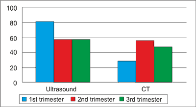
Fig. 1. Comparison of the methods for diagnostic imaging in appendicitis according to the trimester of the pregnancy.
Complications of appendicitis in pregnancy:
1. typical for appendicitis:
– perforation of the appendix,
– abscess/periappendical infiltration,
– acute peritonitis,
– wound infection,
– systemic septic complications,
– ileus,
– pneumonia;
2. associated with pregnancy:
– premature contractions,
– premature birth,
– low birth weight of the baby,
– intrauterine fetal death (44).
Algorithm for the management of pregnant patients with suspected appendicitis (13) (fig. 2).
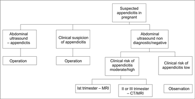
Fig. 2. Algorithm for the management of pregnant patients with suspected appendicitis.
Powyżej zamieściliśmy fragment artykułu, do którego możesz uzyskać pełny dostęp.
Mam kod dostępu
- Aby uzyskać płatny dostęp do pełnej treści powyższego artykułu albo wszystkich artykułów (w zależności od wybranej opcji), należy wprowadzić kod.
- Wprowadzając kod, akceptują Państwo treść Regulaminu oraz potwierdzają zapoznanie się z nim.
- Aby kupić kod proszę skorzystać z jednej z poniższych opcji.
Opcja #1
24 zł
Wybieram
- dostęp do tego artykułu
- dostęp na 7 dni
uzyskany kod musi być wprowadzony na stronie artykułu, do którego został wykupiony
Opcja #2
59 zł
Wybieram
- dostęp do tego i pozostałych ponad 7000 artykułów
- dostęp na 30 dni
- najpopularniejsza opcja
Opcja #3
119 zł
Wybieram
- dostęp do tego i pozostałych ponad 7000 artykułów
- dostęp na 90 dni
- oszczędzasz 28 zł
Piśmiennictwo
1. Wei PL, Keller JJ, Liang HH, Lin HC: Acute appendicitis and adverse pregnancy outcomes: a nationwide population-based study. J Gastrointest Surg 2012; 16(6): 1204-1211.
2. Andersen B, Nielsen TF: Appendicitis in pregnancy: diagnosis, management and complications. Acta Obstet Gynecol Scand 1999; 78: 758-762.
3. Mourad J, Elliott J, Erickson L et al.: Appendicitis in pregnancy: new information that contradicts longheld clinical beliefs. Am J Obstet Gynecol 2000; 182: 1027-1029.
4. Lemieux P, Rheaume P, Levesque I et al.: Laparoscopic appendectomy in pregnant patients: a review of 45 cases. Surg Endosc 2008 [Epub ahead of print]. DOI 10.1007/s00464-008-0201-9.
5. Mazze RI, Kallen B: Appendectomy during pregnancy: a Swedish registry study of 778 cases. Obstet Gynecol 1991; 77: 835-840.
6. Jung SJ, Lee DK, Kim J et al.: Appendicitis during Pregnancy: The Clinical Experience of a Secondary Hospital. Journal of the Korean Society of Coloproctology 06/2012; 28(3):152-159. DOI: 10.3393/jksc.2012.28.3.152.
7. Ueberrueck T, Koch A, Meyer L et al.: Ninety-four appendectomies for suspected acute appendicitis during pregnancy. World J Surg 2004; 28: 508-511.
8. Stone K: Acute abdominal emergencies associated with pregnancy. Clin Obstet Gynecol 2002; 45: 553-561.
9. Sharp HT: The acute abdomen during pregnancy. Clin Obstet Gynecol 2002; 45: 405-413.
10. Yilmaz HG, Akgun Y, Bac B et al.: Acute appendicitis in pregnancy – risk factors associated with principal outcomes: a case control study. Int J Surg 2007; 5: 192-197.
11. Lebeau R, Dianè B, Koffi E et al.: Appendicite aiguë et grossesse : à propos de 21 cas. J Gynecol Obstet Biol Reprod (Paris) 2005; 34: 600-605.
12. Cilo NB, Amini D, Landy HJ: Appendicitis and cholecystitis in pregnancy. Clin Obstet Gynecol 2009; 52(4): 586-596.
13. Freeland M, King E, Safcsak K, Durham R: Diagnosis of appendicitis in pregnancy. The American Journal of Surgery 2009; 198: 753-758.
14. McGory ML, Zingmond DS, Tillou A et al.: Negative appendectomy in pregnant women is associated with a substantial risk of fetal loss. J Am Coll Surg 2007; 205: 534-540.
15. Daniel M, Brent T, Cori-Ann M, Ryan T: Case report and management of suspected acute appendicitis in pregnancy. Hawaii Med J 2011; 70(2): 30-32.
16. Basaran A, Basaran M: Diagnosis of acute appendicitis during pregnancy: a systematic review. Obstet Gynecol Surv 2009; 64: 481-488 [quiz 499].
17. Pates JA, Avendiano TC, Zaretsky MV et al.: The appendix in pregnancy. Obstet Gynecol 2009; 114(4): 805-808.
18. Hodjati H, Kazerooni T: Location of the appendix in the gravid patient: a reevaluation of the established concept. Int J Gynecol Obstet 2003; 8: 245-247.
19. Carlin A, Alfirevic Z: Physiological changes of pregnancy and monitoring. Best Pract Res Clin Obstet Gynecol 2008; 22(5): 801-823.
20. Mourad J, Elliot JP, Erickson L, Lisboa L: Appendicitis in pregnancy: new information that contradicts long-held clinical beliefs. Am J Obstet Gynecol 2000; 182(5): 1027-1029.
21. Bretagnol F, Zappa M, Panis Y: Place de l’imagerie dans le diagnostic d’appendicite aiguë. J Chir (Paris) 2009; 146: 8-11.
22. Douglas CD, Macpherson NE, Davidson PM, Gani JS: Randomised controlled trial of ultrasonography in diagnosis of acute appendicitis, incorporating the Alvarado score. BMJ 2000; 321: 1-6.
23. Pirro N, Berdah SV: Appendicites: coelioscopie ou non? J Chir (Paris) 2006; 143: 3.
24. Lyass S, Pikarsky A, Eisenberg VH: Is laparoscopic appendicectomy safe in pregnant women? Surg Endo 2001; 15: 377-379.
25. Bisharah M, Tulandi T: Laparoscopic surgery in pregnancy. Clin Obstet Gynecol 2003; 46: 92-97.
26. Moreno-Sanz C, Pacual-Pedreno A, Picazo-Yeste JS et al.: Laparoscopic appendectomy during pregnancy: between personal experiences and scientific evidence. J Am Col Surg 2007; 205: 37-42.
27. Chinnusamy P, Muthumarmaran R, Ramakrishnan P: Laparoscopic appendicectomy in pregnancy: a case series of seven patients. J Soc Laparosc Surg 2006; 10: 321-325.
28. Sadot E, Telem DA, Arora M et al.: Laparoscopy: a safe approach to appendicitis during pregnancy. Surg Endosc 2010; 24: 383-389.
29. Bames SL, Shane MD, Shoemann MB et al.: Laparoscopic appendicectomy after 30 weeks pregnancy: report of two cases and description of technique. Am Surg 2004; 70: 733-736.
30. Yilmaz HG, Akgun Y, Bac B, Celik Y: Acute appendicitis in pregnancy: risk factors associated with principal outcomes: a case control study. Int J Surg 2007; 5: 192-197.
31. Nouira M, Jerbi M, Sahraoui W et al.: Appendicite aiguë chez la femme enceinte: à propos de 18 cas. Rev Fr Gynecol Obstet 1999; 94: 486-491.
32. Walsh CA, Tang T, Walsh SR: Laparoscopic versus open appendicectomy in pregnancy: a systematic review. Int J Surg 2008; 6(4): 339-344.
33. Savary D: Appendicitis in the pregnant woman: Be less afraid for the pregnancy than for the consequences of inaction! Journal of Visceral Surgery 2012; 149: e225-e226.
34. Miloudi N, Brahem M, Ben Abid S: Acute appendicitis in pregnancy: Specific features of diagnosis and treatment. Journal of Visceral Surgery 2012; 149: e275-e279.
35. Terasawa T, Blackmore CC, Bent S et al.: Systematic review: computed tomography and ultrasonography to detect acute appendicitis in adults and adolescents. Ann Intern Med 2004; 141: 537-546.
36. Lehnert BE, Gross JA, Linnau KF, Moshiri M: Utility of ultrasound for evaluating the appendix during the second and third trimester of pregnancy. Emerg Radiol 2012, doi:10.1007/s10140-012-1029-0.
37. Rosen MP, Ding A, Blake MA et al.: ACR appropriateness Criteria right lower quadrant pain-suspected appendicitis. J Am Coll Radiol 2011; 8(11): 749-755.
38. Pearl J, Price R, Richardson W, Fanelli R: Society of American gastrointestinal endoscopic surgeons. Guidelines for diagnosis, treatment, and use of laparoscopy for surgical problems during pregnancy. Surg Endosc 2011; 25(11): 3479-3492.
39. Chen MM, Coakley FV, Kaimal A, Laros RK Jr: Guidelines for computed tomography and magnetic resonance imaging use during pregnancy and lactation. Obstet Gynecol 2008; 112(2 Pt 1): 333-340.
40. Kennedy A: Assessment of acute abdominal pain in the pregnant patient. Semin Ultrasound CT MR 2000; 21: 64-77.
41. Lim HK, Bae SH, Seo GS: Diagnosis of acute appendicitis in pregnant women: value of sonography. Am J Roentgenol 1992; 159: 342-359.
42. Lazarus E, Mayo-Smith WW, Mainiero MB et al.: CT in the evaluation of non-traumatic abdominal pain in pregnant women. Radiology 2007; 244: 784-790.
43. Pedrosa I, Levine D, Eyvazzadeh A et al.: MR imaging evaluation of acute appendicitis in pregnancy. Radiology 2006; 238: 891-899.
44. Gilo NB, Amini D, Landy HJ: Appendicitis and Cholecystitis in Pregnancy. Clin Obstet Gynecol. 2009 Dec; 52(4): 586-596. Doi: 10.1097/GRF.0b013e3181c11d10.
45. Moreno-Sanz C, Pascual-Pedreño A, Picazo-Yeste JS et al.: Laparoscopic appendectomy during pregnancy: between personal experiences and scientific evidence. J Am Coll Surg 2007; 205: 37-42.
46. Azuar AS, Bouillet-Dejou L, Jardon K et al.: Laparoscopy during pregnancy: experience of the French university hospital of Clermont-Ferrand. Gynecol Obstet Fertil 2009; 37(7-8): 598-603.
47. Colomb S, Bonnin M, Bolandard F et al.: Pregnant woman anaesthetic management in gynaecologic laparoscopic surgery at the maternity hospital of Clermont-Ferrand. Ann Fr Anesth Reanim 2006; 25(1): 11-16.
48. Walsh CA, Walsh SR: Laparoscopic appendectomy during pregnancy: an evidence-based review. Surg Endosc 2009; 23(3): 671.
49. McGory ML, Zingmond DS, Tillou A et al.: Negative appendectomy in pregnant women is associated with a substantial risk of fetal loss. J Am Coll Surg 2007; 205(4): 534-540.
50. Reedy MB, Kallen B, Kuehl TJ: Laparoscopy during pregnancy: a study of five fetal outcome parameters with use of the Swedish health registry. Am J Obstet Gynecol 1997; 177(3): 673-679.
51. Colomb S, Bonnin M, Bolandard F et al.: Prise en charge anesthèsique de la femme enceinte pour coeliochirurgie gynècologique non obstètricale à la maternitè de Clermont-Ferrant. Ann Fr Anesth Reanim 2006; 25: 11-16.
52. Pearl J, Price R, Richardson W, Fanelli R: Guidelines for diagnosis, treatment, and use of laparoscopy for surgical problems during pregnancy. Surg Endosc 2011; 25: 3479-3492.
53. Halvorsen AC, Brandt B, Andreasen JJ: Acute appendicitis in pregnancy: complications and subsequent management. Eur J Surg 1992; 158: 603-606.