© Borgis - Postępy Nauk Medycznych 12/2014, s. 835-837
Małgorzata Gietka-Czernel1, Marzena Dębska2, Piotr Kretowicz2, Romuald Dębski2
Guz szyi u płodu wymagający różnicowania z wolem
Fetal neck tumor – differentiation from thyroid goiter
1Department of Endocrinology, Centre of Postgraduate Medical Education, Warszawa
Head of Department: prof. Wojciech Zgliczyński, MD, PhD
2Department of Obstetrics and Gynecology, Centre of Postgraduate Medical Education, Warszawa
Head of Department: prof. Romuald Dębski, MD, PhD
Streszczenie
Przedstawiamy przypadek płodu z guzem szyi wykrytym w 22. tygodniu ciąży w trakcie rutynowego badania ultrasonograficznego. Guz wymagał różnicowania pomiędzy wolem, potworniakiem, nerwiakiem niedojrzałym, torbielą przewodu tarczowo-językowego, torbielakiem surowiczym i naczyniakiem. Badanie ultrasonograficzne wykazało litotorbielowaty charakter guza, umiarkowane unaczynienie, gwałtowny wzrost, odgięcie głowy, ucisk przełyku, a w konsekwencji wielowodzie. Obydwoje rodzice byli zdrowi, nie przyjmowali żadnych leków, ocenione stężenia TSH, fT4 i fT3 były prawidłowe, przeciwciała tarczycowe negatywne, obraz ultrasonograficzny tarczycy nie wykazywał nieprawidłowości. Wykonano kordocentezę i stwierdzono prawidłową czynność hormonalną tarczycy płodu, co ostatecznie wykluczyło obecność wola. Obserwowane gwałtowne powiększanie się guza oraz jego obraz ultrasonograficzny nasuwały podejrzenie potworniaka. Nastąpiła terminacja ciąży, a badanie histopatologiczne potwierdziło obecność potworniaka.
Summary
Fetal anterior neck tumor is a rare condition and differential diagnosis includes goiter, teratoma, thyroglossal duct cyst, cystic hygroma, branchial cleft cyst, lymphangioma/hemangioma and neuroblastoma. The case of fetal anterior cervical tumor was diagnosed on routine ultrasonographic assessment on the 22nd week of gestation. Tumor was mixed solid and cystic with moderate vascularization, exhibited rapid growth causing neck hyperextension, esophageal compression and polyhydramnion. The both parents were healthy, their serum thyrotropin, thyroid hormones results and thyroid ultrasonography were normal, antithyroid antibodies negative. There was no history of excessive iodine intake by the mother. Fetal blood sampling was performed and normal fetal thyroid function was documented. Thus the diagnosis of fetal goiter was rejected. The tumor course and its ultrasonographic presentation indicated cervical teratoma. Pregnancy was terminated because of the poor prognosis for the fetus and diagnosis of teratoma was confirmed by pathological examination.

Introduction
Ultrasonographic (US) assessment of the fetus is routinely recommended between 11-14, 18-23 and 30-33 week of gestation. We present a case of a fetus with cervical tumor diagnosed in the 22nd week of gestation which was differentiated from thyroid goiter.
Case study
Fetal tumor localized in left anterior part of the neck was diagnosed on routine US assessment. Its size at the time of diagnosis was 54 x 50 x 47 mm, it was mostly solid with small cystic areas, with moderate vascularization and no calcifications. Normal fetal thyroid was not visible (fig. 1-3). Both parents were healthy with no family nor personal history of thyroid diseases, they had no palpable goiter and they did not take any medications. The mother had no history of excessive iodine intake nor medical procedures with iodine contrast media prior to the pregnancy. Parental serum thyrotropin (TSH), free T3 (fT3), free T4 (fT4) were within normal range, thyroid-peroxidase antibodies (TPOAbs), thyroglobulin antibodies (TGAbs) and TSH receptor antibodies (TRAbs) were negative and their thyroids US performance was normal. Cordocentesis was performed to evaluate fetal thyroid function and exclude fetal goiter. The results of the fetal blood examination were within the normal range for gestational age: TSH 8.4 mU/L (normal: 2.0-9.5 mU/L), fT4 – 8.38 pmol/l (normal: 3.0-9.0 pmol/L), fT3 < 1 pmol/L (normal: < 1.0 pmol/L) (1), TPOAb – 15.6 IU/mL (normal: < 60 IU/mL), TgAb – < 20 IU/mL (normal: < 60 IU/mL), TRAb – 0.8 IU/mL (normal: < 1.8 IU/mL). In the next US examination performed one week later rapid tumor growth was observed: its size enlarged to 68 x 62 x 56 mm causing significant hyperextension of the fetal neck and polyhydramnios resulting from esophageal obstruction. Fetal cervical teratoma was suspected and pregnancy was terminated because of the poor prognosis for the fetus. The pathological examination confirmed initial diagnosis: the tumor consisted of cartilaginous, intestinal and neural tissue, it adhered strictly to the tissues of the oral cavity, compressed the throat and larynx causing lesion of the jaw.

Fig. 1. Fetal examination on 2-dimensional ultrasonography. Giant anterior neck tumor with head hyperextension and polyhydramnion.
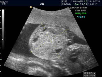
Fig. 2. Fetal neck tumor on 2-dimensional ultrasonography.
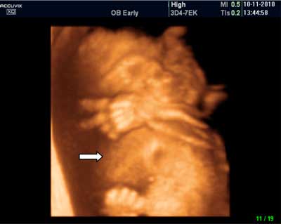
Fig. 3. Fetal examination on 3-dimensional ultrasonography. Giant anterior neck tumor causing head deviation.
Discussion
Powyżej zamieściliśmy fragment artykułu, do którego możesz uzyskać pełny dostęp.
Mam kod dostępu
- Aby uzyskać płatny dostęp do pełnej treści powyższego artykułu albo wszystkich artykułów (w zależności od wybranej opcji), należy wprowadzić kod.
- Wprowadzając kod, akceptują Państwo treść Regulaminu oraz potwierdzają zapoznanie się z nim.
- Aby kupić kod proszę skorzystać z jednej z poniższych opcji.
Opcja #1
24 zł
Wybieram
- dostęp do tego artykułu
- dostęp na 7 dni
uzyskany kod musi być wprowadzony na stronie artykułu, do którego został wykupiony
Opcja #2
59 zł
Wybieram
- dostęp do tego i pozostałych ponad 7000 artykułów
- dostęp na 30 dni
- najpopularniejsza opcja
Opcja #3
119 zł
Wybieram
- dostęp do tego i pozostałych ponad 7000 artykułów
- dostęp na 90 dni
- oszczędzasz 28 zł
Piśmiennictwo
1. Thorpe-Beston JG, Nicolaides KH, McGregor AM: Fetal thyroid function. Thyroid 1992; 2: 207-217.
2. Luton D, Le Gac I, Vuillard E et al.: Management of Graves’ disease during pregnancy: the key role of fetal thyroid monitoring. J Clin Endocrinol Metab 2005; 90: 6093-6098.
3. Peleg D, Cada S, Peleg A, Ben-Ami M: The relationship between maternal serum thyroid-stimulating immunoglobulin and fetal and neonatal thyrotoxicosis. Obstet Gynecol 2002; 99: 1040-1043.
4. Zimmerman D: Fetal and neonatal hyperthyroidism. Thyroid 1999; 9: 727-733.
5. Conelly KJ, Boston BA, Pearce EN et al.: Congenital hypothyroidism caused by excess prenatal maternal iodine ingestion. J Pediatr 201; 161: 760-762.
6. Medeiros-Neto G, Bunduki V, Tomimori E et al.: Prenatal diagnosis and treatment of dyshormonogenetic fetal goiter due to defective thyroglobulin synthesis. J Clin Endocrinol Metab 1997; 82: 4239-4242.
7. Luton D, Fried D, Sibony O et al.: Assessment of fetal thyroid function by colored Doppler echography. Fetal Diagn Ther 1997; 12: 24-27.
8. Agarwal P, Ogilvy-Stuart A, Lees C: Intrauterine diagnosis and management of congenital goitrous hypothyroidism. Ultrasound Obstet Gynecol 2002; 19: 501-505.
9. Van Loon AJ, Derksen JTM, Bos A et al.: In utero diagnosis and treatment of fetal goitrous hypothyroidism, caused by maternal use of propylthiouracil. Prenat Diagn 1995; 15: 599-604.
10. Cohen O, Pinhas-Hamiel O, Sivan E et al.: Serial in utero ultrasonographic measurements of the fetal thyroid: a new complementary tool in the management of maternal hyperthyroidism in pregnancy. Prenat Diagn 2003; 23: 740-742.
11. Nahum Z, Rakower Y, Weiner E et al.: Graves’ disease in pregnancy: prospective evaluation of a selective invasive treatment protocol. Am J Obstet Gynecol 2003; 189: 159-165.
12. Sichel JY, Eliashar R, Yatsiv I et al.: A multidisciplinary team approach for management of a giant congenital cervical teratoma. Int J Pediatr Otorhinolaryngol 2002; 65(3): 241-247.
13. Vujanic GM, Harach HR, Minic PB, Vuckovic N: Thyroid/cervical teratoma in infancy. Immunohisto-chemical studies for specific thyroid epithelial cell markers. Pediatr Develop Pathol 1994; 14: 369-375.