© Borgis - New Medicine 3/2014, s. 92-94
*Konrad Wroński1, 2
Inflammatory breast cancer – case report and review of literature
1Department of Oncology, Faculty of Medicine, University of Warmia and Mazury, Olsztyn, Poland
Head of Department: prof Sergiusz Nawrocki, MD, PhD
2Department of Surgical Oncology, Hospital Ministry of Internal Affairs with Warmia and Mazury Oncology Centre, Olsztyn, Poland
Head of Department: Andrzej Lachowski, MD
Summary
Inflammatory breast cancer is rare breast neoplasm. Because of its rarity, this cancer is often misdiagnosed as generalized dermatitis or mastitis. It is characterized by an aggressive behavior and rapid progression. Five year survival times have increased due to multimodal therapy but they are still much lower than for other breast cancers.
The author of this article presented a case of a 40-year-old Caucasian woman who was admitted to the hospital because of advanced inflammatory breast cancer. The progression of disease was very fast and the patient died before the start of chemotherapy treatment. The author performed a literature review on inflammatory breast cancer diagnosis and treatment.
INTRODUCTION
Inflammatory breast cancer is rare breast neoplasm (1-3). Because of its rarity, this cancer is often misdiagnosed as generalized dermatitis or mastitis (4). It is characterized by an aggressive behavior and rapid progression (2, 4, 5). Five year survival times have increased due to multimodal therapy but they are still much lower than for other breast cancers (1-4).
CASE REPORT
A 40-year-old Caucasian woman was referred to the Department of Surgical Oncology due to swelling and redness of the right breast of two months (fig. 1, 2). For a month she reported increasing swelling of the right half of the face and redness with palpable nodules on the right side of the neck (fig. 3, 4). She reported general weakness and malaise. In the interview, the patient reported no fever. According to the patient the rate of change was very fast.

Fig. 1. Edema and redness of the right breast for 2 months in a 40-year-old Caucasian woman.

Fig. 2. Edema and redness of the right breast.
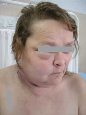
Fig. 3. The patient reported for a month increasing swelling of the right half of the face and redness with palpable nodules on the right side of the neck.
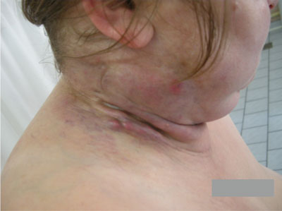
Fig. 4. Redness and enlarge palpable lymph nodes on the right side of the neck.
She had no any other symptoms, there was no history of weight loss and loss of appetite. The patient was not treated for chronically diseases. She had no surgeries and there was no history of carcinoma in patient family. Blood test and other routine hematological examinations and biochemical tests were within normal limits.
In the Department of Surgical Oncology the patient had open surgical biopsy of right breast and enlarged lymph nodes of the right side neck. After biopsy due to condition of the patient, she was transferred for further treatment to a Department of Chemotherapy. Four days after the biopsy, the patient due to the progression of cancer died. The family did not give permission for an autopsy.
Histopathology examination of material showed inflammatory breast cancer and metastases of this cancer into right lymph nodes of the neck.
DISCUSSION
Inflammatory breast carcinoma characterized by rapid progression, therefore accurate diagnosis is very important (1,2). Clinical signs and symptoms of primary inflammatory breast cancer are characteristic for benign breast disease as dermatitis and mastitis (4). The most frequent changes associated with inflammatory breast cancer is erythema (1-4). Sometimes, the erythema is associated with a sensation of heat in the affected breast (1-4). The edema is associated with a characteristic orange peel. The color of breast may be from pinkish to dark red or purple (2-4). It is important to know that despite such symptoms as the erythema and the edema there are not present fever, leukocytosis and localized pain (5).
In majority of patients with inflammatory breast carcinoma there are not palpable tumors during clinical examination. Majority of patients present metastases to the axillary or supraclavicular lymph nodes that may be detected during palpable examination (5).
Dynamic contrast-enhanced (DCE) magnetic resonance imaging (MRI) is the most accurate for detecting primary breast cancer (6, 7). This test may show the presence of angiogenesis in malignant tumor, describe staging and control response to therapy in inflammatory breast cancer (6, 7). Second important imaging technique is positron emission tomography (PET) (8). This test is sensitive and show metabolic changes in breast tissues and distatnt metastases at staging (8). Mammography in inflammatory breast cancer is not successful test. This examination shows breast masses or architectural distortion (9). Ultrasonography may show delineating focal breast parenchymal lesions and subcutaneous edema in breast where is located inflammatory breast cancer (9). Ultrasonography control regional lymph nodes and may affect locoregional therapeutic planning (9).
Surgery plays important role in multimodal therapy in inflammatory breast cancer. To optimally surgically treated patients suffer from inflammatory breast cancer it is necessary neoadjuvant chemotherapy (10, 11). Mastectomy with complete axillary lymph node dissection is gold standard in this disease (10, 11). Sentinel lymph node biopsy is not recommended for patients with inflammatory breast cancer. From 55% to 85% patients with inflammatory breast cancer have metastases in axillary lymph nodes (5). Most patients who required radiation therapy had neoadjuvant chemotherapy and then surgery (12, 13). Radiation therapy has as a target axillary, infraclavicular, supraclavicular and internal mammary lymph nodes and also the chest wall (13).
The treatment of inflammatory breast cancer need multimodal therapy. So that it is important to treat women in high specialized oncological centers.
CONCLUSIONS
1. Inflammatory breast cancer is rare neoplasm.
2. It is often misdiagnosed as generalized dermatitis or mastitis.
3. It is characterized by an aggressive behavior and rapid progression.
4. Five year survival times have increased due to multimodal therapy but they are still much lower than for other breast cancers.
Piśmiennictwo
1. Hance KW, Anderson WF, Devesa SS et al.: Trends in inflammatory breast carcinoma incidence and survival: the surviveillance, epidemiology and results program at the National Cancer Institute. J Natl Cancer Inst 2005; 97; 966-975. 2. Dawood S, Cristofanilli M: Inflamatory breast cancer: what progress have we made? Oncology 2011; 25(3): 264-270. 3. Yang WT, Le-Petross HT, Macapinlac H et al.: Inflammatory breast cancer: PET/CT, MRI, mammography and sonography findings. Breast Cancer Res Treat 2008; 109(3): 417-426. 4. Dawood S, Cristofanilli M: What progress have we made in imaging inflammatory breast cancer? Oncology 2007; 21: 673-679. 5. Walshe JM, Swain SM: Clinical aspects of inflammatory breast cancer. Breast Dis 2005; 22: 35-44. 6. Chow CK: Imaging in inflammatory breast carcinoma. Breast Dis 2005; 22: 45-54. 7. Lee KW, Chung SY, Yang I et al.: Inflammatory breast cancer: imaging findings. Clin Imaging 2005; 29: 22-25. 8. Eubank WB, Mankoff DA: Evolving role of positron emission tomography in breast cancer imaging. Semin Nucl Med 2005; 35: 84-99. 9. Yang WT, Le-Petross HT, Macapinlac H et al.: Inflammatory breast cancer: PET/CT, MRI, mammography, and sonography findings. Breast Cancer Res Treat 2008; 109: 417-426. 10. Curcio LD, Rupp E, Williams WL et al.: Beyond palliative mastectomy in inflammatory breast cancer – a reassessment of margin status. Ann Surg Oncol 1999; 6:249-254. 11. Bristol IJ, Woodward WA, Strom EA et al.: Locoregional treatment outcomes after multimodality management of inflammatory breast cancer. Int J Radiat Oncol Biol Phys 2008; 72: 474-484. 12. Thomas WW Jr, McNeese MD, Fletcher GH et al.: Multimodal treatment for inflammatory breast cancer. Int J Radiat Oncol Biol Phys 1989; 17: 739-745. 13. Arthur DW, Schmidt-Ullrich RK, Friedman RB et al.: Accelerated superfractionated radiotherapy for inflammatory breast carcinoma: complete response predicts outcome and allows for breast conservation. Int J Radiat Oncol Biol Phys 1999; 44: 289-296.