*Konrad Wroński1, Zbigniew Masłowski2, Przemysław Stefaniak2, Leszek Frąckowiak3
Treatment Merkel cell carcinoma of the facial skin – case report
1Department of Oncology, Faculty of Medicine, University of Warmia and Mazury, Olsztyn, Poland
Head of Department: prof. Sergiusz Nawrocki, MD, PhD
2Department of Surgical Oncology, Hospital Ministry of Internal Affairs with Warmia and Mazury Oncology Centre, Olsztyn, Poland
Head of Department: Andrzej Lachowski, MD
3Department of Public Health and Epidemiology, University of Warmia and Mazury, Olsztyn, Poland
Head of Department: prof. Anna Abramczyk, MD, PhD
Summary
Merkel cell carcinoma (MCC) is a rare neuroendocrine malignancy of the skin. It is highly aggressive skin cancer which is observed in patients older than 50 years (more than 95%). Merkel cell carcinoma was first time described by Toker in 1972. Merkel cell carcinoma often presents a rapid growth, reddish-blue dermal papule and can be asymptomatic. Surgery and radiotherapy are the most important in treatment Merkel cell carcinoma. Up to 48% of Merkel cell carcinomas are located in the head and neck region. Recommendations for surgery are based on the clinical size of the primary tumor. Excision margins for tumors < 2 cm should be 1 cm, and for tumors > 2 cm the margins should be 2 cm. Sentinel lymph node biopsy is recommended for all untreated primary tumors at the time of wide local excision. SLN biopsy is important in the staging and prognosis of Merkel cell carcinoma. In this article the authors presented a case of a 60-year-old man, Caucasian race, who was admitted to the hospital because of Merkel cell carcinoma of the facial skin. The patient underwent wide excision of the tumor and in the sixth day after surgery was discharged home.
INTRODUCTION
Merkel cell carcinoma (MCC) is a rare neuroendocrine malignancy of the skin (1, 2). It is highly aggressive skin cancer which is observed in patients older than 50 years (more than 95%) (1, 3). The cell of origin was first time described in 1875 by Friedrich Merkel as epithelial in derivation with neuroendocrine differentiation (3). Merkel cell carcinoma was first time described by Toker in 1972 (3). Merkel cell carcinoma often presents a rapid growth, reddish-blue dermal papule and can be asymptomatic. Surgery and radiotherapy are the most important in treatment Merkel cell carcinoma (1, 2).
CASE REPORT
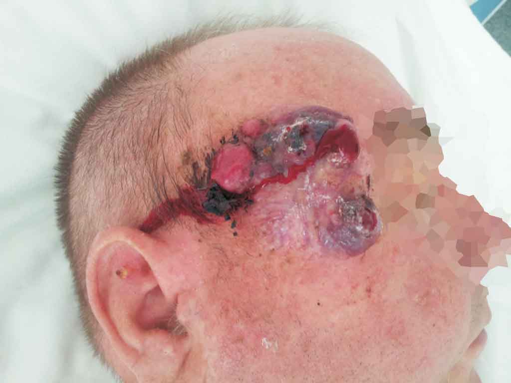
A 60-year-old man, Caucasian race, was admitted to the Department of Surgical Oncology because of Merkel cell carcinoma of the facial skin (fig. 1 and 2). In an interview with the patient, he informed us that the tumor of the facial skin was observed six months earlier and gradually expanded. The patient reported pain in the area of the tumor. Surgical biopsy showed Merkel cell carcinoma.

Fig. 1. Merkel cell carcinoma of the facial skin.

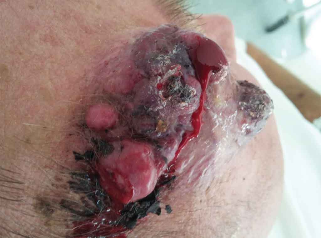
Fig. 2. Bleeding Merkel cell carcinoma of the facial skin.
He had no any other symptoms, there was no history of weight loss and loss of appetite. The patient was not treated chronically diseases. He had no surgeries before and there was no history of carcinoma in patient family. Blood test and other routine hematological examinations and biochemical tests were within normal limits.
On physical examination, there was bleeding tumor size 55 x 65 millimeters. Regional lymph nodes were not enlarged. The patient was qualified for surgery. In general anesthesia has been wide tumor resection of the right temporal area with adequate free margins and free graft skin was taken from the supraclavicular region. The tumor was collected for histopathological examination. Histopathological examination revealed Merkel cell carcinoma. The tumor was excised in its entirety. The duration of surgery was 65 minutes. Patient after surgery felt good and did not complain of pain. The postoperative period was uncomplicated and the patient left the ward in the sixth day after surgery. The patient is in the care of outpatient surgical oncology.
DISCUSSION
Merkel cell carcinoma is rare neoplasm with an approximate incidence of 2.4 cases per 100.000 person-years (1, 4). In the last two decades the number of newly reported cases has increased. UV radiation and immunosuppression play important role in pathogenesis of Merkel cell carcinoma (5). In 2008, Feng et al described a novel polyomavirus (MCPyV) which can play role in viral tumorigenesis (6, 7).
Merkel cell carcinoma tumors clinically present as asymptomatic, solitary nodules with pink or red coloring and may have ulceration or telangiectasia (8, 9). Up to 48% of Merkel cell carcinomas are located in the head and neck region (2).
Surgery and radiotherapy are the most important in treatment Merkel cell carcinoma (1, 2). Recommendations for surgery are based on the clinical size of the primary tumor. Excision margins for tumors < 2 cm should be 1 cm, and for tumors > 2 cm the margins should be 2 cm (10, 11). Sentinel lymph node biopsy is recommended for all untreated primary tumors at the time of wide local excision. SLN biopsy is important in the staging and prognosis of Merkel cell carcinoma. SLN biopsy in the head and neck region can be technically challenging but is also recommended. Merkel cell carcinoma is a radiosensitive neoplasm. It means that radiotherapy plays important role as an adjunct to surgery and as primary therapy in inoperable cases or when the patient refuses surgery (10, 11). The role of chemotherapy is still unclear and is consider as palliative treatment in cases of disseminated disease (12).
National Comprehensive Cancer Network suggest that the patient after treatment Merkel cell carcinoma should undergo whole skin examination and regional lymph nodes examination every 1 to 3 months for the first year and every 3 to 6 months for the second year and annually thereafter.
The authors of this article, having an experience in the treatment of patients with Merkel cell carcinoma believes, that good surgery is „the gold standard” in the treatment of such patients. The authors of this article believes that patients with such rare and an extremely aggressive cutaneous malignancy should be treated only in highly specialized oncological surgery departments.
CONCLUSIONS
1. Merkel cell carcinoma is an extremely aggressive cutaneous malignancy.
2. Radical surgical resection and radiotherpy is the treatment of choice.
3. The role of chemotherapy is still unclear and is consider as palliative treatment.
4. SLN biopsy is recommended in this skin malignancy.
5. Warthin’s tumors should be treated in highly specialized oncological surgery departments.
Piśmiennictwo
1. Lemos B, Nghiem P: Merkel cell carcinoma: more deaths but still no pathway to blame. J Investig Dermatol 2007; 127: 2100-2103. 2. Lemos BD, Storer BR, Iyer JG et al.: Pathologic nodal evaluation improves prognostic accuracy in merkel cell carcinoma: analysis of 5823 cases as the basis of the first consensus staging system. J Am Acad Dermatol 2010; 63: 751-761. 3. Toker C: Trabecular carcinoma of the skin. Arch Dermatol 1972; 105: 107-110. 4. Agelli M, Clegg LX: Epidemiology of primary Merkel cell carcinoma in the United States. J Am Acad Dermatol 2003; 49: 832-841. 5. Miller RW, Rabkin CS: Merkel cell carcinoma and melanoma: etiology similarities and differences. Cancer Epidemiol Biomarkers Prev 1999; 8: 485. 6. Feng H, Shuda M, Chang Y et al.: Clonal integration of a polyomavirus in human Merkel cell carcinoma. Science 2008; 319: 1096-1100. 7. Shuda M, Feng H, Kwun HJ et al.: T antigen mutations are a human tumor-specific signature for Merkel cell polyomavirus. Proc Natl Acad Sci USA 2008; 105: 16272-16277. 8. Heath M, Jaimes N, Lemos B et al.: Clinical characteristics of merkel cell carcinoma at diagnosis in 195 patients: The AEIOU features. J Am Acad Dermatol 2008; 58: 375-381. 9. Kivela T, Tarkkanen A: The Merkel cell and associated neoplasms in the eyelids and periocular region. Surv Ophthalmol 1990; 35: 171-187. 10. Mortier L, Mirabel X, Fournier C et al.: Radiotherapy alone for primary Merkel cell carcinoma. Arch Dermatol 2003; 139: 1587-1590. 11. Veness M, Foote M, Gebski V et al.: The role of radiotherapy alone in patients with Merkel cell carcinoma: Reporting the Australian experience of 43 patients. Int J Radiat Oncol Biol Phys 2010; 78: 703-709. 12. Garneski KM, Nghiem P: Merkel cell carcinoma adjuvant therapy: current data support radiation but not chemotherapy. J Am Acad Dermatol 2007; 57: 166-169.