© Borgis - Postępy Nauk Medycznych 6/2017, s. 299-303
*Magdalena Kwiatkowska1, 2, Baljinder Dhinsa2, Anant Mahapatra2, Jarosław Czubak1
Does the surgery time affect the final outcome of type III supracondylar humeral fractures?
Ocena wyników leczenia złamań nadkłykciowych kości ramiennej typu III w zależności od czasu podjętej interwencji chirurgicznej
1Department of Orthopaedics, Pediatric Orthopaedics and Traumatology, Centre of Postgraduate Medical Education, Gruca Teaching Hospital, Otwock
Head of Department: Associate Professor Jarosław Czubak, MD, PhD
2Department of Orthopedics, Our Lady of Lourdes Hospital, Drogheda, Co. Louth, Ireland
Head of Department: Alan Walsh MBBS, MSc. (Orth), FRCSI, FRCS (Ed)
Streszczenie
Wstęp. Złamania nadkłykciowe są jednym z najczęstszych złamań okresu dziecięcego wymagających interwencji chirurgicznej. Stanowią około 60-70% urazów okolicy łokcia u pacjentów w wieku od 5 do 7 lat. Złamania te są klasyfikowane według opisu Gartlanda jako typ I, II, i III i najczęściej obserwowany jest typ złamania wyprostnego. Jak dotąd brak jest jednoznacznych wytycznych dotyczących czasu podjęcia interwencji chirurgicznej w przypadku wystąpienia złamania typu III bez uszkodzeń nerwowo-naczyniowych. Wielu badaczy uznaje konieczność jak najszybszej interwencji chirurgicznej, zanim narastający obrzęk zwiększy ryzyko śródoperacyjnych powikłań takich jak uszkodzenia nerwów oraz ryzyko rozwoju zespołu ciasnoty przedziałów powięziowych. Celem badania była analiza sposobu leczenia złamań nadkłykciowych oraz ocena wyniku leczenia w zależności od czasu podjętej interwencji chirurgicznej.
Materiał i metodyka. Badanie przeprowadzono w sposób retrospektywny. Analizie poddano 116 historii chorób oraz badań radiologicznych pacjentów hospitalizowanych w okresie od stycznia 2014 do grudnia 2015 z powodu złamania nadkłykciowego, z czego 23 z powodu złamania nadkłykciowego typu III. Pacjenci byli obserwowani klinicznie i radiologicznie przez 12 miesięcy od okresu złamania. Pacjentów podzielono na 3 grupy: grupa pierwsza – czas oczekiwania na zabieg operacyjny poniżej 6 godzin, grupa druga – czas oczekiwania pomiędzy 6 a 12 godzin i grupa 3 – czas oczekiwania powyżej 12 godzin.
Ocenę kliniczną przeprowadzono w oparciu o kryteria proponowane przez Flynna. W ramach oceny radiologicznej dokonano pomiarów kąta Bowmana na ostatnim RTG kontrolnym.
Wyniki. Średni wiek pacjentów wynosił 6 lat i 2 miesiące (od 3 lat i 5 miesięcy do 10 lat i 7 miesięcy), w tym 13 chłopców i 10 dziewcząt. Średni okres od przyjęcia do czasu rozpoczęcia zabiegu operacyjnego wynosił 14 godzin i 28 minut (od 2 godzin i 12 minut do 20 godzin i 23 minut).
Siedmiu z 23 pacjentów wymagało otwartej repozycji złamania. Nie zaobserwoawano powikłań klinicznych i radiologicznych oraz zaburzeń zrostu w populacji zbadanych pacjentów.
Wnioski. Czas podjętej interwencji chirurgicznej nie miał wpływu na ostateczny wynik lecznia pacjentów ze złamaniem nadkłykciowym kości ramiennej typu III niezależnie, czy wymagało ono zamkniętej czy otwartej repozycji.
Wynik badania sugeruje, że pacjenci hospitalizowani z powodu złamania nadkłykciowego typu III, u których nie obserwuje się deficytu nerwowo-naczyniowego, mogą być chirurgicznie zaopatrzeni w okresie 12 godzin bez znaczącego wpływu na ostateczny wynik leczenia.
Summary
Introduction. Supracondylar humeral fractures are common in the pediatric population, with displaced fractures requiring operative intervention.
Aim. The purpose of this study was to look at our practice and assess whether a difference in clinical outcomes and requirement for open reduction was observed if surgery was delayed.
Material and methods. This was a retrospective medical record and plain radiograph review of patients admitted with type III Gartland supracondylar fractures between January 2014 and December 2015. The patients were seen for up to 12 months postoperatively, and clinical assessment was performed at this stage.
Results. There were 116 supracondylar humeral fractures admitted between January 2014 and December 2015, 23 of which were Gartland type III. The mean age of the patients was 6 years, and the mean time from emergency department presentation to surgery was 14 hours. Seven of the 23 patients required conversion to open reduction. There were no reported complications and all fractures demonstrated radiographic union. The length of time to surgery did not increase the number of cases requiring open reduction. The results demonstrated that there was no difference in clinical outcomes found between those that had closed manipulation or required conversion to open reduction, nor the time taken to surgery.
Conclusions. This study suggest that patients who present with type III supracondylar humeral fractures and have no neurovascular deficit, a delay in pinning of 12 hours or more may not result in a significant difference in the need for open reduction or clinical outcomes.
Level of Evidence IV.

Introduction
Supracondylar fractures of the humerus (SCHF) are the most common pediatric elbow injury, accounting for approximately 60 to 70% of all elbow fractures in patients aged between five and seven years of age (1, 2). Extension type supracondylar fractures are the most frequently seen (3-6). These factures were classified into three types by Gartland (7); type I is undisplaced, type II is displaced with an intact posterior cortex, and type III is displaced without cortical contact. Type I fractures are typically treated non-operatively; whilst some type II and almost all type III fractures usually require surgical intervention. This may be closed reduction or open reduction with pinning (1, 2, 8).
There remains a lack of consensus regarding the need for emergent surgical intervention of type III fractures without neurovascular compromise. With many hospitals having a protected daytime trauma theatre list, the demand for out of hours operating has decreased. Despite this, many surgeons advocate emergent surgery before swelling increases the risk of intraoperative complications (such as iatrogenic nerve injury and compartment syndrome) and failure of closed reduction necessitating open reduction. The reported risk of neurovascular injury is as high as 49% (7).
There have been a number of studies from trauma centres reporting no difference in perioperative complications, need for open reduction or clinical results between those that had emergent surgery and those operated on after eight hours post-injury (6, 9-11). A further two studies (12, 13), concurred there was no difference in the risk of perioperative complications between these groups; however, they reported an increased requirement for open reduction in the delayed surgery group.
Aim
This study was proposed to evaluate the current practice in a hospital that is not a major trauma centre, and whether there was a difference in outcomes and requirement for open reduction if surgery was delayed.
Material and methods
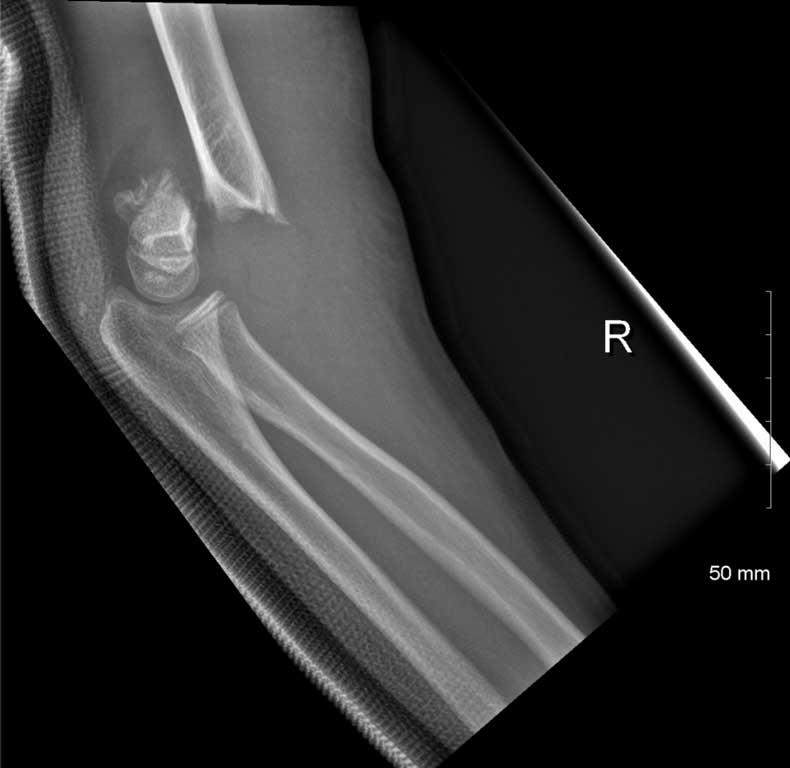
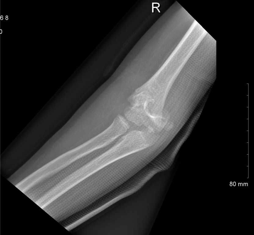
This was a retrospective review of pediatric patients admitted for surgical management of Gartland type III SCHF between January 2014 and December 2015 at Our Lady Of Lourdes Hospital in Drogheda, Ireland. The medical records and plain radiographs were reviewed. Inclusion criteria was a plain radiograph demonstrating a type III SCHF (fig. 1, 2), that this was an isolated injury, there were no open wounds, no neurovascular injury and patients were under sixteen years of age.

Fig. 1. Lateral plain radiograph demonstrating a supracondylar fracture

Fig. 2. Anterior-posterior plain radiograph of a supracondylar fracture
The authors documented sex, side affected, age at time of injury, time from emergency department to theatre, as well as radiographic and clinical assessment at last follow up. Clinical assessment was graded according to criteria described by Flynn et al. (14); with functional restriction based on the flexion-extension arc of movement and appearance judged on the change in carrying angle compared to the contralateral side (tab. 1).
Tab. 1. Criteria for evaluating clinical outcome (14)
| Results | Scaling | Esthetic factor: loss
of angle of alignment (degrees) | Functional factor: loss of movement (degrees) |
| Satisfactory | Excellent | 0-5 | 0-5 |
| Good | 6-10 | 6-10 |
| Moderate | 11-15 | 11-15 |
| Unsatisfactory | Poor | > 15 | > 15 |
Powyżej zamieściliśmy fragment artykułu, do którego możesz uzyskać pełny dostęp.
Mam kod dostępu
- Aby uzyskać płatny dostęp do pełnej treści powyższego artykułu albo wszystkich artykułów (w zależności od wybranej opcji), należy wprowadzić kod.
- Wprowadzając kod, akceptują Państwo treść Regulaminu oraz potwierdzają zapoznanie się z nim.
- Aby kupić kod proszę skorzystać z jednej z poniższych opcji.
Opcja #1
24 zł
Wybieram
- dostęp do tego artykułu
- dostęp na 7 dni
uzyskany kod musi być wprowadzony na stronie artykułu, do którego został wykupiony
Opcja #2
59 zł
Wybieram
- dostęp do tego i pozostałych ponad 7000 artykułów
- dostęp na 30 dni
- najpopularniejsza opcja
Opcja #3
119 zł
Wybieram
- dostęp do tego i pozostałych ponad 7000 artykułów
- dostęp na 90 dni
- oszczędzasz 28 zł
Piśmiennictwo
1. Omid R, Choi PD, Skaggs DL: Supracondylar humeral fractures in children. J Bone Joint Surg Am 2008; 90(5): 1121-1132.
2. Otsuka NY, Kasser JR: Supracondylar fractures of the humerus in children. J Am Acad Orthop Surg 1997; 5(1): 19-26.
3. Carmichael KD, Joyner K: Quality of reduction versus timing of surgical intervention for pediatric supracondylar humerus fractures. Orthopedics 2006; 29(7): 628-632.
4. Mallo G, Stanat SJC, Gaffney J: Use of the Gartland classification system for treatment of pediatric supracondylar humerus fractures. Orthopedics 2010; 33(1): 19.
5. Babal JC, Mehlman CT, Klein G: Nerve injuries associated with pediatric supracondylar humeral fractures: a meta-analysis. J Pediatr Orthop 2010; 30(3): 253-263.
6. White L, Mehlman CT, Crawford AH: Perfused, pulseless, and puzzling: a systematic review of vascular injuries in pediatric supracondylar humerus fractures and results of a POSNA questionnaire. J Pediatr Orthop 2010; 30(4): 328-335.
7. Gartland JJ: Management of supracondylar fractures of the humerus in children. Surg Gynecol Obstet 1959; 109(2): 145-154.
8. Ozkoc G, Gonc U, Kayaalp A et al.: Displaced supracondylar humeral fractures in children: open reduction vs. closed reduction and pinning. Arch Orthop Trauma Surg 2004; 124(8): 547-551.
9. Iyengar SR, Hoffinger SA, Townsend DR: Early versus delayed reduction and pinning of type III displaced supracondylar fractures of the humerus in children: a comparative study. J Orthop Trauma 1999; 13: 51-55.
10. Sibinski M, Sharma H, Bennet GC: Early versus delayed treatment of extension type-3 supracondylar fractures of the humerus in children. J Bone Joint Surg Br 2006; 88: 380-381.
11. Garg S, Weller A, Larson AN et al.: Clinical characteristics of severe supracondylar humerus fractures in children. J Pediatr Orthop 2014; 34(1): 34-39.
12. Walmsley PJ, Kelly MB, Robb JE et al.: Delay increases the need for open reduction of type-III supracondylar fractures of the humerus. J Bone Joint Surg Br 2006; 88: 528-530.
13. Yildirim AO, Unal VS, Oken OF et al.: Timing of surgical treatment for type III supracondylar humerus fractures in pediatric patients. J Child Orthop 2009; 3: 265-269.
14. Flynn JC, Matthews JG, Benoit RL: Blind pinning of displaced supracondylar fractures of the humerus in children. Sixteen years’ experience with long-term follow-up. J Bone Joint Surg Am 1974; 56: 263-272.
15. McCarthy SM, Ogden JA: Radiology of postnatal skeletal development. V. Distal humerus. Sceletal Radiology 1982; 7: 239-249.
16. Wilkins KE: The operative management of supracondylar fractures. Orthop Clin North Am 1990; 21(2): 269-289.
17. Leitch KK, Kay RM, Femino JD et al.: Treatment of multidirectionally unstable supracondylar humeral fractures in children: a modified Gartland type-IV fracture. J Bone Joint Surg Am 2006; 88(5): 980-985.
18. Ramachandran M, Skaggs DL, Crawford HA et al.: Delaying treatment of supracondylar fractures in children: has the pendulum swung too far? J Bone Joint Surg Br 2008; 90(9): 1228-1233. DOI: 10.1302/0301-620X.90B9.20728.
19. Tiwari A, Kanojia RK, Kapoor SK: Surgical management for late presentation of supracondylar humeral fracture in children. J Orthop Surg (Hong Kong) 2007; 15(2): 177-182.
20. Gupta N, Kay RM, Leitch K et al.: Effect of surgical delay on perioperative complications and need for open reduction in supracondylar humerus fractures in children. J Pediatr Orthop 2004; 24(3): 245-248.
21. Eren A, Güven M, Erol B et al.: Delayed surgical treatment of supracondylar humerus fractures in children using a medial approach. J Child Orthop 2008; 2: 21-27.
22. Leet A, Frisancho J, Ebramzadeh E: Delayed treatment of type 3 supracondylar humerus fractures in children. J Pediatr Orthop 2002; 22: 203-207.
23. Mehlman CT, Strub WM, Roy DR et al.: The effect of surgical timing on the perioperative complications of treatment of supracondylar humeral fractures in children. J Bone Joint Surg Am 2001; 83-A: 323-327.
24. Bales J, Spencer H, Wong M: The effect of surgical delay on the outcome on pediatric supracondylar fractures. J Pediatric Orthop 2010; 30: 785-791.
25. Cramer KE, Devito DP, Green NE: Comparison of closed reduction and percutaneous pinning versus open reduction and percutaneous pinning in displaced supracondylar fractures of the humerus in children. J Orthop Trauma 1992; 6: 407-412.
26. Pirone AM, Graham HK, Krajbich JI: Management of displaced extension-type supracondylar fractures of the humerus in children. J Bone Joint Surg Am 1998; 70: 641-650.
27. Sharma A, Walia P, Brar B et al.: Early results of displaced supracondylar fractures of humerus in children treated by closed reduction and percutaneous pinning. Indian J Orthop 2015; 49(5): 529-535.