*Dariusz Szczepanek1, Cezary Grochowski1, 2, Jakub Litak1, Witold Janusz1, Ryszard Maciejewski2, Tomasz Trojanowski1
Moyamoya disease among Polish population: single clinic experience and literature review
Choroba Moyamoya wśród ludności polskiej: doświadczenia jednego ośrodka i przegląd piśmiennictwa
1Chair and Department of Neurosurgery and Paediatric Neurosurgery, Medical University in Lublin
Head of Department: Professor Tomasz Trojanowski, MD, PhD
2Chair and Department of Human Anatomy, Medical University in Lublin
Head of Department: Professor Ryszard Maciejewski, MD, PhD
Streszczenie
Wstęp. Choroba Moyamoya (MMD) jest bardzo rzadką chorobą o nieznanej etologii. Proces patologiczny zachodzący w naczyniach wewnątrzczaszkowych prowadzi do zwężenia lub całkowitego zamknięcia tętnic usytuowanych na podstawie mózgowia. Postępujące zwężenie występuje zwykle w dystalnej części (nadklinowej) tętnicy szyjnej wewnętrznej, bliższej części tętnicy przedniej mózgu lub tętnicy środkowej mózgu, z towarzyszącym mu rozwojem drobnych patologicznych naczyń. Głównym objawem choroby są udary niedokrwienne, głównie u dzieci, lub krwawienia śródczaszkowe u dorosłych.
Cel pracy. Celem pracy jest analiza danych epidemiologicznych choroby Moyamoya wśród pacjentów, którzy byli leczeni w Klinice Neurochirurgii w Lublinie.
Materiał i metody. W latach 2006-2016 przeprowadzono retrospektywną analizę 12 przypadków choroby Moyamoya leczonych w Klinice Neurochirurgii i Neurochirurgii Dziecięcej w Lublinie. Analiza koncentruje się na danych epidemiologicznych: wieku, płci, uszkodzeniach półkul mózgu i tętnic objętych zmianami, objawach klinicznych i liczbie wykonanych otworów trepanacyjnych.
Wyniki. U wszystkich pacjentów rozpoznano chorobę Moyamoya. Wśród 12 pacjentów 83% stanowiły kobiety, a 17% mężczyźni. Stosunek kobiet do mężczyzn wynosił 5. Średni wiek populacji wynosił 11,2 i 8,6 roku, gdy analizowano populację dziecięcą. Wśród wszystkich przypadków najczęstszym objawem był niedowład połowiczy (92%), który wystąpił z równą częstotliwością po obu stronach (50% po stronie lewej i 50% po stronie prawej). Był też jeden przypadek niedowładu czterokończynowego. Wśród naszych pacjentów zaobserwowano także: zaburzenia mowy o typie dyzfazji (50%), upośledzenie funkcji poznawczych (33%), bóle głowy (25%), napady padaczkowe (17%) i drętwienie kończyn (9%). Wszyscy pacjenci byli poddani leczeniu operacyjnemu polegającemu na wielokrotnym nawierceniu otworów trepanacyjnych od 6 do 20 (średnia liczba otworów – 11,5).
Wnioski. Choroba Moyamoya jest bardzo rzadka w populacji polskiej i dotyczy głównie dzieci, zwłaszcza płci żeńskiej. Ważne jest, aby pediatrzy, neurolodzy i neurochirurdzy pamiętali o chorobie Moyamoya podczas leczenia dzieci z udarami niedokrwiennymi.
Summary
Introduction. Moyamoya disease (MMD) is an extremely rare condition with unknown ethology. The pathological process occurring in intracerebral vessels results with stenosis or occlusion of the arteries situated on the base of the scull. The progressive stenosis usually occurs at the distal part (supraclinoid) of the internal carotid artery (ICA) and proximal portion of anterior cerebral artery (ACA) and medial cerebral artery (MCA) and development of an abnormal vascular network is observed. Patients usually suffer from ischemic attacks, mostly among paediatric population or an intracranial haemorrhage most often among adults.
Aim. The aim of this study is to determine the epidemiological data of Moyamoya disease among patients, who underwent treatment in Neurosurgery Department in Lublin.
Material and methods. Retrospective analysis of twelve cases of Moyamoya disease treated in the Department of Neurosurgery and Paediatric Neurosurgery in Lublin between 2006 and 2016 was performed. The analysis is focused on epidemiological features: age, gender, affected hemisphere, treated artery, symptoms and number of burr holes.
Results. All patients were classified with Moyamoya disease. Among 12 patients 83% of them were female and 18% were male. The female-to-male ratio was 5. The mean age of the population was 11.2 years and 8.6 years when only paediatric population is analyzed. Among all cases the most common symptom was hemiparesis (92%), which occurred with equal frequency on both sides (50% on the left side and 50% on the right side). There was also one case of tetraparesis. Among our patients we also observed dysphasia (50%), cognitive impairment (33%), headaches (25%), epilepsy (17%) and numbness of the limbs (9%). All patients underwent multiple burr hole surgery with the number of holes between 6 and 20 (mean hole number – 11.5).
Conclusions. Moyamoya disease is very rare in Polish population and mostly affect paediatric patients. MMD mostly affect women and children. It is very important for the pediatrist, neurologist and neurosurgeon to remember about MMD when dealing with child suffering from ischemic stroke, which are very rare in paediatric population.

Introduction
Moyamoya disease (MMD) is an extremely rare condition with unknown ethology. The pathological process occurring in intracerebral vessels results with stenosis or occlusion of the arteries situated on the base of the scull. The progressive stenosis usually occurs at the distal part (supraclinoid) of the internal carotid artery (ICA) and proximal portion of anterior cerebral artery (ACA) and medial cerebral artery (MCA) and development of an abnormal vascular network is observed (1). MMD mostly occurs in Asian population an among women. In Japanese “Moyamoya” means puff of smoke. Affected vessels as pathologic analysis revealed did not found any arteriosclerotic or inflammatory changes. Assays of dura and scalp has been harvested and analyzed in patients with Moyamoya syndrome. The test revealed elevated basic fibroblast growth factor in those assays as well as in cerebrospinal fluid sampled during a surgery. This founding may suggest systemic process underlying as a cause of the disease (2-6). Although the cause of the disease remains unknown some cases are linked with specific genetic mutations. Studies identified RNF213 in the 17q25-ter region as an important susceptibility gene of MMD among East Asian populations (7) and RNF213/Mysterin may also be considered as a major cause of MMD occurrence among white patients (8).
Patients usually suffer from ischemic attacks, mostly among paediatric population or an intracranial haemorrhage most often among adults. The process and prognosis of the disease is difficult to foreseen because the progression of MMD can be slow and stable with rare disease events or it can cause fast neurological deterioration (9, 10). Suzuki and Takaku radiological scale may help to predict the prognosis and asses permanent neurological deficits.
Aim
The aim of this study is to determine the epidemiology among Polish population treated in Neurosurgery Department in Lublin. This is the first study describing Moyamoya disease epidemiology among Polish patients.
Material and methods
This study was approved by the University in Lublin Ethical Committee. Retrospective analysis of twelve cases of Moyamoya disease treated in the Department of Neurosurgery and Paediatric Neurosurgery in Lublin between 2006 and 2016 was performed. Diagnosis was based on neuroimaging test results: MR angiography and CT angiography and DSA, characteristic for the disease and criteria stated by the Research Committee on Spontaneous Occlusion of the Circle of Willis (Moyamoya disease) in Japan. Embase, Pubmed and Google Scholar bases were searched using key words: “Moyamoya disease”, “MMD epidemiology”. Each patient underwent a burr hole surgery. The analysis is focused on epidemiological features: age, gender, location, traded artery, symptoms and number of burr holes. We used Suzuki and Takaku grading scale with Fukuyama and Umezu modification to determine the progress of the disease.
Results
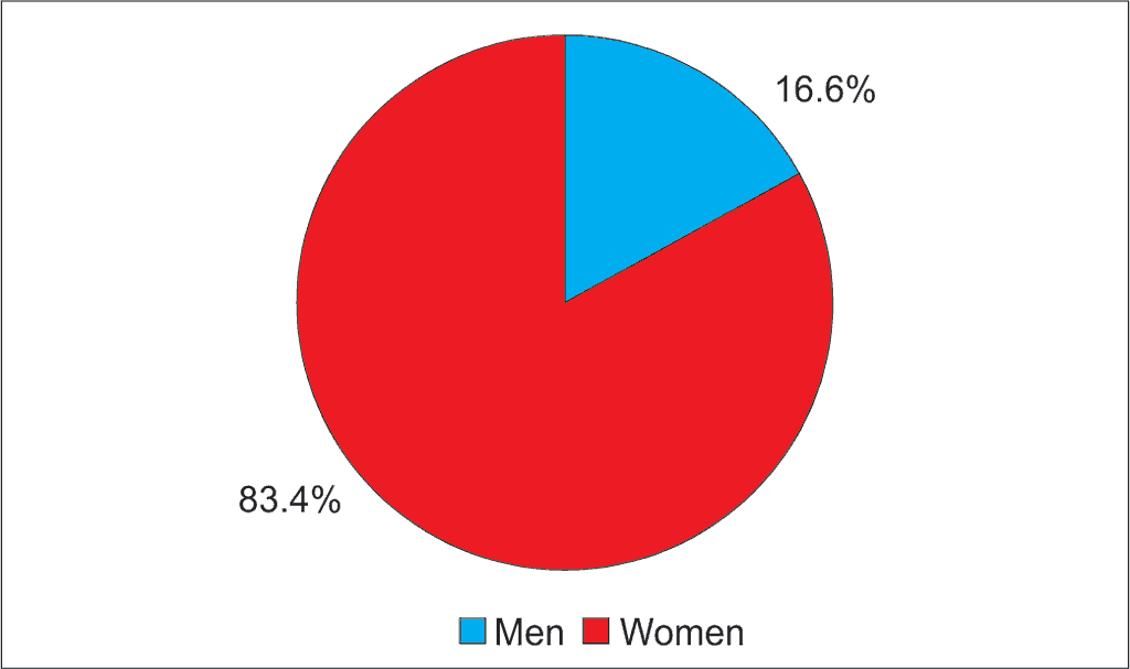
All patients were classified with Moyamoya disease. Among 12 patients 83.4% of them were female and 16.6% were male (fig. 1). The female-to-male ratio was 5. Research was performed mostly on paediatric population (age 1-16), one patient was an adult (39 y.o.). The mean age of the population was 11.2 years and 8.6 years when only paediatric population is analyzed. We diagnosed 8 patients (67%) with ischemic stroke, all of them were female and 4 patients (33%) with transient ischemic attacks (TIA) (fig. 2). The disease was diagnosed unilateral in 17% of all cases and 83% of all cases were bilateral. MMD mostly affected the right hemisphere (57%) when diagnosed unilateral and 43% of all cases considered the left hemisphere. MMD causes vessel stenosis and occlusion. In our research in 100% of all cases we found pathological changes in ICA and MCA and in 92% of all cases in ACA. The pathological changes in internal carotid artery were mostly bilateral (83%) rather than unilateral where we found changes only in 17% of all cases and always in the right ICA. The pathological process in medial cerebral artery was also mostly bilateral (50%), when unilateral mostly in the right MCA (33%) than left (17%). Unlike the previous vessels, the anterior carotid artery was occupied mostly unilaterally. The pathological process included the right ACA in 42% of all cases, the left ACA in 25% of all cases and 33% of all cases were bilateral. Among all cases the most common symptom was hemiparesis (92%), which occurred with equal frequency on both sides (45% on the left side and 45% on the right side). There was also one case of tetraparesis. Among our patients we also observed dysphasia (50%), cognitive processes abnormality (33%), headaches (25%), epilepsy (17%) and numbness of the limbs (9%) (tab. 1). All patients underwent multiple burr hole surgery with the number of holes between 6 and 20 (mean hole number – 11.5). We used Suzuki and Takaku grading scale with Fukuyama and Umezu modification to determine the progress of the disease. Among all patients 50% of them were diagnosed with stage 3c of MMD, 42% with stage 3a and 8% with stage 3b.

Fig. 1. Sex distribution of Moyamoya disease in 12 patients

Fig. 2. Presenting symptoms in 12 Moyamoya patients
Tab. 1. Moyamoya disease: epidemiology: a single Polish centre experience
| Symptom | Patient No. | Percent |
| Hemiparesis | 11 | 92% |
| Speech abnormality | 6 | 50% |
| Cognitive impairment | 4 | 33% |
| Headache | 3 | 25% |
| Epilepsy | 2 | 17% |
| Limb numbness | 1 | 9% |
Discussion
Powyżej zamieściliśmy fragment artykułu, do którego możesz uzyskać pełny dostęp.
Mam kod dostępu
- Aby uzyskać płatny dostęp do pełnej treści powyższego artykułu albo wszystkich artykułów (w zależności od wybranej opcji), należy wprowadzić kod.
- Wprowadzając kod, akceptują Państwo treść Regulaminu oraz potwierdzają zapoznanie się z nim.
- Aby kupić kod proszę skorzystać z jednej z poniższych opcji.
Opcja #1
24 zł
Wybieram
- dostęp do tego artykułu
- dostęp na 7 dni
uzyskany kod musi być wprowadzony na stronie artykułu, do którego został wykupiony
Opcja #2
59 zł
Wybieram
- dostęp do tego i pozostałych ponad 7000 artykułów
- dostęp na 30 dni
- najpopularniejsza opcja
Opcja #3
119 zł
Wybieram
- dostęp do tego i pozostałych ponad 7000 artykułów
- dostęp na 90 dni
- oszczędzasz 28 zł
Piśmiennictwo
1. Suzuki J, Takaku A: Cerebrovascular “Moyamoya” disease. Disease showing abnormal net-like vessels in base of brain. Arch Neurol 1969; 20: 288-299.
2. Hoshimaru M, Takahashi JA, Kikuchi H et al.: Possible roles of basic fibroblast growth factor in the pathogenesis of Moyamoya disease: an immunohistochemical study. J Neurosurg 1991; 75: 267-270.
3. Takahashi A, Sawamura Y, Houkin K et al.: The cerebrospinal fluid in patients with Moyamoya disease (spontaneous occlusion of the circle of Willis) contains high level of basic fibroblast growth factor. Neurosci Lett 1993; 160: 214-216.
4. Suzui H, Hoshimaru M, Takahashi JA et al.: Immunohistochemical reactions for fibroblast growth factor receptor in arteries of patients with Moyamoya disease. Neurosurgery 1994; 35: 20-24; discussion 24-25.
5. Malek AM, Connors S, Robertson RL et al.: Elevation of cerebrospinal fluid levels of basic fibroblast growth factor in Moyamoya and central nervous system disorders. Pediatr Neurosurg 1997; 27: 182-189.
6. Soriano SG, Cowan DB, Proctor MR, Scott RM: Levels of soluble adhesion molecules are elevated in the cerebrospinal fluid of children with Moyamoya syndrome. Neurosurgery 2002; 50: 544-549.
7. Jong S, Kim J: Moyamoya disease: epidemiology, clinical features, and diagnosis. J Stroke 2016; 18(1): 2-11. Published online 2016 Jan 29.
8. Kobayashi H, Brozman M, Kyselová K et al.: RNF213 rare variants in Slovakian and Czech Moyamoya disease patients. PLoS ONE 2016; 11(10): e0164759.
9. Scott RM, Smith JL, Robertson RL et al.: Long-term outcome in children with Moyamoya syndrome after cranial revascularization by pial synangiosis. J Neurosurg Spine 2004; 100: 142-149.
10. Ohaegbulam C, Scott RM: Moyamoya syndrome. [In:] McLone D (ed.): Pediatric neurosurgery. WB Saunders, Philadelphia 2001: 1077-1092.
11. Wakai K, Tamakoshi A, Ikezaki K et al.: Epidemiological features of Moyamoya disease in Japan: Findings from a nationwide survey. Clin Neurol Neurosurg 1997; 99 (suppl. 2): S1-5.
12. Baba T, Houkin K, Kuroda S: Novel epidemiological features of Moyamoya disease. J Neurol Neurosurg Psychiatry 2008; 79: 900-904.
13. Yim SH, Cho CB, Joo WI et al.: Prevalence and epidemiological features of Moyamoya disease in Korea. J Cerebrovasc Endovasc Neurosurg 2012; 14: 75-78.
14. Chen PC, Yang SH, Chien KL et al.: Epidemiology of Moyamoya disease in Taiwan: a nationwide population-based study. Stroke 2014; 45: 1258-1263.
15. Han DH, Kwon OK, Byun BJ et al.: A co-operative study: clinical characteristics of 334 Korean patients with Moyamoya disease treated at neurosurgical institutes (1976-1994). The Korean Society for Cerebrovascular Disease. Acta Neurochir (Wien) 2000; 142: 1263-1273.
16. Yilmaz EY, Pritz MB, Bruno A et al.: Moyamoya: Indiana University Medical Center experience. Arch Neurol 2001; 58: 1274-1278.
17. Hoshing H, Ozawa Y, Suzuki N: Epidemiological features of Moyamoya Disease in Japan Neural Med Chir (Tokyo) 2012; 52: 295-298.
18. Edward R, Smith R, Michael S: Surgical management of Moyamoya syndrome. Skull Base 2005; 15(1): 15-26.
19. Kuriyama S, Kusaka Y, Fujimura M et al.: Prevalence and clinicoepidemiological features of Moyamoya disease in Japan: findings from a nationwide epidemiological survey. Stroke 2008; 39: 42-47.
20. Uchino K, Johnston SC, Becker KJ, Tirschwell DL: Moyamoya disease in Washington state and California. Neurology 2005; 65: 956-958.
21. Kraemer M, Heienbrok W, Berlit P: Moyamoya Disease in Europeans. Stroke 2008; 39: 3193-3200.
22. Saarela M, Mustanoja S, Pekkola J et al.: Moyamoya vasculopathy – patient demographics and characteristics in the Finnish population. Int J Stroke 2016. pii: 1747493016669847.
23. Shoukat S, Itrat A, Taqui AM et al.: Kamal Moyamoya disease: A clinical spectrum, literature review and case series from a tertiary care hospital in Pakistan. BMC Neurology 2009; 9: 15.
24. Endo M, Kawano N, Miyaska Y, Yada K: Cranial burr hole for revascularization in Moyamoya disease. J Neurosurg 1989; 71: 180-185.
25. Kawaguchi T, Fujita S, Hosoda K et al.: Multiple burrhole operation for adult Moyamoya disease. J Neurosurg 1996; 84: 468-476.
26. Sainte-Rose C, Oliveira R, Puget S et al.: Multiple bur hole surgery for the treatment of Moyamoya disease in children. J Neurosurg 2006; 105: 437-443.
27. Oliveira RS, Amato MC, Simao GN et al.: Effect of multiple cranial burr hole surgery on prevention of recurrent ischemic attacks in children with Moyamoya disease. Neuropediatrics 2009; 40: 260-264.
28. Houkin K, Kuroda S, Ishikawa T, Abe H: Neovascularization (angiogenesis) after revascularization in Moyamoya disease. Which technique is most useful for Moyamoya disease? Acta Neurochir 2000; 142(3): 269-276.
29. Kim SK, Seol HJ, Cho BK et al.: Moyamoya disease among young patients: its aggressive clinical course and the role of active surgical treatment. Neurosurgery 2004; 54(4): 840-844.
30. Karasawa J, Touho H, Ohnishi H et al.: Long-term follow-up study after extracranial-intracranial bypass surgery for anterior circulation ischemia in childhood Moyamoya disease. J Neurosurg 1992; 77(1): 84-89.