© Borgis - New Medicine 3/2013, s. 78-83
*Michał Wendt, Piotr Bartkowiak, Zuzanna Klupsch, Paweł Szulc
The effect of american football training on the angles of spinal curvatures and segmental mobility of the spine
Department of Functional Anatomy, University School of Physical Education in Poznań, Poland
Head of Department: Krystyna Cieślik, PhD, Assoc. Prof.
Summary
Aim. To verify the effect of long-term American football training on the physiological curvatures and mobility of the spine.
Material and methods. The study included 35 men (21-23 years of age) who practiced American football in teams from the Polish Top League, and 35 healthy controls of similar age who did not practice any sport discipline. The functional spinal parameters were determined with an aid of Penny & Giles tensometric electrogoniometer.
Results. The analyzed groups differed significantly in terms of the angles of cervical and lumbar lordosis which proved greater in football players, by 1.1° and 2.2°, respectively. A significant reduction of the lumbar spine mobility: anteroflexion (by 6%), extension (by 21.3%), left and right lateral flexion (by 9.3% and 8.7%), and rotation to the left and right (by 17.7% and 14.7%), was revealed in football players, along with an increased mobility of the thoracic segment: anteroflexion (by 5.3%), extension (by 8.3%), and rotation to the right (by 5.6%). No significant differences in the mobility of cervical spine were documented.
Discussion. According to literature, football players are characterized by increased lumbar lordosis and mobility of lumbar spine. Our partially contradictory findings may result from the fact that previous studies involved players from more experienced American league. Significantly reduced mobility of lumbar spine reflects greater compression forces generated in this segment.
Conclusions. Tailored bioregeneration should constitute a vital component of the training programs as it could counterbalance the alterations of functional parameters resulting from the specifics of American football.
INTRODUCTION
Functional parameters of the spine change throughout the entire life. This can be associated with an array of factors, such as age, gender, body weight, or physical activity. Spondylometric parameters, especially reduced segmental mobility, and increased or decreased spinal curvatures, constitute a sensitive indicator of pathological changes (1-3).
A number of published studies dealt with the relationships between practicing various sport disciplines and functional parameters of the spine (2, 4-7). To the best of our knowledge, the number of published studies on changes in the segmental mobility of the spine and the angles of spinal curvatures in American football players is sparse (8).
AIM
The aim of this study was to verify the effect of long-term American football training on the physiological curvatures and segmental mobility of the spine.
MATERIAL AND METHODS
The study included 35 men between 21 and 23 years of age, who practiced American football in teams from the Top League of the Polish American Football Association. Mean training history of the players was 6 years.
The control group comprised 35 healthy men aged between 21 and 23 years, who did not practice any sport discipline. The protocol of the study was approved by the Local Bioethical Committee, and all the subjects gave their written informed consent to participate in the project.
We examined the segmental mobility of the cervical, thoracic, and lumbar spine and determined the angles of physiological spinal curvatures. All the measurements were taken in the same baseline position (standing freely with arms alongside the trunk, equal pressure applied on both feet, and head in the horizontal Frankfurt plane). The spinal mobility was determined in three planes: sagittal, coronal, and transverse. All the measurements were taken in the same room, with air temperature between 22°C and 24°C. Each participant was examined at the same time of the day; the day was free from any other physical exercise and training.
We used the system of Penny & Giles tensometric electrogoniometers, modified according to Boocock in order to eliminate potential measurement bias associated with shifting skin and soft tissues in relation to bones (9). The measurements were taken according to Lewandowski (3). Sensors of the electrogoniometer were placed alongside the long axis of participant’s body and fixed to the skin with an aid of double-sided medical adhesive tape.
Basic statistical characteristics of studied variables: arithmetic means, standard deviations, and minimum and maximum values were determined. As the normal distribution of the studied material was confirmed by means of the Shapiro-Wilk test, the parametric Student t-test was used to verify the significance of intergroup differences. The results documented in our participants were compared with the age-adjusted reference values reported by Lewandowski (3).
RESULTS
The analyzed groups differed significantly in terms of the angles of cervical and lumbar lordosis. The average cervical lordosis in football players was 1.1° greater than in the controls. The increased cervical lordosis of the athletes was confirmed on comparison with the respective reference values. The intergroup differences in the angles of thoracic kyphosis did not prove significant. The most pronounced intergroup differences pertained to lumbar lordosis. Football players were characterized by an increased angle of this curvature, by 2° on average, as compared to the controls and normative values (tab. 1, 2).
Table. 1. Basic statistical characteristics of the physiological spinal curvatures in American football players and in the controls (*p < 0.05).
| Curvature | Football players | Controls | Student t-test |
| N | x (o) | SD | N | x (o) | SD | t | df | p |
| Cervical lordosis | 35 | 36.89 | 2.36 | 35 | 35.77 | 1.86 | 2.19 | 68 | 0.032* |
| Thoracic kyphosis | 35 | 31.60 | 2.34 | 35 | 31.20 | 1.95 | 0.78 | 68 | 0.440 |
| Lumbar lordosis | 35 | 37.91 | 2.89 | 35 | 35.49 | 2.23 | 3.93 | 68 | 0.000* |
t – values read from respective distribution tables; df – degrees of freedom; p – level of statistical significance
Table. 2. Reference values of the physiological spinal curvatures in 22-year-old men, according to J. Lewandowski (3).
| Curvature | x (o) | SD |
| Cervical lordosis | 36.52 | 5.21 |
| Thoracic kyphosis | 32.43 | 4.15 |
| Lumbar lordosis | 36.06 | 4.97 |
The differences in the mobility of cervical spine were not significant. An increased mobility of the thoracic segment: anteroflexion (by 5.3%), extension (by 8.3%), and rotation to the right (by 5.6%), was documented in football players, along with a significant reduction of the lumbar spine mobility: anteroflexion (by 6%), extension (by 21.3%), left and right lateral flexion (by 9.3% and 8.7%), and rotation to the left and right (by 17.7% and 14.7%) (tab. 3, 4, 5).
Table. 3. Basic statistical characteristics of the functional parameters of three spinal segments in American football players.
| Spinal segment | Mobility | N | x (o) | SD | Min (o) | Max (o) |
| Cervical | Flexion | 35 | 61.46 | 3.04 | 53 | 70 |
| Extension | 57.31 | 3.98 | 50 | 71 |
| Left lateral flexion | 50.03 | 8.17 | 48 | 56 |
| Right lateral flexion | 51.11 | 2.65 | 46 | 56 |
| Rotation to the left | 83.66 | 2.80 | 79 | 89 |
| Rotation to the right | 83.94 | 2.87 | 79 | 89 |
| Thoracic | Flexion | 35 | 26.89 | 2.76 | 20 | 31 |
| Extension | 22.89 | 2.34 | 13 | 27 |
| Left lateral flexion | 29.74 | 3.37 | 17 | 34 |
| Right lateral flexion | 30.46 | 3.35 | 18 | 34 |
| Rotation to the left | 31.40 | 2.94 | 21 | 36 |
| Rotation to the right | 31.43 | 3.01 | 26 | 35 |
| Lumbar | Flexion | 35 | 62.63 | 4.71 | 54 | 76 |
| Extension | 28.34 | 2.95 | 24 | 29 |
| Left lateral flexion | 27.74 | 3.57 | 17 | 35 |
| Right lateral flexion | 28.00 | 3.67 | 15 | 34 |
| Rotation to the left | 7.46 | 2.47 | 4 | 12 |
| Rotation to the right | 7.43 | 2.44 | 3 | 13 |
Table. 4. Basic statistical characteristics of the functional parameters of three spinal segments in the controls.
| Spinal segment | Mobility | N | x (o) | SD | Min (o) | Max (o) |
| Cervical | Flexion | 35 | 61.71 | 4.01 | 50 | 69 |
| Extension | 58.66 | 4.21 | 45 | 72 |
| Left lateral flexion | 51.40 | 2.35 | 47 | 56 |
| Right lateral flexion | 51.06 | 2.33 | 46 | 55 |
| Rotation to the left | 84.11 | 2.63 | 79 | 88 |
| Rotation to the right | 83.83 | 2.41 | 79 | 89 |
| Thoracic | Flexion | 35 | 25.54 | 2.39 | 19 | 30 |
| Extension | 21.14 | 2.50 | 11 | 30 |
| Left lateral flexion | 29.00 | 2.74 | 15 | 34 |
| Right lateral flexion | 29.71 | 2.82 | 17 | 33 |
| Rotation to the left | 30.17 | 2.18 | 24 | 35 |
| Rotation to the right | 29.86 | 2.53 | 22 | 35 |
| Lumbar | Flexion | 35 | 66.63 | 3.16 | 61 | 78 |
| Extension | 36.03 | 3.68 | 28 | 45 |
| Left lateral flexion | 30.57 | 1.87 | 28 | 36 |
| Right lateral flexion | 30.66 | 2.18 | 25 | 40 |
| Rotation to the left | 9.06 | 1.53 | 5 | 13 |
| Rotation to the right | 8.71 | 1.60 | 4 | 13 |
Table. 5. Significance of differences in the mobility of various spinal segments in American football players and in the controls (*p < 0.05).
| Spinal segment | Mobility | N | Student t-test |
| t | df | p |
| Cervical | Flexion | 35 | -0.30 | 68 | 0.763 |
| Extension | -1.37 | 68 | 0.174 |
| Left lateral flexion | -0.95 | 68 | 0.343 |
| Right lateral flexion | 0.10 | 68 | 0.924 |
| Rotation to the left | -0.70 | 68 | 0.484 |
| Rotation to the right | 0.18 | 68 | 0.857 |
| Thoracic | Flexion | 35 | 2.17 | 68 | 0.033* |
| Extension | 3.01 | 68 | 0.004* |
| Left lateral flexion | 1.01 | 68 | 0.316 |
| Right lateral flexion | 1.00 | 68 | 0.319 |
| Rotation to the left | 1.99 | 68 | 0.051 |
| Rotation to the right | 2.36 | 68 | 0.021* |
| Lumbar | Flexion | 35 | -4.17 | 68 | 0.000* |
| Extension | -9.65 | 68 | 0.000* |
| Left lateral flexion | -4.16 | 68 | 0.000* |
| Right lateral flexion | -3.68 | 68 | 0.000* |
| Rotation to the left | -3.26 | 68 | 0.002* |
| Rotation to the right | -2.61 | 68 | 0.011* |
Comparison with the reference values published by Lewandowski revealed reduced mobility in all spinal segments. The reduction was most pronounced in the lumbar segment, in the case of which the deviation from normative value ranged between 3.9° and 5.8° (tab 3, 6).
Table. 6. Reference values of the mobility of three spinal segments in 22-year-old men, according to J. Lewandowski (3).
| Spinal segment | Mobility | N | (o) | SD | Min (o) | Max (o) |
| Cervical | Flexion | 35 | 64.06 | 5.33 | 47 | 74 |
| Extension | 60.29 | 5.26 | 50 | 75 |
| Left lateral flexion | 52.88 | 3.30 | 45 | 63 |
| Right lateral flexion | 53.09 | 3.17 | 46 | 62 |
| Rotation to the left | 86.37 | 3.73 | 77 | 97 |
| Rotation to the right | 86.38 | 3.70 | 77 | 96 |
| Thoracic | Flexion | 35 | 27.71 | 3.36 | 21 | 39 |
| Extension | 23.37 | 2.14 | 19 | 29 |
| Left lateral flexion | 31.51 | 3.54 | 25 | 40 |
| Right lateral flexion | 31.43 | 3.32 | 24 | 40 |
| Rotation to the left | 31.75 | 3.79 | 24 | 40 |
| Rotation to the right | 31.61 | 3.68 | 25 | 40 |
| Lumbar | Flexion | 35 | 68.41 | 6.36 | 59 | 90 |
| Extension | 32.22 | 4.76 | 20 | 46 |
| Left lateral flexion | 32.20 | 2.74 | 27 | 40 |
| Right lateral flexion | 32.26 | 2.71 | 26 | 40 |
| Rotation to the left | 11.46 | 1.12 | 9 | 14 |
| Rotation to the right | 11.56 | 1.13 | 9 | 15 |
DISCUSSION
Strains the athlete is exposed to can affect the lordosis or kyphosis. An increase or decrease in spinal curvatures can exert a negative effect on the vertebrae, intervertebral discs, ligaments, and some muscle groups. Maintenance of physiological curvatures in all spinal segments is vital for the normal balance of the musculoskeletal system strain (4, 10).
Nyland analyzed the effect of football training on the lordosis and mobility of the cervical spine in athletes from two age categories. He observed increased cervical lordosis in older athletes, and attributed this finding to adaptive changes associated with the involvement in football training or matches (8). The more intensive the athletic training is, the larger are the spinal curvatures (6). The increase in the cervical and lumbar lordosis was documented in our study as well. The training program of football players includes many athletic components, which could be reflected by larger angles of these curvatures (11). Nyland analyzed solely the mobility of cervical spine and revealed its improvement in football players. In contrast, we did not observe significant changes in the cervical mobility. However, Nyland analyzed players from much more experienced American league. Perhaps, also the methods of training these players were more elaborated, which was reflected by their improved spinal mobility (8).
The greatest reduction of spinal mobility pertained the lumbar segment, which can result from excessive tone of its stabilizing muscles. During a blocking maneuver, the L5-S1 spinal segment is exposed to huge compression forces, up to 8679 N. Such strain can enforce adaptive changes in the musculoskeletal system (12).
Szulc analyzed the spondylometric parameters of 20 rugby players (7). The comparative analysis of football and rugby players suggests that the latter are characterized by greater reduction of spinal mobility and increased curvatures in all segments (fig. 1, 2, 3). The greatest differences in the mobility pertained to the rotatory movements of cervical spine. Compared to the football players, the rugby players showed reduced rotation to the left and right (by 52.2% and 52.8%, respectively; fig. 1). Football players are equipped with pads and helmet, which can attenuate strains specific for this discipline. The lack of helmet and the resultant micro-injuries can be reflected by markedly reduced rotation of cervical spine in rugby players. Furthermore, the lack of protective equipment can be associated with greater axial strain, which negatively affects intervertebral joints, ligaments, and muscles (7, 13, 14).
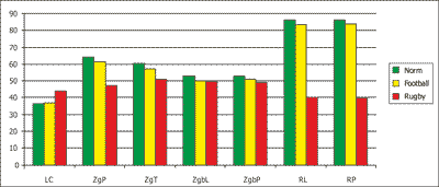
Fig. 1. Comparison between the functional parameters of cervical spine in American football players, controls, and respective reference values (o).
LC – cervical lordosis, ZgP – anteroflexion; ZgT – posterior flexion (extension); ZgbL – left lateral flexion; ZgbP – right lateral flexion; RL – rotation to the left; RP – rotation to the right
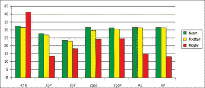
Fig. 2. Comparison between the functional parameters of thoracic spine in American football players, controls, and respective reference values (o).
KTh – thoracic kyphosis; ZgP – anteroflexion; ZgT – posterior flexion (extension); ZgbL – left lateral flexion; ZgbP – right lateral flexion; RL – rotation to the left; RP – rotation to the right
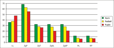
Fig. 3. Comparison between the functional parameters of lumbar spine in American football players, controls, and respective reference values (o).
LL – lumbar lordosis; ZgP – anteroflexion; ZgT – posterior flexion (extension); ZgbL – left lateral flexion; ZgbP – right
We postulate the important role of balanced muscle tone. Bioregeneration should constitute an important component of the training program, which should be enhanced by relaxation techniques and tailored strengthening exercises. Particular attention should be paid to the muscle groups that are responsible for increased lumbar and cervical lordosis (7, 15).
CONCLUSIONS
Due to its athletic components, American football training can result in increased lumbar and cervical lordosis.
The greatest reduction of mobility in the lumbar spine can correspond to the magnitude of strain of this spinal segment.
Comparison with literature data suggests than American football is associated with smaller deficits in the functional parameters of the spine than rugby.
Tailored bioregeneration should constitute a vital component of the training programs for football players as it could counterbalance the alterations of functional parameters resulting from the specifics of this discipline.
Piśmiennictwo
1. Doriot N, Wang X: Effects of age and gender on maximum voluntary range of motion of the upper body joints. Ergonomics 2006; 49: 269-281. 2. Wood KB: Spinal deformity in the adolescent athlete. Clin Sports Med 2002; 21: 77-92. 3. Lewandowski J: Kształtowanie się krzywizn fizjologicznych i zakresów ruchomości odcinkowej kręgosłupa człowieka w wieku 3-25 lat w obrazie elektrogoniometrycznym. AWF Poznań 2006. 4. Lichota M, Plandowska M, Mil P: The shape of anterior-posterior curvatures of the spine in athletes practising selected sports. Pol J Sport Tourism 2011; 18: 112-116. 5. Schiller JR, Eberson CP: Spinal deformity and athletics. Sports Med Arthrosc 2008; 16: 26-31. 6. Wojtys EM, Ashton-Miller JA, Huston LJ, Moga PJ: The association between athletic training time and the sagittal curvature of the immature spine. Am J Sports Med 2000; 28: 490-498. 7. Szulc P, Boch-Kmieciak J, Bartkowiak P, Lewandowski J: The impact of rugby training on the spinal functional parameters. New Medicine 2011; 14: 46-51. 8. Nyland J, Johnson D: Collegiate football players display more active cervical spine mobility than high school football players. J Athl Train 2004; 39: 146-150. 9. Boocock MG, Jackson JA, Burton AK, Tillotson KM: Continuous measurement of lumbar posture using flexible electrogoniometers. Ergonomics 1994; 37: 175-185. 10. Keller TS, Colloca CJ, Harrison DE et al.: Influence of spine morphology on intervertebral disc loads and stresses in asymptomatic adults: implications for the ideal spine. Spine J 2005; 5: 297-300. 11. Baskett D: Confusing conditioning for football speed development. American Football Monthly 2011; 6: 14-15. 12. Gatt CJ Jr, Hosea TM, Palumbo RC, Zawadsky JP: Impact loading of the lumbar spine during football blocking. Am J Sports Med 1997; 25: 317-321. 13. Badgeley MA, McIlvain NM, Yard EE et al.: Epidemiology of 10,000 high school football injuries: patterns of injury by position played. J Phys Act Health 2013; 10: 160-169. 14. Castinel BH, Adam P, Prat C: A stress fracture of the lumbar spine in a professional rugby player. Br J Sports Med 2007; 41: 337-338. 15. Ogurkowska MB: Analysis of radiological characteristics distribution in the vertebral bodies of the lumbosacral spine of competitive rowers. Biol Sport 2010; 27: 213-219.
