*Jacek Wadełek
Septic shock in the course of Fournier’s gangrene – a case report
Wstrząs septyczny w przebiegu zgorzeli Fourniera – opis przypadku
Anaesthesiology and Intensive Care Unit, Saint Anna Traumatology Hospital, Mazowsze Rehabilitation Centre „STOCER”, Warsaw
Head of Department: Elżbieta Kurmin-Gryz, MD
Streszczenie
Zgorzel Fourniera jest piorunującą postacią infekcyjnego martwiczego zapalenia powięzi okolic krocza, zewnętrznych narządów płciowych i okołoodbytniczej, które zwykle występuje u mężczyzn, ale może również dotyczyć kobiet i dzieci. W 2016 roku robocza grupa ekspertów do spraw sepsy wprowadziła nową definicję sepsy, jako zagrażającej życiu dysfunkcji narządu wywołanej rozregulowaną odpowiedzią gospodarza na zakażenie. Autor przedstawia przypadek wstrząsu septycznego z zespołem wielonarządowej niewydolności wywołanego zgorzelą Fourniera, który wymagał postępowania wielodyscyplinarnego i był z powodzeniem leczony w oddziale intensywnej terapii.
Sześćdziesięcioczteroletni pacjent został przyjęty do szpitala z silnym bólem okolicy okołoodbytniczej i wysoką gorączką (39°C). Pacjent leczył się doustnymi lekami hipoglikemizującymi z powodu cukrzycy. Stwierdzono ropień okołoodbytniczy i rozległe zmiany okolicznych tkanek. Postawiono rozpoznanie zgorzeli Fourniera w przebiegu ropnia okołoodbytniczego. U pacjenta zastosowano agresywną płynoterapię dożylną i farmakologiczne wspomaganie krążenia. W trybie pilnym pacjent został poddany rozległej operacyjnej nekrektomii w znieczuleniu ogólnym. Pobrano materiał do badania mikrobiologicznego. Martwicze tkanki skóry i podskórne moszny i okolicy okołoodbytniczej usunięto, a łożysko rany pooperacyjnej pozostawiono do drenażu na otwarto. Przedoperacyjnie podano antybiotyki o szerokim spektrum działania, leczenie antybiotykami zmodyfikowano po uzyskaniu wyników badań mikrobiologicznych. Wyniki posiewów pobrane podczas chirurgicznego opracowania rany potwierdziły obecność mieszanej flory bakteryjnej. Pacjent wymagał jeszcze dwukrotnego chirurgicznego usunięcia martwiczych tkanek. Udało się opanować zakażenie, dobre ziarninowanie rany uzyskano po 5 tygodniach leczenia.
Podstawę leczenia stanowią: wczesne i rozległe chirurgiczne opracowanie rany, drenaż rany na otwarto, szerokospektralna antybiotykoterapia oraz intensywne leczenie wspomagające, takie jak płynoterapia i optymalizacja układu krążenia.
Summary
Fournier’s gangrene (FG) is a fulminant form of infective necrotising fasciitis of the perineal, genital, or perianal regions, which commonly affects men, but can also occur in women and children. In 2016 a task force with expertise in sepsis has introduced a new definition of sepsis as a life-threatening organ dysfunction caused by a dysregulated host response to infection. We present a case of septic shock with multiple organ dysfunction syndrome due to FG which needed interdisciplinary management and was successfully treated in intensive care unit.
A 64-year-old male patient was admitted to our hospital with severe perianal pain and high fever (39°C). The patient had a history of diabetes mellitus under oral hypoglycaemic. Upon physical examination, the patient was hypotensive (80/40 mmHg) with tachycardia (126/min). A perianal abscess was identified. White blood cell count was 17 800/μl, C-reactive protein (CRP) was 340 mg/dl, and blood glucose was 260 mg/dl. A diagnosis of Fournier’s gangrene complicating a perianal abscess was made. The patient underwent aggressive fluid administration and haemodynamic support. He was treated with immediate extensive surgical debridement under general anaesthesia. Tissue cultures were obtained for isolation of the responsible microorganisms. The necrotic skin in the scrotum and the perianal region was evacuated into a wide-open drainage area. Preoperative antibiotic treatment with broad-spectrum antibiotics combinations was initiated and later adjusted to the culture sensitivity of the microbial isolates. According to tissue samples taken during debridement, the microbiological aetiology of FG was polymicrobial. He underwent two subsequent surgical debridements. The infection gradually subsided, gas gangrene resolved completely, and good granulation was present five weeks after surgery.
The mainstay of treatment should be open drainage and early aggressive surgical debridement of all necrotic tissue, followed by broad-spectrum antibiotics therapy and intensive therapy supportive treatment with fluid therapy and cardiovascular optimisation.

Introduction
Fournier’s gangrene (Fournier’s syndrome, multiple bacteria, synergistic Fournier’s gangrene, FG) is an infection-origin disease concerning soft tissues of the perineum, male reproductive organs and perianal soft tissues. As regards classification, it is one of necrotising soft tissue infections (NSTI). It is a type of necrotising fasciitis (NF) with multiple bacteria aetiology. The first description of the disease in literature made by Baurienne in 1764, the more widely known description by Fournier, a Parisian venereologist on the basis of a group analysis of 5 patients dating back to 1883 (1, 2). The disease has a severe, aggressively progressing course and apart from local soft tissue degeneration, it causes direct threat to the life of the patient due to severe inflammation. The new definition of sepsis proposed in 2016 defines sepsis to be a life-threatening organ dysfunction caused by the disturbed systemic response to infection regulation. Its diagnosis criteria were based on the SOFA score (sudden increase in the score by ≥ 2 points in the case of infection or its suspicion) and qSOFA (≥ 2 due to the following symptoms: disorders of consciousness, systolic blood pressure ≤ 100 mmHg, frequency of breathing ≥ 22/min) (3).
Case was presented of a septic shock in the course of Fournier’s gangrene requiring interdisciplinary treatment, in particular treating shock and organ failure under intensive care unit conditions.
Case report
A 64-year-old patient admitted to the Surgery Ward of the local hospital on 5 May 2016 due to a fracture near the right wrist. The patient lives alone, poor hygiene observed, injured at home. On examination, a spread cutaneous necrotic lesion was observed in the perianal and perineal area and on the buttocks, huge inflammatory reaction and developing Fournier’s gangrene. History: fracture within the right wrist fixed with a plaster cast, hypertension, type 2. diabetes, depression and chronic pain in the form of lumbar discopathy, treated at the Pain-treating Outpatient Clinic. Condition following a cervical spine surgery. Medium paresis of the upper limbs due to cervical spine discopathy. Temperature up to 39°C.
Additional examination: blood smear proved increase in leukocytosis up to 17.8 thousand/μl, increase in polymorphism (86%), elevated C-reactive protein (CRP 340 mg/d1). Moreover, low level of albumins in the serum was observed (2.5 g/dl) and low level of total protein (4.8 g/dl) and lowered haemoglobin concentration (9 g/dl). Elevated glucose concentration in blood serum (260 mg/d) and slightly increased creatinine concentration (1.8 mg/dl).
Admitted to the General Surgery Ward to treat necrotic lesions within the perineum and the perianal area. Operated on the first day of stay, wide tissue revision of the perineal tissue and necrectomy (fig. 1). Following the operation, due to anaemia, the patient required having erythrocyte concentrate transfused and due to clotting disturbances of freshly-frozen plasma empirical antibiotics therapy was started from the first day of stay by administering intravenously tazocin in the dose 3 x 4.5 g, clindamycin 2 x 600 mg, metronidazole 3 x 500 mg after consulting the hospital epidemiologist. The supply of fluids and oral nutrition were stopped. Central insertion of a needle to the right subclavian vein was performed and parenteral nutrition was started. Glycaemia was controlled with a continuous intravenous insulin infusion. Deterioration in the general condition of the patient observed on the tenth day of stay. Dyspnoea at rest was observed with deterioration in arterial blood oxygenation despite passive supply of oxygen using a face mask with the flow of 4 l/min, arterial pressure dropped, tachycardia observed, perspiring skin of the patient. Following consultation with an anaesthetist, qualified and admitted to the Anaesthesiology and Intensive Care Unit. Transportation from the Surgery Ward to the Anaesthesiology and Intensive Care Unit (AICU) was performed with the help of the anaesthetists’ team. During admission to AICU, deterioration in general condition was observed with acute respiratory failure, which required endotracheal intubation and mechanical ventilation as well as acute circulatory failure making it necessary to perform intensive fluid therapy and pressor amine treatment. General condition of the patient following admission: serious; nervous system: conscious patient, mentally slow with impeded logical contact; circulatory system failure, arterial pressure measured non-invasively 80/40 mmHg, marmoration of the lower limb skin. Function of the heart rhythmic, accelerated with frequency of 126/min; respiratory system failure; dyspnoea at rest, percutaneous saturation of arterial blood SpO2 74% at through-mask oxygen supply. On auscultation, over the lung field, acute alveolar murmur, rattling sound on both sides in the basal area of the lungs. Abdomen elevated above the thorax, soft, no pathological resistance, peristalsis audible, quiet. Diuresis supported with furosemide in fractions, oliguria. Skin: on the post-surgery skin of the perineum, wide incisions of the skin and subcutaneous tissues dressed. Lower limbs with trophic lesions from the groin to the knees. Having been admitted to AICU, the patient was informed about the proposed plan of treatment. The patient expressed consent to the proposed procedure in the presence of witnesses, was unable to sign the consent for treatment at AICU in person. Following propofol sedation, respiratory tract of the patient in direct laryngoscopy visualised and intubated, then the patient was under a respirator, mechanical ventilation started in the mode of pressure controlled synchronised intermittent mandatory ventilation – SIMV/PC + PSV, at the beginning with 100% oxygen concentration in the inhalation mixture FiO2 1.0, later lowered to FiO2 0.4 under the control of arterial blood gasometry testing. Aspirate from the respiratory tract, urine and peripheral blood collected for culture. Antibiotics therapy modified taking into account the present results of culture. Midazolam and morphine sedation applied. Due to low arterial pressure values, intravenous, continuous norepinephrine infusion applied by dosing the infusion depending on the arterial blood pressure level to reach mean systemic arterial pressure above 65 mmHg. A follow-up radiological image of the thorax performed to make visible the progression of parenchymal lesions in the right lung. After a few hours the circulatory system stabilised, invasive arterial blood pressure measurement at the level of 140/70 mmHg. Furosemide-supported diuresis in a continuous intravenous infusion on the first day of treatment at AICU 2220 ml. Fluid balance – 170 ml. Patient had no pyrexia. In gasometry – high values of pCO2 despite increasing the minute mechanical ventilation. Mechanical ventilation stopped on the 8th day of treatment at AICU. Pharmacological circulation support stopped on the 9th day of treatment at AICU. Intravenous nutrition treatment continued for two weeks of stay at AICU and the following three weeks of treatment at the General Surgery Ward. While treatment at AICU, general condition of the patient improved as well as the local condition of the wound in the perineal and perianal area. Patient in a general good condition, conscious, respiratory and circulatory efficiency, on the 12th day of treatment at AICU transported to the Surgery Ward to continue treatment. Currently, the patient is awaiting a surgery, post-surgery wound reconstruction (fig. 2).

Fig. 1. Necrotic lesions of the perianal and scrotal area tissues
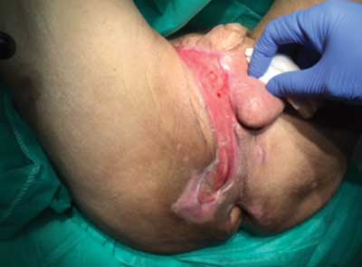
Fig. 2. Wound developing granulation tissue after three necrotic tissue removal procedures
Discussion
Powyżej zamieściliśmy fragment artykułu, do którego możesz uzyskać pełny dostęp.
Mam kod dostępu
- Aby uzyskać płatny dostęp do pełnej treści powyższego artykułu albo wszystkich artykułów (w zależności od wybranej opcji), należy wprowadzić kod.
- Wprowadzając kod, akceptują Państwo treść Regulaminu oraz potwierdzają zapoznanie się z nim.
- Aby kupić kod proszę skorzystać z jednej z poniższych opcji.
Opcja #1
29 zł
Wybieram
- dostęp do tego artykułu
- dostęp na 7 dni
uzyskany kod musi być wprowadzony na stronie artykułu, do którego został wykupiony
Opcja #2
69 zł
Wybieram
- dostęp do tego i pozostałych ponad 7000 artykułów
- dostęp na 30 dni
- najpopularniejsza opcja
Opcja #3
129 zł
Wybieram
- dostęp do tego i pozostałych ponad 7000 artykułów
- dostęp na 90 dni
- oszczędzasz 78 zł
Piśmiennictwo
1. Ochiai T, Ohta K, Takahashi M et al.: Fournier’s gangrene: Report of six cases. Surg Today 2001; 31: 553-556. 2. McCloud JM, Doucas H, Scott ADN, Jameson JS: Delayed presentation of life-threatening perineal sepsis following stapled haemorrhoidectomy: a case report. Ann R Coll Surg Engl 2007; 89: 301-302. 3. Singer M, Deutschman CS, Seymour ChW et al.: The third international consensus definitions for sepsis and septic shock (Sepsis-3). JAMA 2016; 315: 801-810. 4. Eke N: Fournier’s gangrene: a review of 1726 cases. Br J Surg 2000; 87(6): 718-728. 5. Dellinger RP, Levy MM, Rhodes A et al.: Surviving Sepsis Campaign: international guidelines for management of severe sepsis and septic shock, 2012. Intensive Care Med 2013; 39: 165-228.


