© Borgis - New Medicine 4/2007, s. 87-88
*Lechosław P. Chmielik, Romuald Krajewski1
Neurofibroma of the pterygo-palatine fossa in a child
Department of Pediatric Otolaryngology, Medical University of Warsaw, Poland
Head of Department: Prof. Mieczysław Chmielik, MD, PhD
1Head and Neck Cancer Department, Cancer-Center M. Skłodowska-Curie, Memorial Insitute, Warsaw, Poland
Summary
Summary
Tumors in the pterygo-palatine fossa are rarely sun in children. The authors present a case of a boy with a neurofibroma of the pterigo-palatine fossa, which was treated in our clinic.
The neurofibroma is a benign tumor which can be included in the group of genetic syndromes. In the literature we can find opinions that this growth can become malignant.
These tumors arise from nerves, and can be found in different parts of the body. They can occur as a single growth, or many.
They are not often found in children, which is why we are presenting this case; an eight-year-old boy was referred to the clinic because of a growth in the cheek. Computerised tomography, and an MRI scan, showed a growth in the pterygo-palatinum fossa, entering the orbit, and invading the structures of the cheek. A specimen from the growth, and an enlarged node from the area of the mandible, were sent for a histo-pathological examination.
The check showed neurofibroma and the boy was qualified for surgical treatment. The operation was a modification of Fischer C, carried cut on 16.3.2006, protecting the facial nerve. A cut was made in the skin of the head, half-coronary, after which the muscles were cut, allowing inspection of the rim of the orbit and the zygomatic bone, which was removed. In the area of the subtemporal fossa and in the temporal area we could see the mass of the tumor, which was removed, together with the tumor from the subtemporal fossa depression and the check tissue. Ficher C is an efficient method of removing changes in the pterygo –palatine fossa. Patients with neurofibroma require long-term control, and multi-specialist treatment.
INTRODUCTION
Tumors in the pterygo-palatine fossa are rarely sun in children. The authors present a case of a boy with a neurofibroma of the pterigo-palatine fossa, which was treated in our clinic.
The neurofibroma is a benign tumor which can be included in the group of genetic syndromes . In the literature we can find opinions that this growth can become malignant. These tumors arise from nerves, and can be found in different parts of the body. They can occur as a single growth, or many.
CASE REPORT
They are not often found in children, which is why we are presenting this case; an eight-year-old boy was referred to the clinic because of a growth in the cheek. Computerised tomography, and an MRI scan, showed a growth in the pterygo-palatinum fossa, entering the orbit, and invading the structures of the cheek. A specimen from the growth, and an enlarged node from the area of the mandible,were sent for a histo-pathological examination.
The check showed neurofibroma and the boy was qualified for surgical treatment. The operation was a modification of Ficher C, carried cut on 16.3.2006, protecting the facial nerve. A cut was made in the skin of the head, half-coronary, after which the muscles were cut, allowing inspection of the rim of the orbit and the zygomatic bone, which was removed. In the area of the subtemporal fossa and in the temporal area we could see the mass of the tumor, which was removed, together with the tumor from the subtemporal fossa depression and the cheek tissue. After the operation, the forehead was difficult to move on the left side, but this was the only complication in the post-operative period. Rehabilitation and orthodontic control were recommended.
In a CT scan made on 29.1.2007 with an MRI, no relapse symptoms were found in the orbit, or nose, or throat. Tissue showed in a much smaller area of the cheek that before, at the level of the bite, in the cheek muscle and masseter muscle. Changes in post-operational scar tissue caused us to recommend a further MRI scan at between 3 and 6 months.
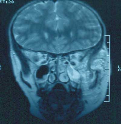
Pic. 1. MRI of the neurofibroma of the pterygo-palatine fossa
CONCLUSIONS
1. Ficher C is an efficient method of removing changes in the pterygo –palatine fossa.
2. Patients with neurofibroma require long-term control, and multi-specialist treatment.
Piśmiennictwo
1. Darrigo Jr LG, et al.: Prevalence of plexiform neurofibroma in children and adolescents with type I neurofibromatosis. J. Pediatr. (Rio J) 2007; Aug 23: 83(6).2. Ferrari A, et al.: Soft-tissue sarcomas in children and adolescents with neurofibromatosis type 1. Cancer 2007; 1: 109(7); 1406-12. 3. Gutarra F, et al.: Ruptured femoropopliteal artery aneurysms in von Recklinghausen neurofibromatosis. J. Vasc. Surg . 2007; 46(4): 808-11. 4. Houreih MA, et al.: A case of fobroblastic low-grande malignant periferal nerve sheath tumor-a neurofibrosarcoma. Ultrastruct Pathol. 2007; 31(5): 347-56.5. Lohmeyer JA, , et al.: Combined manifestation of neurofibroma and a nerve sheath ganglion in the ulnar nerve after radiotherapy in early childhood. J. Plast. Reconstr. Aesthet. Surg. 2007; 60(12): 1338-41.6. Mautner VF, et al.: Clinical relevance of position emission tomography and magnetic resonance imaging in the progression of internal plexiform neurofibroma in NF1. Anticancer Res., 2007; 207(4A): 1819-22.7. Patil K, et al.: Facial plexiform neurofibroma in a child with neurofibromatosis type I: a case report. J. Indian Soc. Pedod. Prev. Dent., 2007 Mar; 25(1): 30-5.8. Ronchetti R, et al.: Nasal Cellularity in 183 Unselected Schoolchildren Aged 9 to 11 Years. Pediatrics 2002; 110: 1137-1142.9. Serletis D, Parkin P.: Massive plexiform neurofibromas in childhood: natural history and management issues. J. Neurosurg., 2007; 106(5 Suppl): 363-7.10. Waqar-uddin; Farooq Ahmad;Anjum Khawar; Recurent neurofibroma in the head and neck. J. Coll. Physicians Surg. Pak., 2007; 17(10): 629-31.
