© Borgis - New Medicine 4/2007, s. 83-86
Eliza Brożek-Mądry, *Lidia Zawadzka-Głos, Mieczysław Chmielik
Long-term experience in the management of choanal atresia
Department of Pediatric Otolaryngology, Medical University of Warsaw, Poland
Head of Department: Prof. Mieczysław Chmielik, MD, PhD
Summary
Summary
Choanal atresia occurs as an isolated congenital malformation or it is associated with other malformations. In Pediatric Otolaryngology Department in Warsaw children with choanal atresia have been operated for many years.
Material and method. Authors present a large material with a group of patients operated due to choanal atresia in Otolaryngology Department, Medical Academy of Warsaw in years 1959-2007. The operations were conducted in these children either by transpalatal or transnasal approach. The patients´ characteristics was presented in children with unilateral and bilateral changes. The operated children were evaluated in terms of reoperation necessity.
Results. between 1959 and 2007 a group of 136 patients was operated due to choanal atresia with a total number of 183 operations including 47 reoperations.
Conclusions. (1) Choice of operating method depends on anatomic conditions and experience of particular operating centre (2). Stent removal – when they achieve good mobility in posterior choanae and can be easily moved to nasopharynx (3). Method gaining much attention in the management of chonal atresia resently is transnasal approach due to the development of endoscopic techniques (4). Children with bilateral choanal atresia require re-dilation more frequently (5). Bilateral choanal atresia is more common in children with co-existing anomalies and with perinatal burden.
INTRODUCTION
Choanal atresia (CA) is a congenital malformation leading up to nasal patency disorders, depending on its unilateral or bilateral character. First descriptions of the disorder in literature date the middle of XVIII century (Roederer) but it was 80 years later when the anatomy of choanal atresia was described (Otto) (1) . The disease prevalence is estimated between 1:5000 and 1:8000 live births, more frequently occurs in females, usually unilateraly and on the right side (2). In 70% patients the atretic plate in choanae is bony-membraneous, while the rest patients (30%) have a bony plate (3). There are four hypotheses leading to choanal atresia formation in embryogenesis: (1) persistence of the naso-buccal membrane of Hochstetter – disturbed perforation process in the 7th intrauterine week (2), persistence of bucco-pharyngeal membrane (3), abnormal adhesions formation in choanae, abnormal mesodermal flow secondary to local factors (4). CA co-exists with other congenital anomalies in CHARGE syndrome (5-6) and VATER syndrome (7). It is also sometimes observed in Down syndrome, Crouson syndrome, Apert syndrome and other. In bilateral CA the surgical treatment is advised as soon as the child´s general condition allows. In unilateral changes the operation is usually performed between 3rd and 5th year of life. Approaches described in surgical treatment include transpalatal, transnasal, transseptal in adults and transmaxillar approach which is no longer used. In recent years many surgeries are proceeded transnasaly due to spectacular development of endoscopic techniques (8,9). There are also introduced some modifications in surgical treatment such as using mitomycin C, KTP laser, or abandoning stent insertion, but no distinct answers on benefits were published (10).
MATERIAL AND METHOD
Documentation considering surgical treatment of CA in children was collected from January 1959 to May 2007 and analysed retrospectively. Patients since 2000 were searched in computer data base and the patients operated earlier were found in operation books of particular years. A group of children operated between 1995 and 2007 were analysed more precisely. The group of patients was divided depending on unilateral or bilateral changes. Sex, age, co-existing diagnosis, symptoms, perinatal history, examination results and radiological findings were discussed. Children were operated in general anesthesia through transnasal or transpalatal approach. Last stage in both methods was stent placement into the nasal cavities, with their proper fixation in front of columella and in choanae. The stents were made of sufficiently sized endotracheal tubes. In postoperative period parents were instructed how to take care of the stents properely. They had to learn to suck the excess discharge, moisurize the air in child´s surroundings, drop the saline into the stents and make inhalations of saline. After the scar stabilisation, the stents were removed. The complications and the necessity of reoperations were noted.
RESULTS
Between 1959 and 2007 there were 136 patients operated in Otolaryngolgy Department of Medical University in Warsaw (96 females and 40 males; F/M ratio ~ 5:2). Total number of operations in this period was 183 including 47 reoperations. Unilateral CA was diagnosed in 77 patients and bilateral CA – in 59 children. The effects in terms of reoperations were presented in table 1.
Table 1. The results of treatment with the respect of reoperations (1959-2007)
| Type of operation | No. of patients | Reoperations | % of reoperations |
| Transnasal | 45 | 19 | 42% |
| Transpalatal | 91 | 28 | 31% |
Since 1996 till 2007 there were 27 patients admitted and surgically treated due to CA (16 females and 11 males). In 18 children the changes were unilateral and in 9 patients the nose was obstructed bilaterally. The group of children with unilateral changes consisted of 11 girls and 7 boys. Mean age of children at the time of operation was 5 years and 6 months (fig. 1). Nasal obstruction was more frequently observed on the right side (61%) than on the left (fig. 2). Four patients had CA diagnosed together with iris coloboma and cleft palate, congenital malformation of lacrimal duct, heart defect or hypospadias. General symptoms were nasal patency disorders, usually without determination of the "worse” side, constant unilateral nasal discharge, recurrent upper respiratory tract infections, snoring, feeding problems and suffles at infancy. In anterior rhinoscopy most children presented with mucous or muco-purulent discharge on one side, rarely on both sides, ocassionally blue, swollen or hypertrophic inferior nasal conchae. Most children in this group were born at term on 10 points in Apgar scale, and delivery was spontaneous. One child with eye malformation and cleft palate was born in 35th week of gestation. Ninety percent children had a computer tomography examination of the facial sceleton (ryc.1), where usually a bony-membraneous atretic plate or abnormal mass filling posterior choanae was described. All patients were operated with transnasal or transpalatal approach (fig. 3). The stents inserted into the nasal cavities in the last phase of the operation were made of endotracheal tubes sized 4,0 to 7,0. The neighbouring cavity was either stented or teflon was inserted into it (5 cases) in order to properely fix the stent in the newly opened cavity. Postoperationally 9 patients (50%) required antibiotic therapy. The choice was either cefuroxym or amoxicillin with clavulonic acid for 7 to 10 days. During the period of stenting two children self removed the stents from the nose and in one patient a decubitous ulcer formation was observed in the area where the the stent touched the skin of anterior choanae. The stents were removed 4 up to 16 weeks after the operation (mean time 6,7 week). Two patients treated in our department, underwent 2-3 operations of CA earlier. Children with unilateral atresia managed surgically in our department between 1996-2007 did not require reoperation so far.
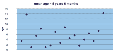
Fig. 1. Characteristics of the age of children operated due to unilateral choanal atresia between 1996 and 2007
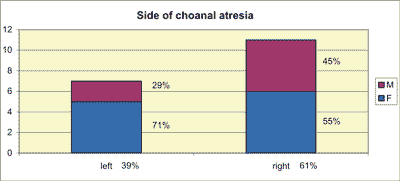
Fig. 2. Unilateral choanal atresia – side and gender
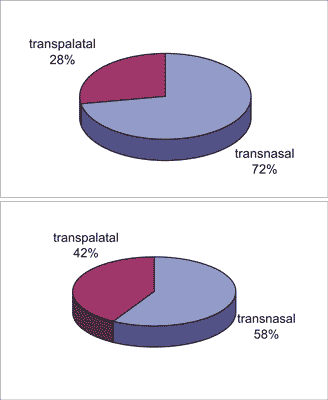
Fig. 3. Surgical approach In choanal atresia treatment a) unilateral, b) bilateral atresia (1996-2007)
The group of patients with bilateral CA consisted of 4 males and 5 females (total 9 patients). First surgical dilations of CA in these children were performed in the first month of life – mean age of children operated in our department was 2 years and 6 months. Patients characteristics in bilateral CA is presented in table 2. The most frequent symptoms beside nasal patency impairment, were feeding difficulty, increased fatiguability, upper respiratory tract infections, and a very difficult perinatal period with increasing respiratory distress, leading in some cases to respirator therapy. Anterior rhinoscopy revealed usually total nasal obstruction on one side and decreased patency on the other side (children previously operated), with mucous or muco-purulent discharge in nasal cavities. First patient described in table 2 was also diagnosed with nasal septum perforation. Seven patients had a perinatal burden (tab. 2). Total number of operations was 12 including 4 reoperations, of which 3 were performed in one patient. Time between subsequent reoperations was 2 to 9 months. Not only had reoperated patients membraneous atretic plate but also bony plate. Among 12 operations five were performed with transpalatal approach and seven with transnasal approach. The nasal cavities were stented with properly sized and fixed endotracheal tubes no. 2,5-4,5. The stents were left in patients´ nasal cavities 4 weeks to 6 months (mean time – approx. 10 weeks). In the postoperational period six patients required antibiotic therapy and the antibiotics such as cefuroxym, ceftriakson or amoxicillin with clavulonic acid were administered for 7 to 14 days. Two children operated in fourth and eighteenth month of life required blood transfusion in postoperational period.
Table 2. Patients´ characteristics in bilateral choanal atresia
| Age (at the time of the operation in our dept.) | Gender | Co-existing abnormalities | No. of earlier operations | No. of operations in our dept. | Perinatal history |
| 4 years 10/12 | M | | 1 | 1 | CC Apgar 6 |
| 5/12 | F | heart defect coloboma of choroid and optic nerve | 2 | 2 | Birth at term Apgar 6 36 Hbd (preterm amniotic fluid discharge) |
| 2/12 | M | imperforate anus heart defect hypothyroidism convergent squint | 2 | 1 | CC Apgar 9 36 Hbd (fetal distress syndrome) |
| 8 8/12 | F | | 4 | 1 | Birth at term Apgar 4 |
| 5 | F | | 1 | 1 | Birth at term |
| 6/12 | M | Physical and mental retardation | 1 | 1 | Lack of data |
| 4/12 | M | Heart defect Physical and mental retardation | - | 4 | Birth at term Apgar 6/7 36 Hbd (intrauterine hypotrophy) |
| 11 | F | | 1 | 4 | Birth at term Apgar 5 31 Hbd |
| 4/12 | F | Down syndrome | 3 | 1 | CC Apgar 3/4/4/6 38 Hbd (fetal distress syndrome) |
CONCLUSIONS
1. Choice of operating method depends on anatomic conditions and experience of particular operating centre.
2. Stent removal – when they achieve good mobility in posterior choanae and can be easily moved to nasopharynx.
3. Method gaining much attention in the management of chonal atresia resently is transnasal approach due to the development of endoscopic techniques.
4. Children with bilateral choanal atresia require re-dilation more frequently.
5. Bilateral choanal atresia is more common in children with co-existing anomalies and with perinatal burden.
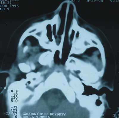
Pic. 1. Computer tomography of a patient with unilateral choanal atresia
Piśmiennictwo
1. Friedman NR, et al.: Management and outcome of choanal atresia correction. Int. J. Pediatr. Otorhinolaryngol 2000; 52: 45-51.2. Chmielik M: Otorynolaryngologia dziecięca. PZWL, Warszawa 2001.3. Brown OE, et al.: Choanal atresia: a new anatomic classification and medical implications. Laryngoscope 1996; 106: 97-101.4. Hengerer AS, et al.: Choanal atresia: a new embryologic theory and its influence on surgical management. Laryngoscope 1982; 92: 913-921.5. Samadi DS, et al.: Choanal atresia: A twenty year review of medical comorbidities and surgical outcomes. Laryngoscope 2003; 113: 254-258.6. Schraff SA, et al.: Management of choanal atresia in CHARGE association patients: A retrospective review. Int. J. Pediatr. Otorhinolaryngol 2006; 70: 1291-1297.7. McLeod IK: Revision choanal atresia repair. Int. J. Pediatr. Otorhinolaryngol 2003; 67: 517-524.8. Khafagy YW: Endoscopic repair of bilateral congenital choanal atresia. Laryngoscope 2002; 112: 316-319.9. Schoem SR: Transnasal endoscopic repair of choanal atresia: why stent? Otolaryngol Head Neck Surg. 2004; 131: 362-366.10. Kubba H, et al.: An update on choanal atresia surgery at Great Ormond Street Hospital for Children: preliminary results with Mitomycin C and the KTP laser. Int. J. Pediatr. Otorhinolaryngol 2004; 68: 939-945.
