© Borgis - New Medicine 4/2007, s. 106-107
Lechosław P. Chmielik1, *Magdalena Frąckiewicz1, Agnieszka Biejat2
Radiological and clinical summary of the pathology of the sphenoid sinus in children. Preliminary report
1Department of Pediatric Otolaryngology, Medical University of Warsaw, Poland
Head of Department: Prof. Mieczysław Chmielik, MD, PhD
2Department of Pediatric Radiology Medical University, Warsaw, Poland
Head of Department: Prof. Andrzej Marciński MD
Summary
Summary
The sphenoid sinus is found in the area of the sphenoid bone. In the close neighborhood of the sphenoid sinus many important structures are located, such as optic nerve, oculomotor nerve, abducent and adjacent nerves, internal carotid artery. Diseases occurring in this area are commonly atypical and include asthenia, apathy, postnasal drip and headaches. Difficulties during making the diagnosis are caused by the very little number of clinical symptoms and also because the sphenoid sinus rarely features in laryngological examination. The authors analyzed 30 child´s cases, on whom functional endoscopic sinus surgery was performed during the period from November 2006 to March 2007. Computed tomography images, intra-operative views and clinical data were compared. Sphenoid sinusitis is a very rare disease among the population of the children. In cases of lesions within the sphenoid sinus, ophthalmic and neurological consultations are needed both pre- and post-operatively. The optimal surgical approach to isolated lesions in the sphenoid sinus in children is by Wigand´s method. The sphenoid sinusitis may occur with insufficiently typical syndromes and may demand a range of tests to reach a correct diagnosis.
OBJECTIVE
The sphenoid sinus is found in the area of the sphenoid bone, bordered by the sella turcica above, and the pituitary gland. Cavernous sinuses with oculomotor and abducent nerves lie on both sides of the sinus, and adjacent to it. On the lateral wall there are impressions of the internal carotid arteries and optic nerves.
Diseases occurring in this area are commonly atypical, and include asthaenia, apathy, postnasal drip, and headaches. Visual disturbances include impaired eye movement, anisocoria, and lesions in the fundus of the eye.
Despite its location within the cerebrocranial cavity, the sphenoid sinus rarely features in laryngological examinations. Our basic method, in locating pathological lesions in the cavity, uses the medical history, and computer tomograhpy of the sinuses in both the coronal and axial planes, as wellas ophthalmological and neurological evaluation.
During endoscopic operation of the sphenoid sinus two approaches are used – that of Wigand, or that of Stammberger.
MATERIAL AND METHOD
We analysed 30 child cases, on whom functional endoscopic sinus surgery was performed during the period 11/2006 to 3/2007. The findings included 5 children who had lesions in the sinus. CT images, intra-operative views and reports of clinical syndromes from patient interviews were compared.

Fig. 1. During FESS of the sphenoid sinus Wigand´s approach is used
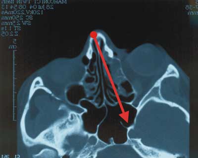
Fig. 2. Stammberger´s method in FESS of the sphenoid sinus.
RESULTS
Headaches were present in 3 children with lesions in sphenoid sinuses, and a post – nasal drip was found in 4. Lesions of the fundus of the eye were seen in 1, and anisocoria was present in 1 case. Intra-operatively oedematous lesions were present in 4 cases, and in 1 case an overall view of the sphenoid sinus was normal.
CONCLUSIONS
1. Sphenoid sinusitis is rare among the child population.
2. In case of lesions within the sphenoid sinus, ophthalmic and neurological consultations are needed both pre and post-operatively.
3. The optimal surgical approach to isolated lesions in the sphenoid sinus in children is byWigand´s method.
4. Sphenoid sinusitis may occur with insufficiently typical syndromes, and may demand a range of tests to reach correct diagnosis.
5. A suspicion of sphenoid sinusitis is often put forward by neurologists during examinations of cerebrocranial area.
Piśmiennictwo
1. Chmielik M, et al.: Operacje endoskopowe zatok (mini FESS ) u dzieci. Otorynolaryngologia; supl. 1, 2004; 38-41.2. Eryilmaz A, Dursun E.: Endoscopic transnasal sphenoidotomy with or without ethmoidectomy. Kulak Burun Bogaz Ihtis Derg. 2007; 17(2): 90-95.3. Stammberger H, Pasawetz W: Functional endoscopic sinus surgery. Concept, indications and results of the Messerklinger technique. Eur. Arch. Otorhinolaryngol., 1990; 247(2).4. Tan HK, Ong YK: Sphenoid sinus: An anatomic and endoscopic study in Asin cadavers. Clin. Anat. 2007; 21.5. Wigand M: Transnasale, Endoskopischer Chirurgie der Nasennebenhohlen bie chronischer sinusitis. II Die endonasale Kieferhhohlen - Operation. HNO 1981; 29: 263-269.
