© Borgis - Postępy Nauk Medycznych 8/2013, s. 577-580
*Małgorzata Kołodziejczak1, Anna Nasierowska-Guttmejer2, 3, Iwona Sudoł-Szopińska1, 4, Anna Wiączek1
Guz Buschke-Lowensteina – trudny problem interdyscyplinarny
Buschke-Loewenstein tumor – a difficult interdisciplinary problem
1Department of General Surgery, Proctology Unit, Solec Hospital, Warsaw
Head of Department: Jacek Bierca, MD, PhD
Head of Unit: Małgorzata Kołodziejczak, MD, PhD
2Pathology Department, Central Clinical, Hospital of Ministry of Interior and Administration, Warsaw
Head of Department: prof. Anna Nasierowska-Guttmejer, MD, PhD
3Faculty of Health Sciences, Department of Pathology, Jan Kochanowski University, Kielce
Head of Faculty: prof. Stanisław Głuszek, MD, PhD
4Department of Radiology, Institute of Rheumatology, Warsaw
Head of Department: prof. Iwona Sudoł-Szopińska, MD, PhD
5Department of Diagnostic Imaging, Second Faculty of Medicine, Medical University, Warsaw
Head of Department: prof. Wiesław Jakubowski, MD, PhD
Streszczenie
Kłykciny olbrzymie, czyli guz Buschke-Loewensteina jest chorobą spowodowaną infekcją wirusem HPV typu 6 i 11, rzadziej 16 i 18, 54 i wg klasyfikacji WHO zaliczany jest do nowotworów brzegu odbytu. Czynnikami ryzyka zachorowania jest zakażenie wirusem HIV, zachowania homoseksualne, immunosupresja oraz choroby obniżające odporność, takie jak cukrzyca czy przewlekły alkoholizm Rozpoznanie opiera się na wywiadzie, badaniu klinicznym, histologicznym, badaniach endoskopowych i badaniach obrazowych. Obraz kliniczny jest bardzo charakterystyczny. Guz ma duże rozmiary (powyżej 10 cm średnicy). W większości przypadków rośnie powoli, przez parę lat, rzadko daje przerzuty wykazując miejscową złośliwość. Może naciekać okoliczne struktury anatomiczne, może też wtórnie powodować przetoki i ropnie. Diagnostyka przedoperacyjna powinna obejmować test na wirus HIV, ultrasonografię transrektalną lub/i MR, która wykazuje stosunek masy guza do mięśni zwieraczy odbytu, CT miednicy, w przypadku naciekania guza na narządy sąsiednie oraz kolonoskopię. Najczęściej stosowaną metodą leczenia jest miejscowe wycięcie guza. Jeżeli występuje naciekanie guza na zwieracze odbytu lub naciekanie guza na sąsiednie narządy – konieczne jest wyprowadzenie stomii, a czasem brzuszno-kroczowe odjęcie odbytnicy. Chemioterapia ma znaczenie drugoplanowe, a skuteczność radioterapii jest dyskusyjna. Konieczne są dalsze multidyscyplinarne badania celem dokładniejszego poznania choroby i określenia skutecznych sposobów leczenia.
Summary
Giant condylomaacuminatum (also known as a Buschke-Lowenstein tumor and giant condyloma of Buschke-Lowenstein tumor) is a disease caused by an infection with HPV type 6 and 11, rarely 16, 18, 54 and according to the WHO, is classified as one of the anal verge neoplasms. Risk factors for the disease are: an infection with HIV, homosexuality, immunosupression and diseases which lower immune system, such as diabetes and chronic alcoholism. Diagnosis is based on clinical examination, histopatological assessment, endoscopic and imaging studies. Clinical presentation is very characteristic. The tumor is large (above 10 cm in diameter). In most cases it is a slow-growing, even up to several years, lesion, which rarely metastases, is locally destructive, may invade surrounding structures, and can also lead to the formation of anal fistulas and abscesses. Preoperative diagnosis should include: HIV testing, anorectalendosonography or MRI, which presents relation of the tumor to anal sphincters, as well as the pelvis, CT, in case of tumor infiltration into the surrounding organs, colonoscopy. The most common method of treatment is a local surgical excission of the tumor. If an infiltration of the tumor to the anal sphincter or tumor invasion into adjacent organs is present – it is necessary to perform a colostomy and sometimes, an abdominoperineal excision of the anus. Chemotherapy is of secondary importance and the effectiveness of radiotherapy is questionable. Multi-disciplinary studies need to be performed to learn more about the disease and to identify effective methods of treatment.

Introduction
Buschke-Lowenstein tumor (BLT) termed the genital giant (giant condyloma), according to the classification of the World Health Organization is a variant of squamous cell carcinoma of the anal canal bank called papillary carcinoma (verrucous carcinoma) (tab. 1) (1). It macroscopically resembles warts (condylomata acuminata), but is larger in size. The rate of BLT in a normal population is low, at 0.1%, while it increases in the male homosexual population. Warts are transmitted sexually and develop as a result of infection with human papillomavirus (HPV). Anal verge cancers are morphologically similar to other tumors of the skin, but are less aggressive than the tumors of anal canal, and more of them vary in terms of histopathology. The incidence of anal verge cancer is about five times smaller than the tumors of the anal canal (2). In most cases patients with Buschke-Lowenstein tumor HPV infection is detected, mainly types 6 and 11, 16 and 18 less frequently and very rarely 54. This may be helpful in differentiating with cancer (squamus cell carcinoma), where mainly virus types 16 and 18 is detected. Currently, over 120 HPV types have been distinguished, which generally can be divided into high risk HPV (e.g., types 16, 18, 33, 34, 45, 52, 56) and low risk HPV (e.g., types 6, 11, 42, 43, 44, 55). Interestingly, the most common ones in patients with HIV infection are warts caused by benign HPV 6 and HPV 11, which are also detected in the Buschke-Loewenstein tumor. The tendency to malignant transformation in Buschke-Loewenstein tumor is much higher than in the case of “typical” warts, approximately 56% (3). The average time of malignant transformation is 5 years (mean 15-100 months) (3).
Table 1. Histological classification of tumors of the anus (21).
| Anal canal |
Squamous cell (cloacogenic) carcinoma
Large cell keratinizing
Large cell nonkeratinizing (transitional)
Basaloid |
Adenocarcinoma
Rectal type
Anal glands/ducts
Within anorectal fistula |
| Small cell carcinoma |
| Undifferentiated |
| Anal margin |
Squamous cell carcinoma
Giant condyloma of Buschke-Loewenstein
Verrucous carcinoma
Basal cell carcinoma
Bowen’s disease
Paget’s disease |
The clinical form of giant condyloma was first described by Buschke and Loewenstein in 1925 (4). The authors present the case of a patient with giant warts located on the penis, describing the change as “invasively growing warts-like tumor”. The perianal location of Buschke-Loewenstein tumor was first described by Peiron in 1931, a 48-year-old patient. Buschke-Loewenstein tumor is more common in men than in women, usually under 50 years of age (the mean 43 years). Trombetta et al. (5) on the basis of the review of scientific literature from the years 1958 to 2000 identified 51 cases of Buschke-Loewenstein tumor. Men got sick more often (2.7:1), the mean age of patients was 43.9 years. Isolated cases of this tumor are also described in children (6). Ambriz-González et al. (6) presented the case of a 12-year-old girl with a Buschke-Loewenstein tumor in the anal area treated effectively with local excision and radiotherapy. Risk factors for developing giant warts are infections with HIV, homosexuality, immunosuppression (e.g. transplant patients after surgery) and immunosuppressive diseases, such as diabetes or chronic alcoholism. In the case of HIV-positive men 92% of HPV infection is recognized around the anus.
Diagnosis is based on history, clinical examination, histological examination, and endoscopic and imaging examinations (transrectal ultrasound, MRI, CT).
Clinical picture is very distinctive. The tumor is large (over 10 cm in diameter) (fig. 1). In most cases, grows slowly, for a few years, rarely metastasizes, exhibits local malice. It may invade surrounding anatomical structures (vagina, urethra, prostate, anal sphincters), it can also cause secondary fistula and abscess. It occasionally invades retroperitoneal space above the anal lifters muscles. Soós et al. (7) described a 63-year-old woman with a Buschke-Loewenstein tumor located on the edge of the anus. The tumor infiltrated the anal sphincter muscle and superalevator retroperitoneal space, causing bilateral hydronephrosis. Patients with a Buschke-Loewenstein tumor may report pain, bleeding, itching and burning of inflamed skin around the anus, and in the case of invasion of the tumor to the anal sphincter, urinary and bowel gas. On the surface of the tumor abscess or anal fistula may occur. According to Trombetta et al. (5) pain in the course of Buschke-Loewenstein tumor is reported by 32% of patients, bleeding in 18% of cases, abscess and fistula can develop in 32% of patients.
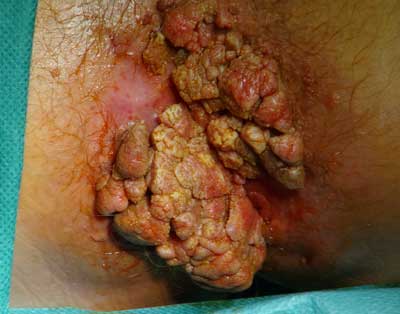
Fig. 1. The clinical picture of Buschke-Loewenstein tumor.
Histopathology
Powyżej zamieściliśmy fragment artykułu, do którego możesz uzyskać pełny dostęp.
Mam kod dostępu
- Aby uzyskać płatny dostęp do pełnej treści powyższego artykułu albo wszystkich artykułów (w zależności od wybranej opcji), należy wprowadzić kod.
- Wprowadzając kod, akceptują Państwo treść Regulaminu oraz potwierdzają zapoznanie się z nim.
- Aby kupić kod proszę skorzystać z jednej z poniższych opcji.
Opcja #1
29 zł
Wybieram
- dostęp do tego artykułu
- dostęp na 7 dni
uzyskany kod musi być wprowadzony na stronie artykułu, do którego został wykupiony
Opcja #2
69 zł
Wybieram
- dostęp do tego i pozostałych ponad 7000 artykułów
- dostęp na 30 dni
- najpopularniejsza opcja
Opcja #3
129 zł
Wybieram
- dostęp do tego i pozostałych ponad 7000 artykułów
- dostęp na 90 dni
- oszczędzasz 78 zł
Piśmiennictwo
1. Bosman FT, Carneiro F, Hruban RH, Thiese ND (eds.): WHO Classification of Tumour of the Digestive System. IARC, Lyon 2010: 183-185.
2. Mik M, Dziki A: Nowotwory odbytu. [W:] Dąbrowski A (red.): Gastroenterologia. Cz. II, Medical Tribune 2011: 398-401.
3. Chu QD, Vezeridis MP, Libbey NP, Wanebo HJ: Giant condyloma acuminatum (Buschke-Loewenstein tumor) of the anorectal and perianal regions. Analysis of 42 cases. Dis Colon Rectum 1994; 37: 950-957.
4. Buschke A, Loewenstein L: Über carcinomahnliche condylomata acuminata des penis. Klin Wochenschr 1925; 4: 1726-1728.
5. Trombetta LJ, Place RJ: Giant condyloma acuminatum of the anorectum: trends in epidemiology and management. Report of a case and review of the literature. Dis Colon Rectum 2001; 44: 1878-1886.
6. Ambriz-González G, Escobedo-Zavala LC, Carrillo de la Mora F et al.: Buschke-Löwenstein tumor in childhood: a case report. J Ped Surg 2005 Sep; 40(9): 25-27.
7. Soós Z, Varga T, Vadinszky P et al.: Verrucous carcinoma of the anal margin. The importance of adequate biopsy technique. Orv Hetil 2011 Feb 27; 152(9): 344-348.
8. Mazurkiewicz W: Choroby przenoszone drogą płciową. [W:] Bielecki K, Dziki A (red.): Proktologia. PZWL, Warszawa 2000: 313-336.
9. Bieniek A, Cisło M, Matusiak Ł et al.: Rak brodawkujący (carcinoma verrucosum) – przegląd objawów klinicznych i histologicznych. Post Dermatol Alergol 2006; XXIII(2): 57-66.
10. Longacre TA, Kong CS, Welton ML: Diagnostic problems in anal pathology. Adv Anat Pathol 2008 Sep; 15(5): 263-278.
11. Renzi A, Giordano P, Renzi G et al.: Buschke-Lowenstein tumor successful treatment by surgical excision alone: a case report. Surg Innov 2006 Mar; 13(1): 69-72.
12. Papiu HS, Dumnici A, Olariu T et al.: Perianal giant condyloma acuminatum (Buschke-Löwenstein tumor). Case report and review of the literature. Chirurgia (Bucur) 2011 Jul-Aug; 106(4): 535-539.
13. De Toma G, Cavallaro G, Bitonti A et al.: Surgical management of perianal giant condyloma acuminatum: Report of three cases. Eur Surg Res 2006; 38: 418-422.
14. Martin JM, Molina I, Monteagudo C et al.: Buschke-Lowenstein tumor. J Dermatol Case Rep 2008 Dec 27; 2(4): 60-62.
15. Tytherleigh MG, Birtle AJ, Cohen CE et al.: Combined surgery and chemoradiation as a treatment for the Buschke-Löwenstein tumour. Surgeon 2006 Dec; 4(6): 378-383.
16. Grochowicz M, Grochowicz P: Kłykciny kończyste. [W:] Bielecki K, Dziki A (red.): Proktologia. PZWL, Warszawa 2000: 313-336.
17. Mudrikowa T, Jaspers C, Ellerbroek P, Hoepelman A: HPV- -related anogenital disease and HIV infection: not always “ordinary” condylomata acuminate. J Med 2008; 66(3): 98-102.
18. Sobrado CW, Mester M, Nadalin W et al.: Radiation-induced total regression of a highly recurrent giant perianal condyloma: report of case. Dis Colon Rectum 2000 Feb; 43(2): 257-260.
19. Picaud A, Faye A, Ogowet-Igumu N et al.: Büschke-Loewenstein tumor during pregnancy. Apropos of 2 cases. Rev Fr Gynecol Obstet 1990 Jun; 85(6): 375-378.
20. Lévy A, Lebbe C: Buschke-Löwenstein tumour: diagnosis and treatment. Ann Urol (Paris) 2006 Jun; 40(3): 175-178.
21. Fenger CF, Frisch M, Marti MC, Parc R: Tumours of anal canal. [In:] Hamilton SR, Aaltonen LA: World Health Organization of Tumours: Pathology and Genetics of Tumours of the Digestive system. IARC Press, Lyon 2000: 145-155.
