© Borgis - New Medicine 2/2011, s. 46-51
*Paweł Szulc, Joanna Boch-Kmieciak, Piotr Bartkowiak, Jacek Lewandowski
The impact of rugby training on the spinal functional parameters
University School of Physical Education in Poznań, Faculty of Functional Anatomy
Department Manager: Jacek Lewandowski, Prof. Ph.D.
Summary
Aim. The aim of the present paper was to determine the impact of many years of rugby training on the formation of spinal curvatures and spinal sectional mobility.
Material and methods. The research material constituted 20 rugby players of the Posnania Sports Club aged 20-25, and the control group of 20 students from the University School of Physical Education in Poznan aged 20-21. The results were also referred to the normative values. In both groups, the measurements of motion ranges of all the spinal sections in three planes, and the measurements of physiological curvatures were carried out using the Penny & Giles tensometric electrogoniometer.
Results. The significant limitations in the cervical spine concerned movements in sagital and transverse plane. The extension motion was limited by 17°, right-sided axial rotation by 29° and the left-sided by 32°.
Similarly, in the thoracic spine, significant mobility limitations occurred in the same planes, however the flexion motion was limited by 12° and axial rotation to both sides by 11°.
There was a statistically significant increase in angular values of spinal physiological curvatures among the rugby players by 10° on average.
Discussion. The obtained results indicate an increase in the angular values of physiological curvatures in rugby players and the limitation of mobility in the cervical and thoracic spine. Other authors indicate the flattening of spinal physiological curvatures. The discrepancies in the obtained study results indicate a more dynamic or static structure of the conducted rugby training.
Conclusions. Early introduction of the compensation exercises in rugby players may limit the unfavourable mobility changes within the functional spinal characteristics.
Introduction
The mobility range in respective spinal sections in people is an eco-sensitive value. This means that the spine, through the impact of the development factors, becomes a unique sensor of changes to which the human body is subjected during the development (1). Besides the ontogenetic factors, the sectional spinal mobility is also affected by the pathologic processes and the specific physical activity during the training (2, 3, 4, 5, 6).
Just as in many other fields, the knowledge of spondylometric parameters is necessary for conducting and applying appropriate training methods. All the team sport games are based on the strictly specified character of sport struggle. It is closely related to the regulations and the nature of the discipline. Taking this into account, rugby may be included in the contact sports (8, 7, 4, 9). The literature on the subject related to the impact of sport training on the functional spinal parameters is not presented in the scientific publications to a great extent (2, 5, 6) On the other hand, there are numerous reports on the motor organ ailments caused by sport training and relevant treatment methods (3, 4, 9). Any changes in spinal functional parameters impact greatly the occurrence of the motor apparatus ailments related to its overburdening (10). Changing functional parameters of the spine result in further consequences in the form of disturbances in the biokinematic chain mechanisms (10). The rugby training influences the strain of the cervical and lumbar spine significantly, as a consequence of positions applied during the game. Above all, this refers to the attack formation player and the jumper during his landing on one leg (6, 11). On top of this, the weight-bearing exercises are a significant component of the training process, leading to the development of muscular mass, causing functional changes within the spine (2). The changes may have a negative impact on the anatomic structures of the spine in individuals practicing rugby (6, 11, 12, 13 ).
Aim of the study
The aim of this paper is to determine the impact of many years of rugby training on the formation of physiological curvatures and sectional spinal mobility.
Material and methods
The research material covered 20 players aged 20-25, whose mean age was 22 years – practicing rugby at the Posnania Sports Club, with the training experience of minimum 5 years. The average training experience of the players was 6 years.
The control group consisted of 20 students of the E. Piasecki University School of Physical Education in Poznan aged 20-21. In order to perform the tests, the consent of the Bioethical Committee was obtained. The results of the conducted studies were referred to the normative values for the same age group presented by Lewandowski (14).
The motion ranges of the cervical, thoracic and lumbar spine and the angular values of its physiological curvatures were measured five times in both groups.
The tests were performed in a room with a stable temperature of 22°-24°C for all the players and the control group, at the same time of the day, during the days which were free of training and physical exercises. No warm-up was performed, before proceeding to the tests. The mean from five measurements for each value constituted the measuring result.
The tests were performed using the electrogoniometric measuring system of the Penny & Giles company (according to Lewandowski’s methodology) in the Boocock’s modification (14, 15). This modification eliminates measuring errors related to the movement of skin and soft tissues in relation to the bones (fig. 1). The electrogoniometric sensors were fixed to the body with a two-sided medical tape. The measurement constituted growth of the joint angle. The starting position for the test was the upright standing position with limbs arranged along the torso and head in the Frankfurt plane (fig. 2).
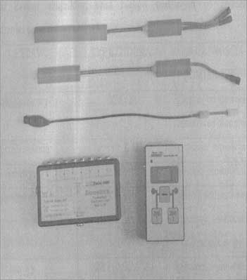
Fig. 1. Penny & Giles electrogoniometric measuring set.
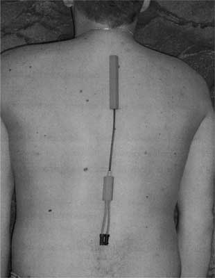
Fig. 2 . The method of fixing the electrogoniometric sensor for the measurement of physiological curvatures and sectional mobility of the spine.
Obtained results of electrogoniometric tests were elaborated by means of the commonly used statistical methods, calculating the arithmetic means, standard deviation X, minimums and maximums. The material distribution was assessed with the use of Shapiro-Wilk test. Due to the absence of the normal material distribution, the non-parametric U Mann-Whitney test was used for determining the significance of differences between the angular values of physiological curvatures and the sectional spinal mobility of the group practicing rugby and the control group.
Results
While analysing the mobility range in the cervical spine in individuals practicing rugby, attention must be paid to a decreased mobility of this section in the majority of the performed movements. The significant limitation concerned the backward bending motion by 17°, right-sided axial rotation by 29° and left-sided by 32°. The players show only higher values of the side flexure to the right and left in relation to the control group and the normative values, whereby these differences are not significant. (tab. 1, 2, 3, 4).
Table 1. Statistical characteristics of spondylometric features (sectional mobility of the spine) in the group of players practicing rugby.
| Spinal section | Mobility | N | x (°) | SD | Min (°) | Max (°) |
| Cervical | Flexion | 20 | 47.4 | 14.12 | 22 | 55 |
| Extension | 51.2 | 7.7 | 40 | 61 |
| Left-sided flexion | 49.6 | 5.55 | 30 | 42 |
| Right-sided flexion | 49 | 7.6 | 30 | 48 |
| Left-sided axial rotation | 40 | 4.06 | 34 | 45 |
| Right-sided axial rotation | 39.6 | 5.38 | 36 | 48 |
| Thoracic | Flexion | 20 | 13.6 | 4.72 | 7 | 18 |
| Extension | 18.2 | 5.89 | 10 | 24 |
| Left-sided flexion | 24.4 | 5.12 | 18 | 30 |
| Right-sided flexion | 24.6 | 4.02 | 20 | 28 |
| Left-sided axial rotation | 15 | 1.3 | 14 | 17 |
| Right-sided axial rotation | 13.2 | 1.3 | 11 | 14 |
| Lumbar | Flexion | 20 | 55.4 | 14.65 | 35 | 73 |
| Extension | 26.4 | 5.8 | 20 | 35 |
| Left-sided flexion | 20.4 | 4.76 | 20 | 33 |
| Right-sided flexion | 20.6 | 1.41 | 19 | 23 |
| Left-sided axial rotation | 6 | 1.3 | 5 | 8 |
| Right-sided axial rotation | 6.6 | 1.78 | 5 | 9 |
Table 2. Statistical characteristics of spondylometric features (sectional mobility of the spine) in the control group.
| Spinal section | Mobility | N | x (°) | SD | Min (°) | Max (°) |
| Cervical | Flexion | 20 | 50.26 | 6.9 | 39 | 63 |
| Extension | 68.47 | 8.66 | 46 | 75 |
| Left-sided flexion | 48.15 | 5.12 | 36 | 55 |
| Right-sided flexion | 46.52 | 5.25 | 41 | 57 |
| Left-sided axial rotation | 69.05 | 4.65 | 56 | 79 |
| Right-sided axial rotation | 71.78 | 5.68 | 49 | 78 |
| Thoracic | Flexion | 20 | 25.73 | 7.77 | 7 | 32 |
| Extension | 23.21 | 6.02 | 13 | 31 |
| Left-sided flexion | 18.15 | 6.11 | 14 | 26 |
| Right-sided flexion | 18.15 | 4.26 | 13 | 24 |
| Left-sided axial rotation | 25.52 | 5.89 | 15 | 34 |
| Right-sided axial rotation | 24.15 | 6.65 | 16 | 32 |
| Lumbar | Flexion | 20 | 55.63 | 11.07 | 35 | 73 |
| Extension | 28 | 9 | 17 | 41 |
| Left-sided flexion | 25.47 | 5.02 | 14 | 42 |
| Right-sided flexion | 25.84 | 5.89 | 12 | 40 |
| Left-sided axial rotation | 9.73 | 2.43 | 5 | 15 |
| Right-sided axial rotation | 10.94 | 2.67 | 5 | 17 |
Table 3. Statistical characteristics of spondylometric features (sectional mobility of the spine) in males aged 22 – normative values from J. Lewandowski’s studies (14).
| Spinal section | Mobility | N | x (°) | SD | Min (°) | Max (°) |
| Cervical | Flexion | 20 | 64.06 | 5.33 | 47 | 74 |
| Extension | 60.29 | 5.26 | 50 | 75 |
| Left-sided flexion | 52.88 | 3.30 | 45 | 63 |
| Right-sided flexion | 53.09 | 3.17 | 46 | 62 |
| Left-sided axial rotation | 86.37 | 3.73 | 77 | 97 |
| Right-sided axial rotation | 86.38 | 3.70 | 77 | 96 |
| Thoracic | Flexion | 20 | 27.71 | 3.36 | 21 | 39 |
| Extension | 23.37 | 2.14 | 19 | 29 |
| Left-sided flexion | 31.51 | 3.54 | 25 | 40 |
| Right-sided flexion | 31.43 | 3.32 | 24 | 40 |
| Left-sided axial rotation | 31.75 | 3.79 | 24 | 40 |
| Right-sided axial rotation | 31.61 | 3.68 | 25 | 40 |
| Lumbar | Flexion | 20 | 68.41 | 6.36 | 59 | 90 |
| Extension | 32.22 | 4.76 | 20 | 46 |
| Left-sided flexion | 32.20 | 2.74 | 27 | 40 |
| Right-sided flexion | 32.26 | 2.71 | 26 | 40 |
| Left-sided axial rotation | 11.46 | 1.12 | 9 | 14 |
| Right-sided axial rotation | 11.56 | 1.13 | 9 | 15 |
Table 4. The significance of differences in the sectional mobility of the spine between the group of players and the control group, performer by means of the U Mann – Whitney test (*p ≤ 0.05, **p ≤ 0.1).
| N | Mobility | Cervical section-test value | Thoracic section-test value | Lumbar section-test value |
| 20 | Flexion | 0.374 | 0.035* | 0.490 |
| Extension | 0.026* | 0.189 | 0.386 |
| Left-sided flexion | 0.442 | 0.264 | 0.238 |
| Left-sided flexion | 0.353 | 0.258 | 0.235 |
| Axial rotation L | 0.031* | 0.047* | 0.212 |
| Axial rotation R | 0.022* | 0.046* | 0.226 |
The significant mobility limitations appeared also in the thoracic spine and concerned the forward bending motions by 12° in relation to the control group, and the axial rotations to both sides by 11° (tab. 1, 2, 3, 4).
The lumbar spine is characterised by the decreased mobility in the transverse and frontal plane, however these changes are not statistically significant, and remaining values approximate the normative values (tab. 1, 2, 3, 4).
The statistically significant angular changes in spinal physiological curvatures were observed among the rugby players.
The angular value of the physiological curvatures in all three spines is higher by 10° on average in reference to the control group and to the normative values (tab. 5, 6, 7, 8).
Table 5. Statistical angular values in three spinal sections in the group practicing rugby.
| N | Curvature | x (°) | SD | Min (°) | Max (°) |
| 20 | Cervical lordosis | 44 | 7.43 | 32 | 50 |
| Thoracic kyphosis | 41.24 | 6.27 | 34 | 50 |
| Lumbar lordosis | 47.75 | 4.42 | 42 | 55 |
Table 6. Statistical angular values of curvatures in three spinal sections in the control group.
| N | Curvature | x (°) | SD | Min (°) | Max (°) |
| 20 | Cervical lordosis | 35.88 | 6.74 | 31 | 51 |
| Thoracic kyphosis | 32.07 | 6.03 | 27 | 37 |
| Lumbar lordosis | 37.61 | 7.68 | 30 | 48 |
Table 7. Statistical angular values of curvatures in three spinal sections for the group with normative values given by J. Lewandowski [Lewandowski J. 2006].
| N | Curvature | x (°) | SD | Min (°) | Max (°) |
| 20 | Cervical lordosis | 36.52 | 5.21 | 26 | 56 |
| Thoracic kyphosis | 32.43 | 4.15 | 24 | 42 |
| Lumbar lordosis | 36.06 | 4.97 | 24 | 52 |
Table 8. The significance of differences in angular values of physiological curvatures of the spine between the group of players practicing rugby and the control group performed by means of the U Mann – Whitney test (*p ≤ 0.05, **p ≤ 0.1).
| N | Spinal curvature | U Manna - Whitney’s test value |
| 20 | Cervical lordosis | 0.047* |
| Thoracic kyphosis | 0.043* |
| Lumbar lordosis | 0.039* |
Discussion
There are different views documented by the results of studies with regard to the formation of physiological spinal curvatures in rugby players. Uetake in his paper presents results of studies of the physiological spinal curvatures in 380 sportsmen of various disciplines, including rugby players, made with the Moire’s method. According to this author, as a result of practicing running and jumping, the physiological curvatures become greater, whereas in body-builders and sportsmen practicing weight lifting and rugby, they become flattened as a result of assuming the forward leaning body position similarly to the case of grass hockey players (6). The iliocostalis muscle and the longissimus dorsi muscle is tensed, which flattens the lumbar lordosis and thoracic kyphosis. The application of axial loads in rugby players at insufficient strength of the erector spinae may deepen the physiological spinal curvatures, which contributes to degenerative changes in intervertebral discs, compression fractures and fatigue fractures of vertebral bodies and stenoses as a result of ligament overgrowth, which takes over the muscle function (3, 4, 13, 16). The deepening of the physiological spinal curvatures was observed in own material, which proves the greater component of dynamic exercises – i.e. running and jumping, and insufficient strength of the muscles stabilising the spine during axial loads e.g. attack formation (3, 11). Therefore it may be concluded that not only does the type of practiced sport discipline determine the deepening and decreasing of the spinal physiological curvatures, but, above all, the structure of the conducted training (2 ,5, 6).
The decreased respective spinal sections mobility, particularly in the sagital and transversal plane of the thoracic and cervical spine may follow from the ligament apparatus overgrowths of the spine, which does not provide protection against increasing and deepening of kyphoses and lordoses, but limits the motion ranges (13). Additionally, attention must be paid to the fact that in the course of deepening of the physiological curvatures, the erector spinae is stretched, especially the medial band (17). The stretched group of muscles reacts with a contraction, which also limits mobility in the direction opposite to the action of these muscles in the concentric contraction. This explains the mobility limitations in the sagital and transversal plane of the top part of the spine observed in own material (17). Uetake in his paper ascertains the flattening of the physiological spinal curvatures in rugby players, which proves the advantage of the low positions, with torso leaned forward and a great component of weight-bearing exercises applied in the training cycle. Such structure of sport activities causes the formation of functional characteristics of the studied motor organ which is different than the one observed in own material (6). The training should consist of many exercises strengthening the muscular apparatus of the spine, which serves as a protection against the deepening of the physiological curvatures of the studied organ. It should take into account the symmetrical exercises, post isometric relaxation in order to eliminate the effects of axial loads and excessive muscular tensions resulting from rugby playing (18).
The lack of compensation of axial loads may lead to a decrease in the sectional spinal mobility and an increase in the physiological curvatures, which, as a consequence, may cause degenerative changes of the studied motor organ (7, 16). The early introduction of compensation exercises may have a positive impact on the decrease in the described negative changes in mobility and curvatures of the spine (19, 20).
The above-mentioned observations should be a precious clue which the coaching team must consider while arranging the training program.
Conclusions
1. The results of the paper determine the tendency of changes in spinal physiological curvatures in rugby players tested by the research team. The depth of physiological curvatures is affected by the dynamic character of loads in the practiced sport discipline.
2. Significant axial loads cause limitations in mobility in the cervical and thoracic spine in the transversal and sagital plane. This is caused by an increase in the muscular tensions during the performance of tactical elements of the game.
3. A relatively high stability of the lumbar spine mobility is caused most probably by the high mass of muscles stabilising this spinal section.
4. In the view of the above results, we have to remember that the early introduction of compensation exercises in rugby players may limit the negative changes in mobility within the functional characteristics of the spine.
Piśmiennictwo
1. Kapandji IA: Funktionelle Anatomie der Gelenke. Bd3 Rumpf Und Wirbelsaule. Ferdinand Enke Verlag 1985; 106-11. 2. Aggrawal N, Kaur R, Kumar S, Mathur D: Study of changes in the spine in weight lifters and other athletes. Preview.ritish Journal of Sports Medicine 1979; 13 (2): 58-61. 3. Berge J, Marque B, Vital JM et al.: Age- related changes in the cervical spines of front-line rugby players. Am J Sports Med 1999; 27 (4): 422-9. 4. Gatt CJ Jr., Hosea TM, Palumbo RC, Zawadsky JP: Impact loading of the lumbar spine during football blocking. Am J Sports Med 1977; 25: 317-321. 5. Nachbauer W, Nigg B: Effects of arch height of the foot on ground reaction forces in running. Medicine & Science in Sports & Exercise 1992; 24 (11): 1264-1269. 6. Uetake T, Ohtsuki F, Tanaka H, Shinodo M: The vertebral curvature of sportsmen. J. of Sports Sci. 1998; 16 (7): 621-628. 7. Bohu YJ, Bagate M, Peyrin C et al.: Cervical spinal injuries in rugby. Rugby - NY 15 Aug 1994; 20: 20-21. 8. Castinel BB, Turblin CH, Milburn PH, Quarrie P: Mecanismes des traumatismes cervicaux dans le jeu de rugby: biomecanique, physiopathologie des blessures, epidemiologie. Journal de Traumatologie du Sport mars 2005; 22 (1): 541. 9. Vital JM; Les traumatismes discoligamentaires du rachis cervical bas dans la pratique du rugby. Journal de Traumatologie du Sport mars 2005; 22 (1): 54-60. 10. Janiszewski M, Wadowski A: Ocena skuteczności terapii manualnej w zaburzeniach czynnościowych łańcucha biokinematycznego: kręgosłup lędźwiowy ,miednica, stawy krzyżowo-biodrowe. Medycyna Manualna 2001; 31-32. 11. Munro CF, Miller DI, Fuglevand AJ: Ground reaction forces in running. Journal of Biomechanics 1987; 20, 147-155. 12. Matzdorff I: Contributions on the external lumbar lordotic curvature of the vertebral column. Journal of the Anthropological Society of Nippon 1972; 80, 113-150. 13. Paterson S, Castinel B, Newsham-West RJ Milburn PD: Spinal canal encroachment demonstrated by a novel MRI technique of an axial loaded cervical spine - a case study. New Zealand Journal of Sports Medicine Autumn 2007; 34 (2): 23-26. 14. Lewandowski J: Kształtowanie się krzywizn fizjologicznych i zakresów ruchomości odcinkowej kręgosłupa człowieka w wieku 3-25 lat w obrazie elektrogoniometrycznym. AWF Poznań 2006. 15. Boock MG, Jackson JA: Continuous measurement of lumbar posture Rusing flexible electrogoniometers. Ergonomics 1994; 37 (1): 175-185. 16. Castinel BH, Prat A, Christophe A stress fracture of the lumbar spine in a professional rugby player. British Journal of Sports Medicine May 2007; 41 (5): 337-338. 17. Richardson C, Houdges P, Hides J: Mechanizm przykręgosłupowy w dolegliwościach bólowych dolnego odcinka kręgosłupa. Kinezyterapia w stabilizacji kompleksu lędźwiowo-miedniczego. Elsevier Urban & Partner 2009; 187-199. 18. Bompa OT: Teoria i metodyka treningu. Resortowe Centrum Metodyczno- Szkoleniowe Kultury Fizycznej i Sportu Warszawa. Warszawa 1990 19. Sinibaldi, Kevin S.1Smith, Dana R.1 Prevention of Spinal Injuries in Rugby. Strength & Conditioning Journal 2007; 29 (4): 18 7p. 20. Quarrie, Kenneth L, Gianotti, Simon M. Hopkins, Will G.3Hume, Patria A: Effect of nationwide injury prevention programme on serious spinal injuries in New Zealand rugby union: ecological study. BMJ: British Medical Journal 2007; 334 (7604): 1150-1153.
