*Csaba Szekrenyesi1, Gabor Sandor2, Andrea Gyenes2, Huba Kiss2, Tamas Filkorn1, 2, Zoltán Z. Nagy1, 2
The change of corneal surface temperature during surface refractive surgical procedures performed using excimer lasers with different repetitive frequencies
1Faculty of Health Sciences, Semmelweis University, Budapest, Hungary
Head of Faculty: Professor Nagy Zoltán Zsolt, MD, PhD
2Department of Ophthalmology, Semmelweis University, Budapest, Hungary
Head of Department: Professor Nagy Zoltán Zsolt, MD, PhD
Summary
Intoduction. Refractive surgery is still one of the most frequently performed procedures in ophthalmology. Millions of patients every year are operated with surface lasers as well as with deep or lamellar lasers. The operational technique and tools are constantly improving.
Aim. To examine the changes in the temperature of the corneal surface during refractive surgery procedures performed using different types of excimer lasers.
Material and methods. 150 eyes of 111 patients were included in the study. The corneal surface temperature was measured with a calibrated EBRO TLC730 infrared thermometer (WTW GmBH, Germany) before photorefractive keratectomy (PRK), after epithelial removal and immediately after finishing the procedure. The average myopic correction was -3.9 ± 1.9 Dpt (ranging from -2.5 Dpt to -6.5 Dpt). The average age of the patients was 25 ± 3.3 years.
Results. Using ANOVA variance analysis, a statistically significant difference in the change of the temperature was found between different laser platforms. With the post hoc Bonferroni correction, a significant difference between the MEL 80 (Carl Zeiss Meditec, Jena, Germany) and the two other analyzed excimer lasers, MEL 70 (Carl Zeiss Meditec, Jena, Germany) and Wavelight Allegretto 400 (Alcon Wavelight, Erlangen, Germany), was found. There was no statistically significant difference between the Wavelight Allegretto and MEL 70 excimer lasers.
Conclusions. The moderate cooling effect of the different excimer laser platforms might be related to different airflow systems used in the models. It seems that the rise in the surface temperature is not a causative factor of postoperative subepithelial corneal haze when the procedure is performed with lasers using frequencies lower or equal to 400 Hz.
Introduction
Refractive surgery is still one of the most frequently performed procedures in ophthalmology. Millions of patients every year are operated with surface lasers (PRK, PTK, LASEK, epi-LASIK) as well as with deep or lamellar lasers (LASIK, femto-LASIK). The operational technique and tools are constantly improving, for example, large beam delivery systems are now replaced by small-beam, flying-spot lasers (1, 2). Modern excimer lasers operate with the beam diameter smaller than 1.0 mm. The repetition frequency increased from 35 Hz in the first lasers ever used to 1000 Hz today (3, 4). In lamellar procedures, mechanical microkeratomes were superseded by femtolasers, that provide a more even and predictable stromal bed (5).
Many studies showed that any cut in the deeper layers of the cornea results in the increase of the higher-order aberrations, leading to visual symptoms, such as glare and halo phenomena. These complications tend to have a smaller prevalence in patients who underwent a procedure on the corneal surface (6, 7).
Femtolaser LASIK is frequently used at the depth of 90 μm. A slogan “back to the surface” was even coined among the surgeons, as nowadays, a maximally thin flap is suggested in order to preserve more untreated stromal tissue (8, 9).
Corneal wound healing was a big issue at the dawn of the refractive surgery. Significant subepithelial haze frequently complicated the procedure, resulting from a large beam delivery system, longer surgery time due to less experienced surgeons, long time of the active laser procedure itself, smaller optical zone, variable depth of ablation. The diopter range and the ablation depth related to the outcomes of the treatment were not as clearly stated as today at that time (10, 11). Poor corneal wound healing was frequently discussed on medical congresses consecrated to refractive surgery. Early publications on the refractive surgery extensively discussed the changes in surface temperature of the cornea. According to the literature (12), the changes in the corneal surface temperature vary between 4 to 96°C depending on the technique of the measurement. There are many techniques from thermal camera to thermal radiometry with very different range of temporal and spatial resolution, which can result in significant differences in the temperature measured. Therefore, the technique of the measurement is very important, but the repetition rate also plays a significant role in changes of the corneal surface temperature.
Formerly, excimer lasers used short-wavelength ultraviolet (UV-C; 193 nm) and a frequency of 35 Hz, while modern excimer lasers are much faster, achieving the frequency of up to 1050 Hz. Again, the warming of the corneal surface becomes an important issue, due to the fact that the higher the frequency, the higher the change in the temperature of the cornea (13, 14). Excessive warming may induce metabolic changes in keratocytes, leading to healing problems, reticular or floccular haze and consequent refractive regression as well as thickening of the cornea (15, 16).
During this study, the authors examined the changes in the corneal surface temperature during surgical refractive procedures performed with an older flying spot excimer laser, MEL 70 (Carl Zeiss Meditec GmBH, Jena, Germany) that operates with the frequency of 35 Hz, and newer models, MEL 80 (Carl Zeiss Meditec GmBH, Jena, Germany) that uses the frequency of 250 Hz and the Wavelight Allegretto (Wavelight Inc., Erlangen, Germany) that operates with the frequency of 400 Hz.
Aim
The aim of this study was to determine whether an increase in the repetitive frequency causes any immediate changes in the corneal surface temperature, and whether there is a difference in postoperative wound healing between patients who underwent surgery performed with different excimer lasers.
Material and methods
150 eyes of 111 patients (35 men and 76 women) were included into the study. The average age of the patients was 25.0 ± 3.3 years. Inclusion criteria were as follows: myopic or myopic astigmatic refractive error between -2.5 and -6.5 Dpt of the spherical equivalent refractive range, astigmatism of up to -2.0 cyl. Dpt.
Treatment groups:
Group 1: MEL 70 flying spot excimer laser (Carl Zeiss Meditec GmBH, Jena, Germany): n = 50 (17 men and 33 women, average age: 25.5 ± 2.7 years).
Group 2: MEL 80 flying spot excimer laser (Carl Zeiss Meditec GmBH, Jena, Germany): n = 50 (15 men and 35 women, average age: 24.6 ± 3.2 years).
Group 3: Allegretto flying spot excimer laser (Wavelight Inc., Erlangen, Germany): n = 50 (18 men and 32 women, average age: 24.8 ± 3.9 years).
The groups did not differ from each other significantly in age, sex and preoperative refractive error.
During the postoperative follow-up time, no difference in corneal wound healing was noted between the groups.
Procedures
All of the patients underwent medical examination before being qualified to refractive surgery. Preoperative assessment included: automated refractometry, uncorrected near and far visual acuity, best corrected far and near visual acuity with both normal and dilated pupil, corneal topography (the assessment of the anterior and posterior corneal surface) and corneal thickness measured and recorded with the Pentacam Scheimpflug device. Intraocular pressure was measured with Goldmann tonometry. Fundus examination on dilated pupil was performed.
Patients received three doses of oxybuprocaine hydrochloride before refractive surgery. Corneal surface temperature was measured using a highly accurate infrared thermometer (EBRO TLC 730, WTW GmBH, Germany) before the procedure, after epithelial removal and directly after refractive treatment. The time elapsed between the measurements was also noted, enabling the calculation of the speed of temperature change in °C/min.
The thermometer was calibrated. Its accuracy was 0.1°C. All the measurements were collected 3 seconds after finishing the surgical procedure, from the distance of 8 cm. The distance (D) was proportional to the measured spot size (S), with spot size amounting 1 cm, D/S = 8/1. The thermometer was equipped with integrated red light laser pointers to help identify the surface whose temperature is to be measured. The thermometer was stored in the operating room, enabling its adjustment to the environmental temperature.
After the third measurement, that was taken after the procedure, a cold (+ 4°C) BSS fluid was applied to the corneal surface for 15 seconds in order to achieve better postoperative analgesia. Afterwards, a soft contact lens was applied into the operated eye. Finally, antibiotic eye drops (tobramycin) were instilled and the patients were dismissed.
Gas and suction facilities of the excimer lasers may have a cooling effect on the corneal surface. Because of that, the air velocity on the working plane of the lasers was measured with the TESTO 405-V1 anemometer (accuracy: ± 1 digit - ± (0.1 m/s ± 5% of the measured value) (in the range 0 to 2 m/s), ± (0.3 m/s ± 5% of the measured value) (in the range 2.1 to 10 m/s)). The measured air velocity was 0.4 m/s for the MEL70 laser, 2.2 m/s for the MEL80 laser and 0.15 m/s for the Allegretto laser.
Environmental conditions in the operating room were stable, with temperature set to 22°C and humidity measuring 35% ± 5% (17).
Results
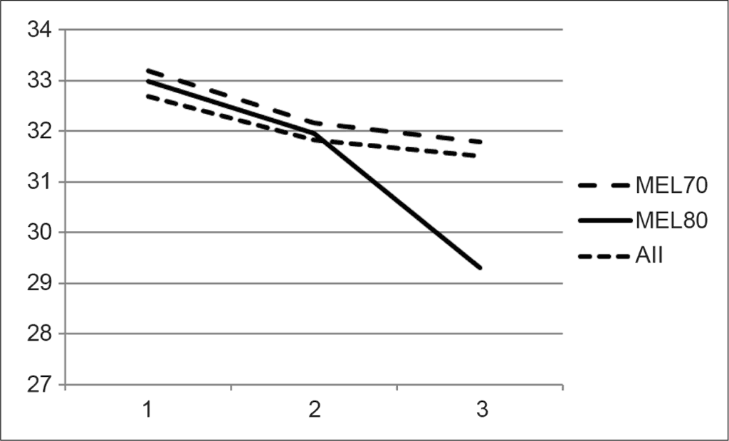
The results for different treatment groups before and after epithelial removal as well as after the treatment are represented in table 1 and figure 1 (in degrees Celsius). At the figure, the measurements taken before the epithelial removal are represented by number 1, after epithelial removal – 2, and after treatment – 3.
Tab. 1. The average measured temperature for the three lasers
| Temperature | MEL 80 | MEL 70 | Allegretto |
| Before epithelial removal | 33.0°C | 33.2°C | 32.6°C |
| After epithelial removal | 32.0°C | 32.2°C | 31.8°C |
| After the procedure | 29.3°C | 31.8°C | 31.5°C |

Fig. 1. The change of the temperature for the three lasers used
The changes in the temperature were subsequently calculated. After dividing the by through the time of the procedure, the speed of change in the temperature in °C/min was obtained.
The speed of the temperature change during the epithelial removal was -1.0°C/min for the MEL70 laser as well as for the MEL80 laser and -0.9°C/min for the Allegretto laser.
The speed of the temperature change during the treatment was -0.48°C/min for the MEL70 laser, -5.0°C/min for the MEL 80 laser, and -1.37°C/min for the Allegretto laser (tab. 2).
Tab. 2. The average change in the temperature for the three laser devices
| Speed of the temperature change (Average (Deviation)) | MEL 80 | MEL 70 | Allegretto |
| During epithelial removal | -1.0 (0.6)°C/min | -1.0 (0.6)°C/min | -0.9 (0.5)°C/min |
| During treatment | -5.0 (2.02)°C/min | -0.48 (0.69)°C/min | -1.37 (1.37)°C/min |
After performing the one-way ANOVA variance analysis, a statistically significant difference was found between the temperature changes during the procedures using different excimer lasers. Using the post hoc Bonferroni procedure, a statistically significant difference was established between the MEL 80 excimer laser and the two other types of excimer lasers (MEL 70 and Allegretto 400) with p < 0.05. No difference was found between the Allegretto and MEL 70 excimer lasers.
There was no statistically significant difference in the average postoperative refractive error between the treatment groups. Similar findings in postoperative subepithelial corneal haze were found between the groups 1 and 6 months after refractive surgery (tab. 3).
Tab. 3. Pre- and postoperative refraction and haze of the patients
| | MEL 80 | MEL 70 | Allegretto |
| Preoperative average refraction | -4.2 ± 1.9 Dpt | -3.6 ± 1.7 Dpt | -3.9 ± 2 Dpt |
| Postoperative average refraction | -0.025 ± 0.5 Dpt | 0.09 ± 0.46 Dpt | 0.05 ± 0.49 Dpt |
| Postoperative corneal haze (1 months) | 0.24 | 0.22 | 0.24 |
| Postoperative corneal haze (6 months) | 0.12 | 0.14 | 0.12 |
Discussion
Changes in the temperature of the corneal surface are of clinical importance to the processes of corneal wound healing. An excessive increase of the temperature may result in unwanted metabolic changes in keratocytes, causing excessive collagen synthesis resulting in floccular or reticular subepithelial opacities and consequent regression of achieved refractive correction (15, 16). Therefore, it may be concluded that the undercorrection of the refractive error may be the consequence not only of alterations of corneal surface due to the wound healing process, but also of the changes in the corneal surface temperature. Therefore, it is important to maintain and monitor controlled environment (including constant temperature, humidity) in the operating room.
In the earlier studies, the maximal temperature rise following photorefractive keratectomy was 7.5°C (18, 19). According to Maldonado-Codina et al. (14), the temperature rises by 7.35 ± 1.13°C during refractive surgery. A positive correlation was found between the attempted depth of the photoablation and the temperature rise in the cornea. Other studies reported that cooling of the cornea results in the reduction of postoperative subepithelial haze after PRK (20, 21). Thermal preconditioning was also reported to have a beneficial effect on the postoperative corneal wound healing. Heat shock proteins, activated by heating or cooling of the cornea, might play an important role in wound healing processes (22). The authors of the study found that certain types of heat shock proteins may contribute to faster wound healing. However, the study was conducted on a mice model and has no confirmation on human subjects so far. Other studies suggest that repetition rate does not influence the final outcomes of PRK (23).
In our study, the surface temperature of the cornea decreased by on average -1.0°C with no statistical difference between the treatment groups (MEL 80, MEL 70 and Allegretto excimer lasers). Using the post hoc Bonferroni procedure, a statistically significant difference was established between the MEL 80 excimer laser and the two other types of excimer lasers (MEL 70 and Allegretto 400) with p < 0.05. No difference was found between the Allegretto and MEL 70 excimer lasers.
Different air velocity of the lasers may be a possible explanation for this observation. The MEL 80 gas suction facility enables air velocity of approximately 2.2 m/s on the working plane. On the other hand, the MEL 70 and the Allegretto gas suction facility provided a lower air velocity, 0.4 and 0.15 m/s, respectively. Therefore, no statistically significant difference was found between the two lasers.
Conclusions
Based on our results, we conclude that the MEL 80 excimer laser, which has been able to produce the highest air velocity, resulted in the highest temperature decrease during the procedure. The MEL 70 excimer laser, the use of which was related to the longest treatment time and had the air velocity higher than the Allegretto excimer laser, had the lowest decrease in the corneal surface temperature.
Our study has some limitations. The sensitivity of the equipment used during our examination is lower than the sensitivity of thermal radiometry, which is able to measure with temporal resolution between 0.001 and 1000 msec.
With the methods used, we could only detect temperature change of the corneal surface as big as on average 4°C between the MEL80 and Allegretto lasers. According to our clinical experience, the temperature rise seems not to be a causative factor of postoperative subepithelial corneal haze and refractive regression when using excimer lasers operating below 400 Hz.
Piśmiennictwo
1. Müller B, Boeck T, Hartmann C: Effect of excimer laser beam delivery and beam shaping on corneal sphericity in photorefractive keratectomy. J Cataract Refract Surg 2004 Feb; 30(2): 464-470. 2. Fiore T, Carones F, Brancato R: Broad beam vs. flying spot excimer laser: refractive and videokeratographic outcomes of two different ablation profiles after photorefractive keratectomy. J Refract Surg 2001 Sep-Oct; 17(5): 534-541. 3. Pettit GH: The ideal excimer beam for refractive surgery. J Refract Surgery 2006; 22: S969-S972. 4. Khoramnia R, Lohmann CP, Wuellner C et al.: Effect of 3 excimer laser ablation frequencies (200 Hz, 500 Hz, 1000 Hz) on the cornea using a 1000 Hz scanning-spot excimer laser. J Cataract Refract Surg 2010 Aug; 36(8): 1385-1391. 5. Ratkay-Traub I, Ferincz IE, Juhasz T et al.: First clinical results with the femtosecond neodynium-glass laser in refractive surgery. J Refract Surg 2003 Mar-Apr; 19(2): 94-103. 6. Villa C, Gutièrrez R, Jimènez JR, González-Mèijome JM: Night vision disturbances after successful LASIK surgery. Br J Ophthalmol 2007 Aug; 91(8): 1031-1037. Epub 2007 Feb 21. 7. Sharma M, Wachler BS, Chan CC: Higher order aberrations and relative risk of symptoms after LASIK. J Refract Surg 2007 Mar; 23(3): 252-256. 8. Kouassi FX, Blaizeau M, Buestel C et al.: Comparison of Lasik with femtosecond laser versus Lasik with mechanical microkeratome: predictability of flap depth, corneal biomechanical effects andoptical aberrations. J Fr Ophtalmol 2012 Jan; 35(1): 2-8. Epub 2011 Jun 14. 9. Kaiserman I, Maresky HS, Bahar I, Rootman DS: Incidence, possible risk factors, and potential effects of an opaque bubble layer created by a femtosecond laser. J Cataract Refract Surg 2008; 34(3): 417-423. 10. Kwon Y, Bott S: Postsurgery corneal asphericity and spherical aberration due to ablation efficiency reduction and corneal remodelling in refractive surgeries. Eye (Lond) 2009 Sep; 23(9): 1845-1850. Epub 2008 Dec 5. 11. Lackerbauer CA, Grüterich M, Ulbig M et al.: Correlation between estimated and measured corneal ablation and refractive outcomes in laser in situ keratomileusis for myopia. J Cataract Refract Surg 2009 Aug; 35(8): 1343-1347. 12. Ishihara M, Arai T, Sato S et al.: Measurement of the Surface Temperature of the Cornea During ArF Excimer Laser Ablation by Thermal Radiometry With a 15-Nanosecond Time Response. Lasers Surg Med 2002; 30(1): 54-59. 13. Betney S, Morgan PB, Doyle SJ, Efron N: Corneal temperature changes during photorefractive keratectomy. Cornea 1997 Mar; 16(2): 158-161. 14. Maldonado-Codina C, Morgan PB, Efron N: Thermal consequences of photorefractive keratoectomy. Cornea 2001; 20: 509-515. 15. Tomás-Juan J, Murueta-Goyena Larrañaga A, Hanneken L: Corneal Regeneration After Photorefractive Keratectomy: A Review. J Optom 2015 Jul-Sep; 8(3): 149-169. 16. Chen X, Stojanovic A, Liu Y, Chen Y et al.: Postoperative Changes in Corneal Epithelial and Stromal Thickness Profiles After Photorefractive Keratectomy in Treatment of Myopia. J Refract Surg 2015 Jul; 31(7): 446-453. 17. Dantas PE, Martins CL, de Souza LB, Dantas MC: Do environmental factors influence excimer laser pulse fluence and efficacy? J Refract Surg 2007 Mar; 23(3): 307-309. 18. Bende T, Seiler T, Wollensack J: Corneal thermal gradients. Graefes Arch for Clin Exp Ophthalmol 1988; 226: 277-280. 19. Langenbucher A, Seitz B, Kus MM, Naumann GOH: Thermal effects in excimer laser trephination of the cornea. Graefes Arch for Clin Exp Ophthalmol 1996; 234 (suppl.): S142-148. 20. Tsubota K, Toda I, Itoh S: Reduction of subepithelial haze after photorefractive keratectomy by cooling the cornea. Am J Ophthalmol 1993; 115: 820-821. 21. Park WC, Tseng SCG: Temperature cooling reduced keratocyte death in excimer laser ablated corneal and skin wounds. INVEST Ophthalmol Vis Sci 1988; 39 (suppl.): 2062. 22. Kim MJ, Kim JC, Park WC et al.: Effect of thermal preconditioning before excimer laser photoablation. J Korean Med Sci 2004; 19: 437-446. 23. Kymionis GD, Diakonis VF, Kounis G et al.: Effect of excimer laser repetition rate on outcomes after photorefractive keratectomy. J Cataract Refract Surg 2008 Jun; 34(6): 916-919.