*Małgorzata Daszkowska1, Paulina Habrat2, Joanna Szczepańska1
Natal and neonatal teeth – a report of five cases
Zęby wrodzone i noworodkowe – opis pięciu przypadków
1Department of Paediatric Dentistry, Medical University of Łódź
Head of Department: Professor Joanna Szczepańska, DMD, PhD
2Paediatric Dentistry Outpatient Clinic, Central Teaching Hospital, Medical University of Łódź
Specialty supervisor: Małgorzata Daszkowska, DMD, PhD
Streszczenie
Występowanie zębów wrodzonych i noworodkowych jest zjawiskiem rzadkim, a jego etiologia nadal nie została do końca poznana i wyjaśniona. Celem pracy było przedstawienie przypadków pięciu pacjentów leczonych w Zakładzie Stomatologii Wieku Rozwojowego UM w Łodzi, u których zaobserwowano obecność zębów przedwcześnie wyrzniętych (wrodzonych lub noworodkowych). W latach 2013-2016 pięciu pacjentów (czterech chłopców i jedna dziewczynka), u których zdiagnozowano ww. zaburzenie wyrzynania zębów, zostało objętych opieką stomatologiczną. W każdym z zaobserwowanych przez nas przypadków z wywiadu wynikało, że matki nie chorowały podczas ciąży i dzieci urodziły się zdrowe. Noworodki z zębami wrodzonymi urodziły się od 2 do 3,5 tygodnia przed planowanym terminem porodu i miały niższą masę urodzeniową niż noworodki z pojedynczymi zębami noworodkowymi. Zależność występowania rodzinnego odnotowano tylko u jednej pacjentki. Najczęstszymi zębami przedwcześnie wyrzynającymi się są dolne zęby sieczne centralne. Jest niezwykle ważne, aby pediatrzy oraz stomatolodzy potrafili zdiagnozować ich obecność, różnicując je np. z guzkami Bohna czy torbielami, a następnie podjąć odpowiednie postępowanie lecznicze.
Summary
Incidence of natal and neonatal teeth is low, and its etiology remains unexplained so far. The aim of this study was to present five cases of patients treated at the Department of Paediatric Dentistry of the Medical University of Łódź, in whom the presence of prematurely erupted (natal or neonatal) teeth was observed. In years 2013-2016, five patients (four boys and one girl) diagnosed with either natal or neonatal teeth were provided with dental care at our Department. In each of the cases reported above, there was no history of disease during pregnancy, and all the children were born healthy. The infants with natal teeth were all born 2-3.5 weeks early, and showed lower weight at birth than the infants with neonatal teeth in our study. Only one of our patients (a girl) had family history of natal teeth. The most common prematurely erupted teeth are lower central incisors. It important that pediatricians and dentists be able to diagnose their presence, differentiating them from Bohn’s nodules or cysts, and pursue appropriate therapeutic measures.

Introduction
Deciduous teeth normally erupt at approximately 6 months of age. Natal teeth are teeth found at birth, while neonatal teeth are ones that erupt within the first month of life. They are nearly always prematurely erupted deciduous teeth, with maturity degree corresponding with the infant’s age. This means that approximately 5/6 of the crown is developed, while the root is not yet formed or its development is incomplete or defective, resulting with their mobility (1). Both types are rare occurrences, yet the earliest known reports date back to antiquity. Titus Livius, a Roman historian from the 1st century BC, commented on the cases of infants born with erupted teeth as an omen of future calamities (2), while his contemporary, Plinius the Elder forecast fortune awaited boys born with teeth, whereas misfortune – girls (3, 4). Famous historical figures who are known to have been born with natal teeth include Hannibal, Richard III, Louis XIV, cardinal Richelieu and Napoleon Bonaparte (2).
The reported incidence of natal and neonatal teeth varies largely depending on the type of studies and the size of the studied group, ranging from 1:2000 to 1:4000 (2). Massler et Savara have estimated the incidence of natal teeth at 1 per 2000 births (5), Bodenhoff et Gofin at 1 per 3000 (3), while Szymańska-Jachimczak based on her 10-year observation period found it to be 1 per 4844 neonates delivered in Warsaw hospitals (6). The majority of authors cite higher incidence of natal and neonatal teeth in girls. The teeth are most commonly prematurely erupted deciduous teeth, not supernumerary or predeciduous teeth, which in fact account for less than 10% of all natal and neonatal teeth (7). Typically, they are mandibular central incisors, however there have also been reports of prematurely erupted molars (8).
The exact aetiology of the phenomenon in question remains elusive. Superficial positioning of the tooth germ on the alveolar bone, increased osteoblastic activity in the area of the tooth germ associated with bone remodelling processes, endocrine diturbances such as hyperactive thyroid or pituitary gland, family history and inheritance pattern of an autosomal dominant trait, vitamin deficiency, malnutrition/undernourishment, PCB exposure, infections, injuries and febrile episodes suffered during pregnancy have all been suggested as possible underlying causes (2, 9-12). The presence of natal and neonatal teeth may be found in many multiple defect syndromes and congenital defects, including Ellis-van-Creveld syndrome, Gorlin syndrome, Hallermann-Streiff syndrome, Pierre Robin syndrome, Struge-Weber syndrome, Jadassohn-Lewandowski syndrome, Kabuki syndrome, Meckel-Gruber syndrome, Opitz syndrome, Pallister-Hall syndrome, Pfeiffer syndrome, Rubinstein-Taybi syndrome, Sotos syndrome and Wiedemann-Rautenstrauch syndrome (2, 9, 10). They are also sometimes present in children with uni- and bilateral orofacial clefts, in which case the teeth are lateral incisors in the region of the cleft (13, 14).
The aim of the study was to discuss the cases of natal and neonatal teeth treated at the Department of Paediatric Dentistry of the Medical University of Łódź.
Case reports
In the years 2013-2016, 5 infants (including 4 boys and 1 girl) were treated at our Department for natal (3 patients) or neonatal (2 patients) teeth. This paper discusses management of each of the cases.
Case 1
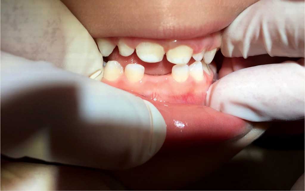
On 8.02.2013, parents with a male neonate 10 days old were admitted for consultation. The boy had been delivered 3 weeks ahead of his due date, at 3300 g. The reason for the dental appointment was the presence of natal teeth, which were mandibular central incisors 71 and 81 (ISO 3950 notation), tooth mobility (TM) Grade 2. The mother reported nursing problems due to sore nipples and the baby’s reluctance to suckle. Soft tissues around the teeth and on the ventral surface of the tongue showed inflammation. Because of the risk for swallowing or aspirating the teeth into the respiratory tract, as well as the problems with proper breastfeeding and the evident inflammation of oral tissues, extraction was necessary. The procedure was performed in topical anaesthesia administered with lidocaine spray placed on a sterile cotton roll. The teeth had no roots, the crowns were underdeveloped, and the enamel was hypomineralized. A follow-up examination on 24.04.2016 revealed presence of primary dentition complete except for the removed natal teeth 71 and 81 (fig. 1). The patient was referred for orthodontic treatment.
Fig. 1. Intraoral photograph: a 3-year old patient, presentation of the oral cavity with removed natal teeth 71 and 81
Case 2
Powyżej zamieściliśmy fragment artykułu, do którego możesz uzyskać pełny dostęp.
Mam kod dostępu
- Aby uzyskać płatny dostęp do pełnej treści powyższego artykułu albo wszystkich artykułów (w zależności od wybranej opcji), należy wprowadzić kod.
- Wprowadzając kod, akceptują Państwo treść Regulaminu oraz potwierdzają zapoznanie się z nim.
- Aby kupić kod proszę skorzystać z jednej z poniższych opcji.
Opcja #1
29 zł
Wybieram
- dostęp do tego artykułu
- dostęp na 7 dni
uzyskany kod musi być wprowadzony na stronie artykułu, do którego został wykupiony
Opcja #2
69 zł
Wybieram
- dostęp do tego i pozostałych ponad 7000 artykułów
- dostęp na 30 dni
- najpopularniejsza opcja
Opcja #3
129 zł
Wybieram
- dostęp do tego i pozostałych ponad 7000 artykułów
- dostęp na 90 dni
- oszczędzasz 78 zł
Piśmiennictwo
1. Cameron AC, Widmer RP: Stomatologia dziecięca. Wyd. III. Elsevier Urban & Partner, Wrocław 2015: 273.
2. Owczarek K, Mielnik Błaszczak M: Zęby wrodzone i noworodkowe – przegląd piśmiennictwa. Nowa Stomatol 2011; 2: 63-66.
3. Bodenhoff J, Gorlin RJ: Natal and neonatal teeth: folklore and fact. J Pediatrics 1963; 32: 1087-1093.
4. Cunha RF, Boer FA, Torriani DD et al.: Natal and neonatal teeth: review of the literature. Pediatr Dent 2001; 23: 158-162.
5. Massler M, Savara BS: Natal and neonatal teeth. A review of twenty four cases reported in the literature. J Pediatr 1950; 36: 349-359.
6. Szymańska-Jachimczak EJ: Dentes natales et neonatales – występowanie i umiejscowienie. Czas Stom 1965; 11: 1299-1303.
7. Zhu J, Kind D: Natal and neonatal teeth. ASDC J Dent Child 1995; 62: 123-128.
8. Kumar A, Grewal H, Verma M: Posterior neonatal teeth. J Indian Soc Pedod Dent 2011; 29: 68-70.
9. Moura LFA, Moura MS, Moura Lima MD et al.: Natal and Neonatal Teeth: A rewiew of 23 cases. J Dent Child 2014; 81(2): 107-111.
10. Malki GA, Al-Badawi EA, Dahlan MA: Natal teeth: a case report and reappraisal. Case Rep Dent 2015; 2015: 147580.
11. Sachdeva A, Punhani N, Bala M, Singh Sethi H: Peek-A-Boo. Natal Teeth: A report of 4 cases. Indian J Dent Sci 2015; 5(7): 96-98.
12. Sethi HS, Munjal D, Dhingra R et al.: Natal tooth associated with fibrous hyperplasia – a rare case report. J Clin Diag Res 2015; 9(4): 18-19.
13. Cabete HF, Gomide MR, Costa B: Evaluation of primary dentition in cleft lip and palate children with and without natal/neonatal teeth. Cleft Palate Craniofac J 2000; 37(1): 406-409.
14. Yilmaz RB, Cakan DG, Mesgarzadeh N: Prevalence and management of natal/neonatal teeth in cleft lip and palate patients. Eur J Dent 2016; 10(1): 54-58.
15. Senanayake MP, Karunaratne I: Persistent lingual ulceration (Riga-Fede disease) in an infant with Down syndrome and natal teeth: a case report. J Med Case Rep 2015; 8: 283.
16. Rao RS, Mathad SV: Natal teeth: case report and review of literature. J Oral Maxillofac Pathol 2009; 13(1): 41-46.
17. Tomizawa M, Yamada Y, Tonouchi K et al.: Treatment of Riga-Fede’s disease by resin-coverage of the incisal edges and seven cases of natal and neonatal teeth. Shoni Shikagaku Zasshi 1989; 27(1): 182-190.