*Michał Michalik
The innovative laryngological cannula facilitates the precise collection of highly diagnostic material from the nose and sinuses in order to improve the diagnostic and therapeutic process
Zastosowanie innowacyjnej kaniuli laryngologicznej w celu precyzyjnego pobrania materiału wysokodiagnostycznego z nosa i zatok – usprawnienie procesu diagnostyczno-terapeutycznego
Medical Center MML, Warsaw
Streszczenie
Pomimo stosowania najnowocześniejszych metod leczenia farmakologicznego i chirurgicznego pacjenci z przewlekłym zapaleniem zatok cierpią na nawracające infekcje, które nie reagują na leczenie. Obecność bakterii w strukturach zatok jest potwierdzana badaniami mikrobiologicznymi. Przedmiotem pracy jest zestaw trzech kaniul przeznaczonych do pobierania materiału biologicznego o wysokiej wartości diagnostycznej pochodzącego z zatok przynosowych.
Pobranie próbek w sposób tradycyjny wiąże się z ryzykiem zanieczyszczenia, wynikającym z obecności losowo pobranych mikroorganizmów. Zastosowana antybiotykoterapia opiera się wówczas na zidentyfikowanych przypadkowych mikroorganizmach, odwracając uwagę od właściwego czynnika wywołującego chorobę.
Zastosowanie wynalazku zgłoszonego do Urzędu Patentowego oznaczonego numerem: P.435850 pozwala na nieinwazyjne i szybkie pobranie próbek wysokodiagnostycznych do badań mikrobiologicznych. Dedykowana końcówka umożliwia precyzyjne dotarcie do naturalnych ujść zatok lub ujść zatok powstałych podczas zabiegu chirurgicznego, bez kontaktu z innymi tkankami. Próbki uzyskane dzięki zastosowaniu wynalazku odzwierciedlają stan mikrobiologiczny zatok. Wymaz z powierzchni błony śluzowej zatok to nieprawidłowo pobrany materiał, który może źle ukierunkować terapię. Zastosowanie zaawansowanego modelu kątowego ujścia zatok przynosowych skutecznie ogranicza takie błędy. Ponadto umożliwia precyzyjne pobranie materiału do badań, bez artefaktów lub zanieczyszczenia mikroorganizmami obecnymi w innych strukturach poza zatokami. Zastosowanie w kolejnym etapie celowanej antybiotykoterapii opartej na wyniku antybiogramu eliminuje bakterie wnikające w błony śluzowe, przestrzenie międzykomórkowe czy tkanki głębokie.
Summary
Despite the use of state-of-the-art methods of pharmacological and surgical treatment, patients with chronic sinusitis suffer from recurrent infections which do not respond to treatment. The presence of bacteria in the sinuses structures is confirmed by microbiological tests. The subject of the application is a set of three cannulas dedicated to collecting microbiological samples, a biological material of high-diagnostic value originating from paranasal sinuses.
Traditionally the collection of samples is associated with a risk of contamination resulting from presence of randomly collected microorganisms. Sometimes antibiotic therapy is based on the random microorganisms, driving attention away from the proper causative factor of the disease.
The application of the invention allows for a non-invasive and quick collection of highly diagnostic samples for proper microbiological testing. The dedicated tip makes it possible to precisely reach natural sinus ostium or sinus ostium created during surgery without contacting other tissues.
The samples obtained through the application of the invention reflect the microbiological condition of the sinuses. An incorrectly collected smear from the sinus mucosa surface may misdirect the therapy. The use of advanced model will effectively reduce such errors. In addition, it will contribute to the precise collection of material for testing, without artifacts or contamination by microorganisms present in other structures outside the sinuses.
Therefore in the next step application of targeted antibiotic therapy based on antibiogram profile eliminates bacteria that penetrates mucous membranes, intercellular space or deep tissues.
Introduction
Sinusitis is a disease that affects about 20% of human population (1). This disease is not only a socioeconomic burden on the community, but also significantly reduces the quality of life of those effected (2). Chronic sinusitis is defined as an infection of the sinuses, which last for 12 weeks or more, a medical diagnosis which is made based on evidence of inflammation confirmed by rhinoscopy and imaging studies complemented by nasal and sinus bacterial cultures, plus allergy testing, and has a multifactorial etiology (3-5). The management of chronic sinusitis involves a combination of antimicrobial treatment and surgery (3, 4).
The factor that correlates to high frequency of sinusitis occurrence in the population is disordered nasal patency. The patients have a deviated nasal septum, swelling and hypertrophy of turbinates, anomalies in the structure of the lateral nose wall, including narrowing of the natural sinuses. In addition, pathologies such as polyps and papilloma may be located in the nasal cavity.
A blockade of unobstructed connection between the sinuses and the nasal cavity forms an ideal environment for colonization and growth of bacteria as it hinders air exchange in narrow spaces. Additionally, the mucosal-ciliary activity decreases, which weakens the natural protective barrier of the host and contributes to stasis of secretions (6). The secretion accumulated in the sinuses becomes a nourishment for bacteria, which leads to bacterial overload and inflammation becoming chronic. The development of pathogenic bacteria causes inflammatory symptoms and strong pain. The most common ailments are: headaches, facial pains, purulent nasal discharge, dripping of the discharge on the back wall of the throat, as well as malaise (7).
In course of the disease a secondary bacterial infection is reported not in all cases. Acute sinusitis occurs most frequently due to a viral infection while bacteria are responsible for chronic sinusitis.
A sinus cavity is physiologically colonized by bacterial microbiota which similarly to intestinal tracts maintain the environment and conductives to efficient functions of the respiratory system. An elimination of bacterial microbiota or imbalance between particular species, i.e. dysbiosis, leading to reduced microbial diversity reported in patients with chronic sinusitis. Another opinion indicates that correct balances between microorganisms present in local microbiom, as well as between microorganisms and host, have significant immunomodulatory effect. Furthermore, an occurrence of inflammation is affected by lack of particular microbiological balance. Such pathogens as Staphylococcus aureus, Streptococcus pyogenes, Str. pneumoniae, Str. viridans, Pseudomonas aeruginosa, Haemophilus influenzae, and coagulase-negative staphylococci (CoNS) are isolated from patients who have suffered from acute or chronic sinusitis (8, 9). Some pathogenic microorganisms from the species mentioned above such Str. pyogenes, Str. pneumoniae, CoNS, H. influenzae, and additionally Moraxella catarrhalis, Stenotrophomonas maltophilia and Enterobacter species were also found in healthy cavities which indicates opportunistic properties of the bacteria, and endogenic character of the infections (10, 11).
Recently a presence of CoNS was reported more frequently in chronic sinusitis (12, 13). It is a heterogeneous group of Gram-positive bacteria colonizing skin and mucous membranes of healthy humans or animals. They are known as skin staphylococci, and were identified as accompanying bacteria that contaminate diagnostic samples. Because of that, their presence was trivialized, marginalized, and overlooked in diagnostics or then in antibiotic therapy. Although CoNS are typical saprophytes under the physiological conditions, they change their properties under additional conditions known as infection facilitating factors, and become pathogens (14, 15). There are many factors facilitating infection but surgical or invasive diagnostic wounds, which are a gate for bacterial invasion, are of particular significance.
A surgical intervention is necessary in the cases of permanent lesions in nose or sinus defects (16). In addition to classic, more invasive sinus surgery methods, minimally invasive methods of accessing the nose and sinuses through natural orifices are increasingly common, such as Functional Endoscopic Sinus Surgery (FESS), or the more target-oriented MIST technique (Minimally Invasive Sinus Technique). A treatment is carried out through that method on the basis of removal of pathologically altered tissues, while saving healthy sections of mucosa, which synchronically creates a better connection between the sinuses and nasal cavity (all the paranasal sinuses may have to be involved in treatment, even in the first surgical intervention).
The laryngologist has access to the sinuses through a natural ostium in the side wall of the nose, as well as through the lacrimal recess. The application of endoscopic projection allows precise surgical removal of the pathologically affected tissue.
Description of the laryngological cannula
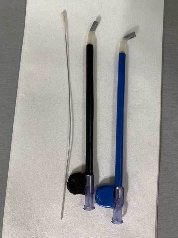
The subject of the patent is a set of three cannulas designed for collecting microbiological samples of high-diagnostic material from sick paranasal sinuses. A subject of the communication is a set of cannulas designed for collecting microbiological samples of high diagnostic material from sick paranasal sinuses (fig. 1). The set includes three cannulas of different angles, properly profiled at angles: 15, 45, and 120 degrees (fig. 2-4).
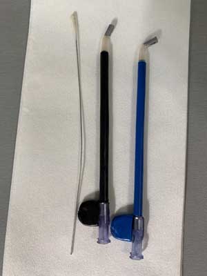
Fig. 1. Set for collecting highly diagnostic material from the paranasal sinuses

Fig. 2. Cannula profiled at an angle of 15 degrees, allows access to the sphenoid sinus
Fig. 3. Cannula profiled at an angle of 45 degrees, allows access to the frontal sinus
Fig. 4. Cannula profiled at an angle of 120 degrees, allows access to the maxillary sinus
The innovative character of the idea is based on a non-invasive, target-oriented approach to the selected ostium of paranasal sinus, collection of biological samples, and additionally avoiding contamination risk of swab stick introduced through the cannula. Thus the obtained reliable samples are of the highest quality for testing and receiving authoritative results. Every individual cannula is sterile, single use, made of transparent plastic, and enables constant monitoring to control the manipulation of the swab stick. The length of each cannula is 12 centimeters with a diameter of 3 millimeters. In proximal part of the cannula there is a handle that allows dependable, safe and precise navigation. In the distal part there is a colorful green marker that facilitates orientation in anatomical structures of the side of the nose wall. The tip of the cannula has atraumatic, gently ended edges. A swab stick dedicated to collecting highly diagnostic samples is plastic, soft and universal. The plasticity enables easy deformation, which allows targeted changes of the cannula shape during penetration of particular sinus and non-invasive collection of the samples. After use, the swab with sample is inserted into a standard transport medium and sent to a diagnostic laboratory.
Application of the invention
Under control of the endoscope used for nose and sinus surgery, natural ostia of the paranasal sinuses are visible. An appropriate angular cannula is introduced depending on the examined sinus. The dedicated tip present at the end of the cannula makes it possible to precisely reach natural sinus ostium or sinus ostium created during the operation without any contact to other tissues.
The swab stick with the collected sample is led out through cannula from inside of the sinus under strict control of the endoscope. Therefore the sample may be collected quickly and non-invasively. The sample collected that way is placed in standard tube with particular transport medium.
The use of an angular cannula during surgery is shown in figures 5 and 6.
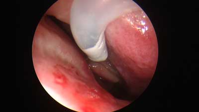
Fig. 5. The use of an angular cannula during surgery

Fig. 6. The use of an angular cannula during surgery
Conclusions
A collection of samples obtained by using traditional methods was associated with the risk of error resulting from collection of random microorganisms including natural commensal organisms present in upper respiratory tracts or from air. Sometimes these random microorganisms were the basis for antibiotic therapy, and diverted attention away from the proper causative factor of the disease. The application of advanced cannula model uses angular paranasal sinuses ostium and effectively eliminates errors mentioned above. In addition, the use of innovative cannula in practice will contribute to the precise collection of samples for microbiological tests without any artifacts or contamination by microorganisms present in other structures outside the sinuses.
Careful collection of diagnostic samples is of high significance for proper microbiological diagnostics, and consequently, for proper antibiotic targeted therapy, allowing eradication of disease-causing microorganisms and sustained recovery of the patient.
Confirmation
Patent Office of the Republic of Poland states that at 2020-11-02 the application for granting a patent for the following invention was accepted in electronic form: Highly diagnostic material collection kit.
Notification is numbered: P.435850 [WIPO ST 10/C PL435850].
Applicant: Michał Michalik, Warsaw, Poland.
Piśmiennictwo
1. de Lima CO, Devito KL, Baraky Vasconcelos LR et al.: Correlation between Endodontic Infection and Periodontal Disease and Their Association with Chronic Sinusitis: A Clinical-Tomographic Study. J Endod 2017; 43(12): 1978-1983.
2. Halawi AM, Smith SS, Chandra RK: Chronic rhinosinusitis: epidemiology and cost. Allergy Asthma Proc 2013; 34(4): 328-334.
3. Ivanchenko OA, Karpishchenko SA, Kozlov RS et al.: The microbiome of the maxillary sinus and middle nasal meatus in chronic rhinosinusitis. Rhinology 2016; 54(1): 68-74.
4. Manes RP, Batra PS: Etiology, diagnosis and management of chronic rhinosinusitis. Expert Rev Anti Infect Ther 2013; 11(1): 25-35.
5. Oakley GM, Curtin K, Orb Q et al.: Familial risk of chronic rhinosinusitis with and without nasal polyposis: genetics or environment. Int Forum Allergy Rhinol 2015; 5(4): 276-282.
6. Szyfter W, Kruk-Zagajewska A, Bartochowska A, Borucki Ł: Intracranial complications from sinusitis. Otolaryngologia Pol 2015; 69(3): 6-14.
7. Fowler KC Duncavage JA, Murray JJ et al.: Chronic Rhinosinusitis and intravenous antibiotic therapy: resolution, recurrent and adverse events. J Allergy Clin Immunol 2003; 111: 85.
8. Anderson M, Stokken J, Sanford T et al.: A systematic review of the sinonasal microbiome in chronic rhinosinusitis. Am J Rhinol Allergy 2016; 30(3): 161-166.
9. Długoszewska J, Leszczynska M, Lenkowski M et al.: The pathophisiological role of bacterial biofilms in chronic sinusitis. Eur Arch Otorhinolaryngol 2016; 273(8): 1989-1994.
10. Ramakrishnan VR, Feazel LM, Gitomer SA et al.: The microbiome of the middle meatus in healthy adults. PLoS One 2013; 8(12): 1-10.
11. Thanasumpun T, Batra PS: Endoscopically-derived bacterial cultures in chronic rhinosinusitis: A systematic review. Am J Otolaryngol Head Neck Med Surg 2015; 36: 686-691.
12. Bachert C, Pawankar R, Zhang L et al.: ICON: chronic rhinosinusitis. World Allergy Organ J 2014; 7(1): 25.
13. Zhang Z, Adappa ND, Lautenbach E et al.: Coagulase-negative staphylococci culture in chronic rhinosinusitis. Int Forum Allergy Rhinol 2015; 5(3): 204-213.
14. Manes RP, Batra PS: Bacteriology and Antibiotic Resistance in Chronic Rhinosinusitis. Facial Plast Surg Clin N Am 2012; 20: 87-91.
15. Michalik M, Samet A, Podbielska-Kubera A et al.: Coagulase-negative staphylococci (CoNS) as a significant etiological factor of laryngological infections: a review. Ann Clin Microbiol Antimicrob 2020; 19: 26.
16. Cullen MM, Bolger WE: Maximal Medical management of chronic rhinosinusitis. Curr Opin Otolaryngol Head Neck Surg 2000; 8: 7-10.