*Małgorzata Kołodziejczak1, 2, Przemysław Ciesielski1, 2
Obstetric anal sphincter injuries – management guidelines
Poporodowe uszkodzenia zwieraczy odbytu – zasady postępowania
1Warsaw Proctology Center, St. Elisabeth Hospital, Mokotów Medical Center, Warsaw
2Department of General Surgery, County Hospital in Ostrów Mazowiecka
Streszczenie
Nietrzymanie gazów i stolca jest poważnym kalectwem, znacznie upośledzającym jakość życia pacjenta w sferze osobistej, zawodowej i społecznej. Ocenia się, że nawet około 15% populacji ludzi dorosłych cierpi na problemy dotyczące obniżenia kontynencji, jakkolwiek problem wydaje się nadal epidemiologiczne niedoszacowany. U kobiet uraz porodowy jest najczęstszą przyczyną inkontynencji, nazywanej „cichą chorobą”, gdyż nie mówią o niej pacjentki, a lekarze też w sposób aktywny nie pytają się pacjentek po porodzie o inkontynencję. Zbyt niska refundacja operacji rekonstrukcji zwieraczy przez Narodowy Fundusz Zdrowia sprawia, że małe jest zainteresowanie na oddziałach chirurgicznych wykonywaniem operacji naprawczych zwieraczy, a trudność zabiegu operacyjnego wymagającego dużego doświadczenia, a także ryzyko powikłań po tej operacji powodują, że zabiegi te równie niechętnie wykonuje się w sektorze prywatnym. Doraźne, prawidłowe zaopatrzenie uszkodzenia zwieraczy po porodzie sprawia, że u pacjentki nie występują powikłania czynnościowe w postaci inkontynencji gazów i stolca. Planowe, odległe rekonstrukcje obarczone są większym odsetkiem powikłań niż rekonstrukcje doraźne, dlatego też powinny być poprzedzone diagnostyką obrazową i czynnościową. W artykule omówiono problem inkontynencji poporodowej, jednocześnie przedstawiając algorytmy postępowania diagnostyczno-terapeutycznego w doraźnej i planowej rekonstrukcji zwieraczy.
Summary
Gas and stool incontinence is a serious disability, which significantly impairs the patient’s quality of life in the personal, professional and social sphere. Although it is estimated that continence problems affect up to 15% of the adult population, the problem still seems to be epidemiologically underestimated. In women, obstetric trauma is the most common cause of incontinence, referred to as “the silent affliction” as the patients do not talk about it, and doctors do not actively ask patients about incontinence after delivery. Insufficient reimbursement of sphincter repair by the National Health Fund means that there is little interest in performing sphincter repair in surgical departments, while the difficulty of the surgery requiring extensive experience, as well as the risk of postoperative complications make these procedures unattractive for the private sector. Proper emergency treatment of postpartum sphincter damage prevents functional complications in the form of gas and faecal incontinence. Elective, distant reconstructions have a higher complication rate than emergency reconstructions, and therefore should be preceded by imaging and functional diagnosis. The paper discusses the problem of postpartum incontinence, presenting the diagnostic and therapeutic algorithms for emergency and elective sphincter repair.

Introduction
Gas and stool incontinence is a serious disability, which significantly impairs the patient’s quality of life in the personal, professional and social sphere. It is estimated that continence problems affect up to 15% of the adult population. It should be mentioned that this problem also affects men and it has been increasingly better diagnosed and noticed in this group in recent years (1). Obstetric trauma, including neurogenic, morphological and mixed injuries, remains the most common cause of incontinence. Anal sphincter damage is a serious complication after vaginal delivery. Rapid diagnosis and proper emergency sphincter suturing prevent functional complications in the form of gas and faecal incontinence. The available epidemiological data indicate that the incidence of obstetric anal sphincter injuries (OASIS) is 0.25 to 6%, but they do not lead to incontinence in all patients. The rate of sphincter damage is higher in primigravidas, i.e. from 1.4 up to 16% compared to 0.4 to 2.7% in multiparas. The risk of second sphincter injury during another delivery is higher, ranging from 5.1 to 10.7%, in women who suffered sphincter injury during their first labour (2). Other risk factors include instrumental delivery, advanced maternal age, macrosomia, mid-perineal incision, breech delivery and prolonged second stage of labour (3). According to our practical observations based on cases of patients presenting with postpartum incontinence, instrumental delivery, forceps delivery in particular, and prolonged second stage of labour in the case of neurogenic injuries, are the most common risk factor for sphincter damage. This is consistent with literature reports (4).
It should be emphasised that neurogenic damage is a more common cause of postpartum incontinence than morphological damage to the sphincter muscles, and not every patient reporting symptoms of postpartum incontinence actually has damaged sphincter muscles. Long-term observational studies show that impaired stool and gas continence after perinatal sphincter injury occurs in 35 to 60% of patients (2). In patients with isolated neurogenic damage to the sphincters, treatment is mainly based on rehabilitation (sphincter gymnastics, electrostimulation).
Diagnosis of sphincter damage
The diagnosis of obstetric sphincter damage usually poses no difficulty. The lack of “anal tightness”, a gaping wound and visible stumps of the torn muscle within the wound are typical signs of postpartum sphincter damage. Bimanual rectovaginal examination allows to detect any defects in the rectovaginal septum and to diagnose rectovaginal fistula at an early stage. Additional tests, such as transrectal ultrasound, magnetic resonance imaging or anorectal manometry are practically not used in fresh sphincter damage. These diagnostic tests are dedicated to patients qualified for elective reconstructive surgeries. Furthermore, they are practically unavailable during the on-call time, and are of purely academic importance, as torn sphincters are clearly visible in most cases.
Emergency repair of obstetric sphincter injury
The repair should be performed up to 6 hours after the injury; however, an experienced coloproctologist may undertake the reconstruction even between 10 and 20 hours after the injury, still obtaining a satisfactory outcome.
It is important from both medical and legal point of view that the most experienced doctor in the team on duty performs or at least assists in the repair. If possible, the procedure should take place in the operating theatre, under anaesthesia. This, however, is not always possible. With no access to the operating suite in the hours following delivery, the procedure is performed as soon as possible in the delivery suite. Postpartum injuries are easier to treat than e.g. those after traffic accidents, as postpartum perineal tissues are usually not crushed, the wound looks like a “an even cut with a knife”, as well as is clean.
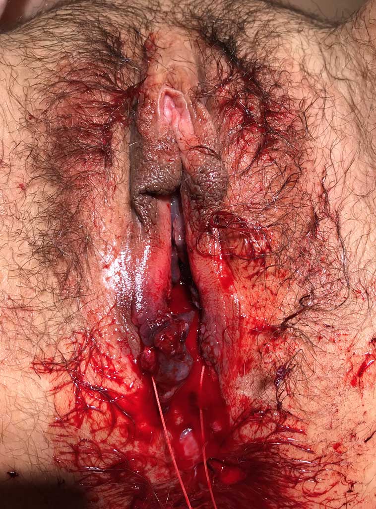
Stages of reconstruction (5) (figs. 1, 2)

Fig. 1. Perinatal perineal trauma
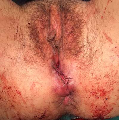
Fig. 2. Status after emergency sphincter repair
Surgical wound debridement, removal of haematomas, and accurate haemostasis.
1. Identification of stumps of damaged muscles; electrical stimulus may be used to induce muscle contraction in doubtful cases.
2. For grade 4 sphincter tear (with rectal involvement), the repair begins with placing sutures on the rectal mucosa to provide support for sphincter repair.
Powyżej zamieściliśmy fragment artykułu, do którego możesz uzyskać pełny dostęp.
Mam kod dostępu
- Aby uzyskać płatny dostęp do pełnej treści powyższego artykułu albo wszystkich artykułów (w zależności od wybranej opcji), należy wprowadzić kod.
- Wprowadzając kod, akceptują Państwo treść Regulaminu oraz potwierdzają zapoznanie się z nim.
- Aby kupić kod proszę skorzystać z jednej z poniższych opcji.
Opcja #1
29 zł
Wybieram
- dostęp do tego artykułu
- dostęp na 7 dni
uzyskany kod musi być wprowadzony na stronie artykułu, do którego został wykupiony
Opcja #2
69 zł
Wybieram
- dostęp do tego i pozostałych ponad 7000 artykułów
- dostęp na 30 dni
- najpopularniejsza opcja
Opcja #3
129 zł
Wybieram
- dostęp do tego i pozostałych ponad 7000 artykułów
- dostęp na 90 dni
- oszczędzasz 78 zł
Piśmiennictwo
1. Bharucha AE, Dunivan G, Goode PS et al.: Epidemiology, pathophysiology, and classification of fecal incontinence: State of the Science Summary for the National Institute of Diabetes and Digestive and Kidney Diseases (NIDDK) Workshop. Am J Gastroenterol 2015; 110(1): 127-136.
2. Thubert T, Cardaillac C, Fritel X et al.: Definition, epidemiology and risk factors of obstetric anal sphincter injuries: CNGOF Perineal Prevention and Protection in Obstetrics Guidelines. Gynecol Obstet Fertil Senol 2018; 46(12): 913-921.
3. Sharma A, Rao SSC: Epidemiologic Trends and Diagnostic Evaluation of Fecal Incontinence. Gastroenterol Hepatol (N Y) 2020; 16(6): 302-309.
4. Ali M, Migisha R, Ngonzi J et al.: Risk Factors for Obstetric Anal Sphincter Injuries among Women Delivering at a Tertiary Hospital in Southwestern Uganda. Obstet Gynecol Int 2020; 2020:6035974.
5. Kołodziejczak M (red.): Leczenie chorób proktologicznych w ciąży i okresie porodu. Wydawnictwo Borgis, Warszawa 2011.
6. Sudoł-Szopińska I, Santoro GA, Kołodziejczak M et al.: Magnetic resonance imaging template to standardize reporting of anal fistulas. Tech Coloproctol 2021; 25(3): 333-337.
7. Jorge JM, Wexner SD: Etiology and management of fecal incontinence. Dis Colon Rectum 1993; 36: 77-97.