*Marek Mazur1, Kacper Bolesta1, Wiktoria Wieczorek1, Dominika Portka1, Maja Olborska1, Piotr Regulski2, Marcin Aluchna3
Comparison of micro-CT and CBCT in the preparation of teeth for the role of samples in in vitro research
Porównanie zastosowania micro-CT i CBCT w przygotowaniu zębów do roli próbek w badaniach in vitro
1Studenckie Koło Naukowe, Zakład Stomatologii Zachowawczej, Warszawski Uniwersytet Medyczny
2Zakład Radiologii Stomatologicznej i Szczękowo-Twarzowej, Warszawski Uniwersytet Medyczny
Kierownik Zakładu: prof. dr hab. n. med. Kazimierz Szopiński
3Zakład Stomatologii Zachowawczej, Warszawski Uniwersytet Medyczny
Kierownik Zakładu: prof. dr hab. n. med. Agnieszka Mielczarek
Streszczenie
Wstęp. Grubość zębiny stanowi ważny czynnik wpływający na wyniki otrzymywane w badaniach eksperymentalnych. Odpowiednio precyzyjna i powtarzalna metoda jego pomiaru ma kluczowe znaczenie w zapewnieniu rzetelności i wiarygodności pracy badawczej.
Cel pracy. Celem niniejszego badania było porównanie zastosowania tomografii komputerowej wiązką stożkową i mikrotomografi w przygotowaniu zębów do roli próbek w badaniach medycznych.
Materiał i metody. Spośród 100 usuniętych zębów wyselekcjonowano 4 o odpowiednich cechach budowy potwierdzonych na obrazach pochodzących z CBCT. Zostały one zatopione w masie silikonowej i przecięte w płaszczyźnie horyzontalnej w taki sposób, by płaszczyzna przecięcia była równoległa do płaszczyzny wyznaczonej przez szczyty rogów miazgi. Następnie próbki zostały poddane badaniom tomografii komputerowej wiązką stożkową oraz mikrotomografii. Dwóch autorów niezależnie wykonywało pomiary odległości pomiędzy płaszczyzną przecięcia a szczytami rogów miazgi na otrzymanych przekrojach czołowych i strzałkowych. Otrzymane wartości pomiarów zostały opracowane statystycznie.
Wyniki. Wartości pomiarów wykonywanych na przekrojach CBCT były większe niż wartości uzyskane z pomiarów wykonywanych w micro-CT. W przypadku dwóch z czterech próbek uzyskano istotne statystycznie różnice w wartościach pomiarów.
Wnioski. Badanie CBCT nie jest wystarczającym narzędziem w precyzyjnym przygotowaniu zębów do roli próbek badawczych.
Summary
Introduction. The thickness of dentine is an important factor affecting the results of experimental studies. An adequately precise and repeatable method for its measurement is of key importance for ensuring the reliability and credibility of research.
Aim. The aim of this study was to compare conical beam computed tomography (CBCT) and microtomography (micro-CT) for the preparation of teeth as specimens in medical research.
Material and methods. From among 100 extracted teeth, 4 teeth with appropriate structural features confirmed in CBCT images were selected. They were embedded in silicone mass and cut in a horizontal plane in such a way that the intersection plane was parallel to the plane defined by the pulp horns. Next, the samples were subjected to conical beam computed tomography and microtomography. Two authors independently measured the distance between the plane of intersection and the pulp horns on the obtained frontal and sagittal sections. The obtained measurements were statistically processed.
Results. The values obtained with CBCT were higher than those obtained with micro-CT. Statistically significant differences were found for two of four samples.
Conclusions. CBCT is not sufficient for precise preparation of teeth for the role of research specimens.
Słowa kluczowe: mikrotomografia rentgenowska, tomografia komputerowa wiązką stożkową, dokładność pomiarów przestrzennych, grubość zębiny
Key words: X-Ray Microtomography, Cone-Beam Computed Tomography, Dimensional Measurement Accuracy, Dentine Thickness

Introduction
Extracted teeth are often used for in vitro studies. If prepared properly, they can simulate the conditions during the preparation and filling of cavities in clinical practice with high reliability. When using extracted teeth for research purposes, precise and reproducible preparation of specimens is needed (1). Obtaining a specific and repeatable dentine thickness for all samples is particularly important for research results. Factors such as thermal conductivity and chemical permeability are directly related to the thickness of the insulating dentine layer. It is one of the key factors that affect pulpal vitality during preparation and filling of cavities (2).
In the available publications, researchers usually specify the depth of preparation of a given cavity. This does not allow for accurate determination of dentinal thickness between the surface of the cavity floor and the pulp chamber. Analysis of microscopic images is the gold standard of measurement in scientific research. Unfortunately, this method cannot be used for preparing samples, as it involves destruction of objects under investigation. Several non-invasive methods to determine residual dentinal thickness may be found in the literature. The most popular and most frequently used methods are based on radiography. Periapical radiograph, which is the simplest tool for imaging the mineralised dental tissues, is a summation image and therefore may be unreliable as it represents an overlapping image of all structures along the radius, including the pulp horns. Cone beam computed tomography (CBCT) offers much greater possibilities in this regard. After reconstruction, it produces a three-dimensional image. However, a relatively low resolution is a limitation of this tool. Currently, high hopes are placed on micro-CT (X-ray microtomography). This tool has been successfully used in many scientific studies and allows for precise measurements comparable to those obtained with a stereoscopic microscope (3).
Aim
The aim of this study was to verify whether CBCT meets the required imaging quality criteria to obtain an accurate measurement of residual dentine thickness when preparing specimens for scientific research.
Material and methods
A total of 100 third molars extracted for periodontal, orthodontic and surgical reasons were collected over a period of two months. The teeth were stored in a 1% thymol solution for 1 month at room temperature. Of all the teeth, 15 non-carious teeth with no enamel developmental defects were selected. The selected teeth were cleaned of residual soft tissue using a 2% sodium hypochlorite solution and a piezoelectric scaler (Woodpecker, China), and then carefully polished using a pumice slurry and a contra-angle brush.
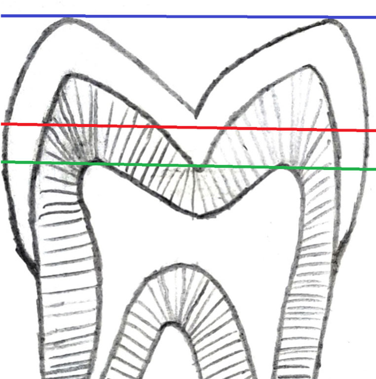
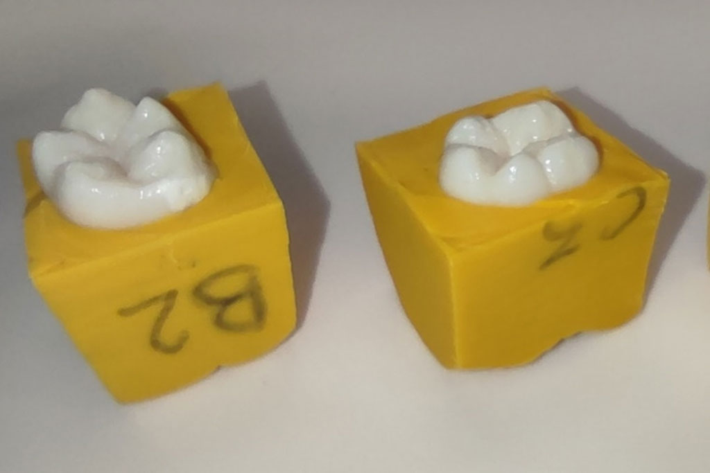
Next, the roots of selected teeth were fully embedded in a silicone mass (3M Express XT Putty Soft, USA) up to the level of the cement-enamel junction (CEJ). Rows were marked with letters of the Latin alphabet (A-D), and columns with Arabic numerals (1-4) on the silicone block (fig. 1). Finished silicone blocks were subjected to CBCT (Vatech Pax-i 3D, South Korea). Baseline CBCT was aimed at selecting teeth with a specific anatomical structure, enabling such setting of the horizontal plane during analysis that it passed through the tips of exactly three selected cusps. Once established, the plane was moved parallel and apically until the pulp horns were exposed. Only those teeth were selected in which the plane passing through the tips of selected cusps was parallel to the plane passing through the tips of all pulp horns. Based on the analysis of CBCT images, 4 teeth meeting the above assumptions were selected. Spatial relationships are shown in figure 2.
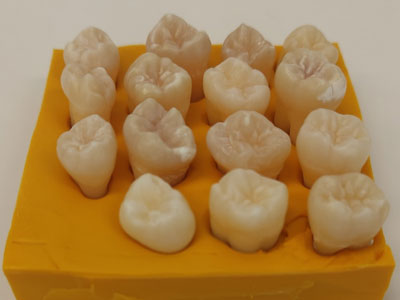
Fig. 1. Teeth prepared for initial CBCT
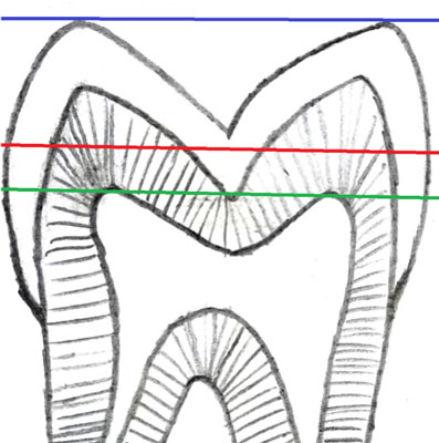
Fig. 2. Spatial relations of planes determined in the specimens
Selected teeth were removed from the silicone. Cusps defining the horizontal plane were identified (the highest line in figure 2). Then, each tooth was placed on a flat glass plate in such a way that the marked cusps contacted its surface. Individual silicone indexes were made (3M Express XT Putty Soft, USA) in a predetermined spatial position. The flat surface of the index was the same as the plane defined by cusp tips and parallel to the plane defined by the tips of the pulp horns.
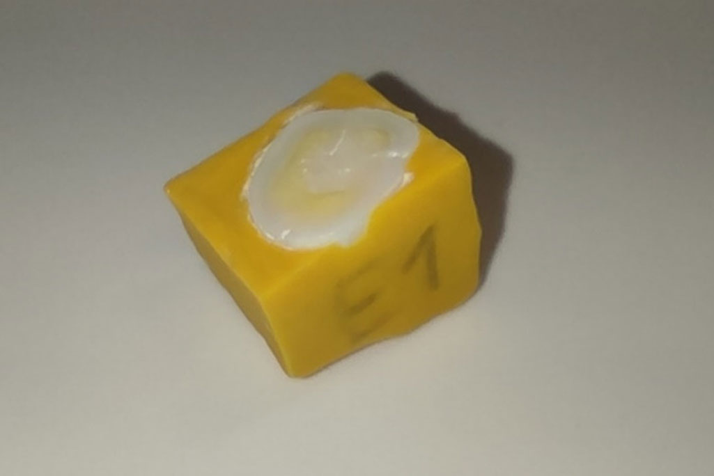
The distance between the plane defined by pulp horn tips and the plane defined by the tops of selected cusps (the highest and the lowest line in figure 2) was measured on CBCT images. Then, an additional plane was determined 2 mm above pulp horn tips in each tooth (middle line in figure 2), and the distance between the obtained plane and the plane passing through the tips of the selected cusps was measured. Any excess material was cut away from the silicone indexes with a Scalpel blade number 11 (SwannMorton, England), after measuring the predetermined distance from the flat surface of the individual silicone index to the previously determined additional plane with a caliper (Magtoto, China). Trimming of the indexes exposed part of the clinical crown of the tooth (fig. 3). It was removed with diamond burs (MDT, Israel) mounted on a dental turbine (W&H, Austria) in such a way as to obtain a flat surface. The surface was then polished with Sof-Lex abrasive discs (3M ESPE, USA) (fig. 4). The distance between the tips of the pulp horns and the cut surface of the specimens should be approximately 2 mm.

Fig. 3. Trimmed individual silicone indices
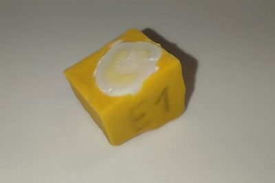
Fig. 4. A specimen prepared for scanning
Powyżej zamieściliśmy fragment artykułu, do którego możesz uzyskać pełny dostęp.
Mam kod dostępu
- Aby uzyskać płatny dostęp do pełnej treści powyższego artykułu albo wszystkich artykułów (w zależności od wybranej opcji), należy wprowadzić kod.
- Wprowadzając kod, akceptują Państwo treść Regulaminu oraz potwierdzają zapoznanie się z nim.
- Aby kupić kod proszę skorzystać z jednej z poniższych opcji.
Opcja #1
29 zł
Wybieram
- dostęp do tego artykułu
- dostęp na 7 dni
uzyskany kod musi być wprowadzony na stronie artykułu, do którego został wykupiony
Opcja #2
69 zł
Wybieram
- dostęp do tego i pozostałych ponad 7000 artykułów
- dostęp na 30 dni
- najpopularniejsza opcja
Opcja #3
129 zł
Wybieram
- dostęp do tego i pozostałych ponad 7000 artykułów
- dostęp na 90 dni
- oszczędzasz 78 zł
Piśmiennictwo
1. Fernandez-Estevan L, Millan-Martinez D, Fons-Font A et al.: Methodology in specimen fabrication for in vitro dental studies: Standardization of extracted tooth preparation. J Clin Exp Dent 2017; 9(7): e897-e900.
2. Yasa E, Atalayin C, Karacolak G et al.: Intrapulpal temperature changes during curing of different bulk-fill restorative materials. Dent Mater J 2017; 36(5): 566-572.
3. Capar ID, Gok T, Uysal B, Keles A: Comparison of microcomputed tomography, cone beam tomography, stereomicroscopy, and scanning electron microscopy techniques for detection of microcracks on root dentin and effect of different apical sizes on microcrack formation. Microsc Res Tech 2019; 82(10): 1748-1755.
4. Mizutani R, Suzuki Y: X-ray microtomography in biology. Micron 2012; 43(2-3): 104-115.
5. Sousa-Neto MD, Silva-Sousa YC, Mazzi-Chaves JF et al.: Root canal preparation using micro-computed tomography analysis: a literature review. Braz Oral Res 2018; 32(suppl 1): e66.
6. Mangione F, Meleo D, Talocco M et al.: Comparative evaluation of the accuracy of linear measurements between cone beam computed tomography and 3D microtomography. Ann Ist Super Sanita 2013; 49(3): 261-265.
7. Tayman MA, Kamburoglu K, Kucuk O et al.: Comparison of linear and volumetric measurements obtained from periodontal defects by using cone beam-CT and micro-CT: an in vitro study. Clin Oral Investig 2019; 23(5): 2235-2244.
8. Ferrare N, Leite AF, Caracas HC et al.: Cone-beam computed tomography and microtomography for alveolar bone measurements. Surg Radiol Anat 2013; 35(6): 495-502.
9. Demirel A, Demirci O, Okte Z: Accuracy of In Vitro Radiographs in Determining the Remaining Dentin Thickness below Deep Dentin Caries in Deciduous Molars. Balkan Journal of Dental Medicine 2020; 24 (3): 18-153.
10. Suassuna FCM, Maia AMA, Melo DP et al.: Comparison of microtomography and optical coherence tomography on apical endodontic filling analysis. Dentomaxillofac Radiol 2018; 47(2): 20170174.
11. Krause F, Kohler C, Ruger C et al.: Visualization of the pulp chamber roof and residual dentin thickness by spectral-domain optical coherence tomography in vitro. Lasers Med Sci 2019; 34(5): 973-980.
12. Majkut P, Sadr A, Shimada Y et al.: Validation of Optical Coherence Tomography against Micro-computed Tomography for Evaluation of Remaining Coronal Dentin Thickness. J Endod 2015; 41(8): 1349-1352.
13. Savas S, Botsali MS, Kucukyilmaz E, Sari T: Evaluation of temperature changes in the pulp chamber during polymerization of light-cured pulp-capping materials by using a VALO LED light curing unit at different curing distances. Dent Mater J 2014; 33(6): 764-769.