© Borgis - New Medicine 3/2005, s. 34-36
Lidia Zawadzka-Głos, Mieczysław Chmielik, Dorota Kowalczys
Acute laryngeal trauma in children
Department of Paediatric Otorhinolaryngology, Medical University of Warsaw, Poland
Head: Prof. Mieczysław Chmielik MD, PhD
Summary
Laryngeal trauma may be life-threatening when airways are compromised. Early diagnosis of laryngeal trauma and appropriate management are necessary to save lives and to prevent delayed airway complications. The present paper is a clinical analysis of seven patients treated for laryngeal trauma.
Introduction
External laryngeal trauma occurs rarely compared to overall trauma incidence. It accounts for less than 1% of the trauma cases seen at major trauma centres. Laryngeal trauma may be life-threatening when airways are compromised.
The larynx is protected against external injury by the mandible from the top, the sternocleidomastoideus muscle from the sides and by the clavicle from the bottom. Several unique anatomical features characterize the paediatric larynx. The larynx of a child differs from that of an adult in terms of its size, shape, position, and consistency. Because of these anatomical differences, the effect of trauma on the paediatric larynx is also different.
In addition to being smaller in actual size, the larynx of a child has also relatively smaller dimensions. In this regard, a similar injury will cause significantly greater functional problems in children than in adults. The consistency of tissues forming the lining of the paediatric larynx predisposes this structure to unfavourable outcomes from blunt trauma. Blunt laryngeal trauma in children is not an uncommon injury but it often remains unrecognized (3, 7). Traumatic injuries of the larynx may be classified according to three criteria:
1. a type of wound – blunt or closed versus penetrating or open
2. a point of application of the wounding force - external or internal
3. an anatomic site of the injury - supraglottic, glottic, subglottic or combined.
Penetrating wounds of the airways are obvious and surgical exploration is usually performed immediately. Blunt trauma creates a different situation, because the extent of injury may not be appropriately assessed so that the patient fails to receive prompt treatment. Internal airway injuries may be the result of endotracheal intubation, laryngoscopy, bronchoscopy or presence of a foreign body. External injuries usually occur as the result of accidents, in which an impact on the anterior neck compresses the larynx against the rigid vertebral column, thereby fracturing the cartilages and contouring or lacerating the mucosal lining.
In every injury involving trauma of the extended anterior neck, laryngeal fracture must be suspected and ruled out. The relatively lower observed incidence of laryngeal fractures in children may reflect the resilience of the young cartilages.
The signs and symptoms of acute laryngeal trauma are airway obstruction, subcutaneous emphysema, hoarseness or aphonia, hemoptysis, odynophagia and loss of the normal external contour of the larynx, arytenoids dislocation, anteroposterior collapse of the larynx and vocal cord paralysis (2, 7).
Material and method
Seven patients treated for acute laryngeal trauma at the ENT Department of the Medical University of Warsaw, were evaluated between 2004 and 2005. These cases were primarily treated by otolaryngologists. All diagnoses of laryngeal trauma were confirmed by radiography and by laryngoscopic examination. The study group consisted of 4 boys and 3 girls with ages ranging from 5 to 9.
Symptoms ranged from mild neck pain to hoarseness and dyspnoea. The presenting signs and symptoms in this group were: hoarseness (5 cases), neck subcutaneous emphysema (2 cases), haemoptysis (4 cases), dyspnoea (3 cases), and mediastinal air (1 case).
Indirect or direct laryngoscopy and plain neck and chest X-rays were routinely performed.
Treatment modalities varied, depending on the severity of the injuries. Six cases were managed conservatively by medical treatment only. These patients were principally victims of simple arytenoids swelling, vocal fold swelling or mucosal lacerations without airway compromise. One case was treated by tracheotomy to ensure adequate airway patency. This patient was victim of combined (supragllotic and glottic) injury with airway compromise.
Discussion
Acute supraglottic injuries are usually associated with fracture of the thyroid cartilage and hyoid bone. The thyroid cartilage fracture is generally horizontal. This may result in inspiratory stridor and airway obstruction. With disruption of the thyroid membrane and thyroepiglottic ligament, a laryngeal fistula may be created through the false cords into the anterior neck, producing subcutaneous emphysema. Dysphagia and aspiration may be prominent features.
Glottic injuries usually accompany vertical, transverse or cruciform fractures of the thyroid cartilage. The voice is poor and airway obstruction is produced by vocal cords oedema. These patients frequently evolve into circumferential scarring and laryngeal stenosis.
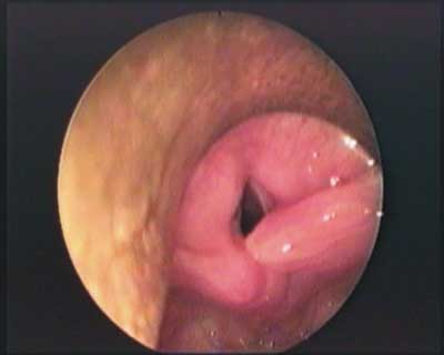
Fig. 1. Dislocation of the right arytenoid.
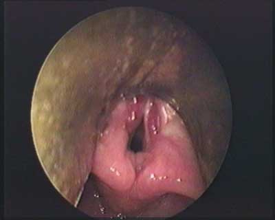
Fig. 2. Haematoma of the vocal folds.
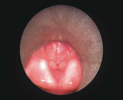
Fig. 3. Complication of supraglottic trauma.
Acute subglottic injury involves the cricoid cartilage and it is often associated with injury to the cervical trachea. As subglottic oedema increases, airway obstruction rapidly develops. Hoarseness is not marked unless recurrent laryngeal nerves have been injured.
Combined injuries (supraglottic, glottic and subglottic) are the most difficult to manage. Failure to treat this type of injury immediately results in fibrosis and airway distortion that is extremely difficult to reconstruct at a later time.
The diagnostic steps in the investigation of blunt laryngeal trauma should always involve detailed history, physical examination, radiographic studies and laryngoscopy. While all patients should undergo radiography of the chest and cervical spine, a CT scan is usually not necessary (4, 5).
Depending on the severity of the injury, patients underwent either medical, surgical treatment or both. Medical treatment included steroids and antibiotics (3, 6).
Schaeffer proposes a classification system based on tissue trauma severity that defines four groups of injuries:
1. group 1 – minor haematoma or lacerations, without fracture – conservative management is recommended.
2. group 2 – oedema, haematoma, lacerations without exposed cartilage – direct laryngoscopy and oesophagoscopy are indicated with tracheotomy if needed. Exploration of the larynx may be deferred if CT shows non evidence of displaced fractures.
3. groups 3 and 4 – massive oedema, mucosal tears, exposed cartilage and cord immobility – tracheotomy, laryngoscopy, oesophagoscopy, and laryngeal exploration are recommended with careful repair of all lacerations.
There is no single surgical technique to reconstruct the traumatized larynx (1, 2). The surgeon must have many techniques at his disposal for every individual case. Accurate surgical technique and knowledge of surgical principles will improve results.
Piśmiennictwo
1. Bryce D.P.: The surgical management of laryngotracheal injury. The Journal of Laryngology and Otology 1972, vol 86: 547-587. 2. Cherian T.A. et al.: External laryngeal trauma: analysis of thirty cases. The Journal of Laryngology and Otology 1993,vol 107: 920-923. 3.Fuhrman G.M., Stieg F.H., Buerk C.A.: Blunt laryngeal trauma: classification and management protocol. Journal of Trauma 30: 87-92, 1990. 4.Manusco A.A., Hanafee W.N.: Computed tomography of the injured larynx. Radiology 133: 139-144, 1979. 5. Myer C.M., Orobello P., Cotton R.T. et al.: Blunt laryngeal trauma in children. Laryngoscope 97:September 1987. 6.Nahum A.M.: Immediate care of acute blunt laryngeal trauma. Journal of Trauma 9: 112-115,1969. 7.Sofferman R.A.: Management of laryngotracheal trauma. Am. Journal Surgery 141: 412-417, 1981.
