© Borgis - Nowa Stomatologia 2/2017, s. 73-79
*Ewa Krasuska-Sławińska1, Tomasz Drewniak2, Bogumiła Koeber1
Mucous retention cysts of minor salivary glands in pediatric patients. Surgical procedures. Report of 3 cases
Torbiele małych gruczołów ślinowych u dzieci. Postępowanie chirurgiczne. Opis trzech przypadków klinicznych
1Dental Surgery Clinic for Adults and Children, Children’s Memorial Health Institute, Warsaw
Head of Clinic: Agnieszka Pieniak, MSc
2Surgical Clinic, Children’s Memorial Health Institute, Warsaw
Head of Clinic: Tomasz Drewniak, MD
Streszczenie
Torbiel z wynaczynienia śluzu jest jednym ze schorzeń małych gruczołów ślinowych, które licznie występują w błonie śluzowej całej jamy ustnej. Obecna jest głównie na wardze dolnej, ale może pojawić się także na wardze górnej, błonie śluzowej policzków, podniebieniu, brzusznej powierzchni języka oraz dnie jamy ustnej. Klinicznie objawia się jako miękkie, chełboczące, niebieskoszare obrzmienie błony śluzowej. Torbiel wypełniona jest przejrzystym, żółtobrązowym, galaretowatym płynem o charakterze śluzowym. Zazwyczaj torbiele z wynaczynienia śluzu są niebolesne, a ich średnica nie przekracza 1 cm. Leczeniem z wyboru jest usunięcie torbieli i wysłanie pobranego materiału do badania histopatologicznego. W przypadku zrostu torebki torbieli z otoczeniem oraz torbieli dużych rozmiarów wykonuje się zabieg marsupializacji, czyli wycięcia przedniej ściany torbieli i połączenia jej światła z jamą ustną. Zastosowanie znajdują lasery i noże elektrochirurgiczne. W określonych przypadkach zalecona jest obserwacja zmiany. Torbiele z wynaczynienia śluzu należy różnicować z włókniakami, naczyniakami, tłuszczakami, gruczolakami wielopostaciowymi.
Celem pracy jest przedstawienie metod leczenia chirurgicznego torbieli małych gruczołów ślinowych u pacjentów pediatrycznych. Uzyskano pisemną zgodę rodziców pacjentów/opiekunów prawnych pacjentów.
Summary
Mucous retention cysts are one of diseases of minor salivary glands located in oral cavity. They occur on the inner side of the lower lip in most cases, but they can also be found on the upper lip, buccal mucosa, palate, ventral surface of the tongue and fundus of the oral cavity. They manifest as soft, bluish grey swelling of the oral mucosa. Usually they are painless and not larger than 1 cm. They are filled with lucid, yellow, mucous fluid. The treatment of mucocele is surgical excision of the lesion followed by histopatological examination. Marsupialization – surgical technique of cutting a slit into cyst – can be performed in large cyst or in cases of adhesion to underlying tissue. Laser and electrosurgery can be used. Some cases may require watchful waiting. Differential diagnosis should include fibromas, hemangiomas, lipomas, pleomorphic adenomas.
The aim of this study was to present methods of surgical treatment of minor salivary glands cysts in pediatric patients. Written consent from the subject’s parents/subject’s legal guardians was obtained.

Introduction
Numerous salivary glands are found in the oral mucosa: mucous, serous and mixed mucous-serous glands. A condition affecting minor salivary glands is mucocele, classified by Pindborg and Kramer as a cyst of the soft tissues of the oral cavity, face and neck.
There are two histological types of mucocele. The first one is mucous retention cyst, also known as ductal cyst, which is formed as a result of a blockage in an excretory duct (1, 2). It is a lesion lined with ductal epithelial cells. Such cysts are found in minor salivary glands, almost never in the lower lip, in individuals over 50 years of age. This type of cysts has been classified by Kaczmarzyk as mucous retention cyst (MRC). It is usually located in the floor of the mouth and the mucosa of the tongue (3).
The second type of cyst called extravasation cyst is a non-ductal cyst which does not contain epithelial lining, but granulation tissue, which makes it a pseudocyst. It is formed as a result of traumatic severance of an excretory duct, which leads to the accumulation of mucus in the connective tissue beyond the lumen of the duct (1, 2). This cyst has been classified by Kaczmarzyk as mucous extravasation cyst (MEC) – mucocele. This type of cyst is usually located in the lower lip (80%) (3), most often in the second decade of life.
Clinical picture
A retention cyst is clinically manifested as a soft, fluctuating, blue and grey oedematous area of usually no more than 1 cm in diameter. It is well delimited from the surrounding area and the mucosa covering the lesion is tensed (fig. 1). The cyst is filled with a clear, yellow and brown, jelly-like fluid. Retention cysts are usually painless.
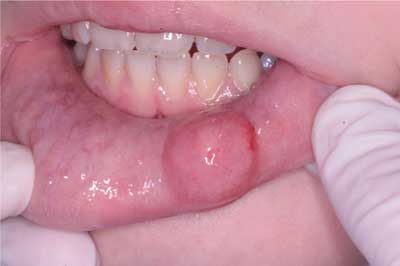
Fig. 1. Clinical picture of a salivary retention cyst (case 1)
Treatment
The treatment of choice is surgical removal of the cyst’s capsule. If the cyst is fused with the surrounding tissue or if it is large, marsupialisation is performed, i.e. the excision of the anterior wall of the cyst and connecting its lumen with the oral cavity, which is a method used primarily for major salivary gland cysts (1, 2).
Preparation for the procedure
Before starting the procedure the surgical field should be disinfected. The procedure is performed under local anaesthesia or, in the case of small children or uncooperative patients, under general anaesthesia.
When administering local anaesthesia one needs to bear in mind that infiltration anaesthesia may deform the tissue bed. Therefore, conduction anaesthesia is recommended and if it is impossible, infiltration anaesthesia at least 1 cm away from the lesion can be performed. The administration of an anaesthetic combined with a vasoconstrictor ensures smaller bleeding during the procedure. An approx. 10-minute interval should be allowed between the administration of anaesthesia and the beginning of the procedure.
For cysts of the lips immobilisation of the tissue bed is required, which is achieved by bilateral turning of the lip inside out by an assistant. At the same time compression of the labial arteries is achieved to reduce bleeding. In addition, appropriate instruments may be used such as retractors or stay sutures – sutures placed deep in the tissues at some distance from the operation site (2).
Surgical management
Powyżej zamieściliśmy fragment artykułu, do którego możesz uzyskać pełny dostęp.
Mam kod dostępu
- Aby uzyskać płatny dostęp do pełnej treści powyższego artykułu albo wszystkich artykułów (w zależności od wybranej opcji), należy wprowadzić kod.
- Wprowadzając kod, akceptują Państwo treść Regulaminu oraz potwierdzają zapoznanie się z nim.
- Aby kupić kod proszę skorzystać z jednej z poniższych opcji.
Opcja #1
29 zł
Wybieram
- dostęp do tego artykułu
- dostęp na 7 dni
uzyskany kod musi być wprowadzony na stronie artykułu, do którego został wykupiony
Opcja #2
69 zł
Wybieram
- dostęp do tego i pozostałych ponad 7000 artykułów
- dostęp na 30 dni
- najpopularniejsza opcja
Opcja #3
129 zł
Wybieram
- dostęp do tego i pozostałych ponad 7000 artykułów
- dostęp na 90 dni
- oszczędzasz 78 zł
Piśmiennictwo
1. Krasuska-Sławińska E, Koeber B, Pronicki M et al.: Torbiele zastoinowe małych gruczołów ślinowych u dzieci w materiale własnym Poradni Chirurgii Stomatologicznej dla Dzieci Instytutu Centrum Zdrowia Dziecka w Warszawie. Mag Stomatol 2014; 24(11): 62-65.
2. Peterson LI, Ellis E, Hupp J, Tucker M: Chirurgia stomatologiczna i szczękowo-twarzowa. Wydawnictwo Czelej, Lublin 2001: 517-539, 543-566.
3. Kaczmarzyk T (red.): Torbiele obszaru szczękowo-twarzowego. Wydawnictwo Kwintesencja, Warszawa 2015: 192-199.
4. Bednarz W, Olrchel-Bednarz M: Torbiel śluzowa gruczołu ślinowego (mucocele) – diagnostyka, metodologia postępowania leczniczego. Opis przypadku. E-Dentico 2009; 1(21): 65-72.
5. Levine R, Vitruc P: Laser mucocele removal in pediatric patients. Dentaltown.com (continuing education future) September 2016: 104-109.
6. Jankowska-Bugaj M, Dudko A, Kurnatowska A: Zastosowanie lasera diodowego w chirurgii tkanek miękkich jamy ustnej. e-Dentico 2012; 3(37): 8-20.
7. Ratajek-Gruda M, Szczepkowska A, Osica P et al.: Mucocele operowana nożem elektrycznym – opis przypadku. Journal of Education, Health and Sport 2016; 6(6): 6.

