© Borgis - New Medicine 2/2009, s. 40-42
*Ewa Ogłodek1, Danuta Mo?2, Aleksander Araszkiewicz1
Does exposure to extremely low frequency magnetic fields produce morphological changes in rat thyrocytes?
1Department of Clinical Psychiatry of the Nicolaus Copernicus University in Toruń, Collegium Medicum in Bydgoszcz, Poland
2Individual medical practice
Summary
Aim. The aim of this research was to evaluate the influence of extremely low-frequency magnetic fields on the morphology of thyroid epithelial cells in female rats.
Material and method. 24 female Wistar rats, Hannover substrain, aged 10 months, weighing 300+/-50 g each were included in the experiment. The rats were divided into 2 groups: group ”40”, exposed to extremely low-frequency magnetic fields of frequency 40 Hz; and control group ”C” with sham exposure.
Results. In the preparations, the thyrocytes, nuclei and nuclear-cytoplasmic ratio were evaluated.
Conclusions. The obtained results suggest that the cause of change in morphology of the epithelium of thyrocytes was stimulation of the metabolism with the activity of extremely low-frequency magnetic fields.
INTRODUCTION
The thyroid gland is one of the endocrine system organs that receives stress stimuli very strongly. Extremely low-frequency magnetic fields (ELF-MF) influence the nervous fibres of thyroid gland follicles localized in the neighbourhood of basement membrane (1, 3, 10). This influence is described as imposing the rhythm of the magnetic field on nerve endings and causing the depolarization of nerve cells and changes in the cell membrane of thyrocytes (6, 7, 9). The long-term exposure of an organism to extremely low-frequency magnetic fields intensifies the process of membrane transmission, resulting in morphological changes in the epithelial cell follicles of the thyroid gland (2, 6, 8).
AIM
The aim of this research was to evaluate the influence of extremely low-frequency magnetic fields on the morphology of thyroid epithelial cells in female rats.
MATERIAL AND METHOD
24 female Wistar rats, Hannover substrain, aged 10 months, weighing 300+/-50 g each, were included in the experiment. The rats were divided into 2 groups: group ”40”, exposed to extremely low-frequency magnetic fields of frequency 40 Hz; and control group ”C” with sham exposure. Parameters of ELF-MF: induction of 10 mT, sinusoidal waveform impulse. Rats was exposed to ELF-MF for 1 hour per day, 7 days a week, for 4 months. During the post-mortem examination the thyroid glands were removed and after formalin fixing preparations were made out of them which, in turn, underwent microscopic evaluation.
The following methods were used for the purpose of the statistical analysis: ANOVA for a single variable was performed using the following tests: Cochran-Cox test and investigation of significance of differences between the mean values (post-hoc analysis) by means of Tukey´s test.
RESULTS
In the preparations, the thyrocytes, nuclei and nuclear-cytoplasmic ratio were evaluated.
In table 1 also are presented the results of statistical analyses for comparison of measurement features – axis of the nucleus height, axis of the nucleus base, surface area of nucleus and the nuclear-cytoplasmic ratio – between the groups of analyzed rats. The results of particular parameters were significantly different depending on the type of the field volume.
Table 1. Examination parameters: Height of thyroid gland cells (H), surface area of cytoplasm of thyroid (SAC), axis of the nucleus height (ANH), axis of the nucleus base (ANB), surface area of nucleus (SAN), nuclear-cytoplasmic ratio (SAN/SAC).
| | Examined groups of rats X +/- SD | Examined groups of rats X +/- SD | Level of significance | Cochran-Cox Test | Tukey Test |
| ´40´ | ´C´ | ´40´ and ´C´ | ´40´ and ´C´ | ´40´ and ´C´ |
| H [ľm] | 4.57 (+/-0.05) | 2.02 (+/-0.08) | p<0.0001 | 0.630 | 0.00002 |
| SAC [ľm2] | 6.71 (+/-0.08) | 7.24 (+/-0.07) | p<0.0001 | 0.490 | 0.00002 |
| ANH [ľm] | 3.17 (+/-0.06) | 1.78 (+/-0.05) | p<0.0001 | 0.709 | 0.00002 |
| ANB [ľm] | 1.65 (+/-0.08) | 3.51 (+/-0.08) | p<0.0001 | 0.492 | 0.00002 |
| SAN [ľm2] | 5.17 (+/-0.08) | 3.27 (+/-0.09) | p<0.0001 | 0.462 | 0.00002 |
| SAN/SAC | 0.80 (+/-0.06) | 0.46 (+/-0.07) | p<0.0001 | 0.934 | 0.00002 |
In the present research it was demonstrated that the exposure to the magnetic field (40 Hz) was accompanied by increase of the height of the nucleus axis, increased surface area of the nucleus and decrease of the basal axis of thyrocytes. Also, increase of the nuclear-cytoplasmic ratio of thyrocytes was noticed (fig. 1).
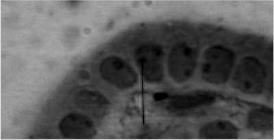
Fig. 1. Rat thyroid gland after exposure of extremely low-frequency magnetic fields (40 Hz).
DISCUSSION
A significant increase in the height of the axis of thyrocytes, height of the nucleus axis, nuclear-cytoplasmic ratio of thyrocytes, and surface area of the nucleus corresponding to the exposure of extremely low-frequency magnetic fields was observed.
According to Naarala J et al. (6) the outer magnetic field influences metabolic processes by change of the energetic state and functions in cell membranes of endocrine glands including the thyroid gland: this is proved by results of investigations performed by Katola VM et al. (7), who applied a magnetic field of the frequency of 50 Hz with the exposure time of rats being 7 hours daily, 5 days per week.
Evidence of the influence of the field intensity on the morphology of thyrocytes was shown by research of Funk RH et al. (4) in which after the application of extremely low-frequency magnetic field of the value of 0.5 Hz and a markedly shorter time of activity, morphological changes of thyroid gland epithelium were observed.
Zagorskaia EA (10) also observed changes of the location of the nucleus in the thyrocytes as an effect of activity of extremely low-frequency magnetic fields. But these authors did not present any detailed morphometric characteristics of the location of nuclei in the thyrocytes.
According to the literature on the subject, 40 Hz magnetic fields have therapeutic properties. Exposure to 40 Hz ELF-MF for several months results in growth stimulation of the thyroid glands, and increased production of the thyroid gland hormones.
CONCLUSIONS
1. The obtained results suggest that the cause of change in morphology of the epithelium of thyrocytes was stimulation of the metabolism with the activity of extremely low-frequency magnetic fields.
2. The parameters of the magnetic field accepted in the investigation corresponded to the typical values applied for magnetic field therapy in humans.
3. Exposure to the magnetic field may be treated as a specific stress situation, which in consequence leads to an increase of cell metabolism of neurotransmitters.
4. The magnetic field affects the activity of the thyroid gland without correlation with a regulatory mechanism of the pituitary-thyroid axis, and exerts a stronger effect on thyroid gland secretion in patients sensitive to its action.
5. Precautions in magnetotherapy and magnetostimu-lation are necessary to minimize the risk of thyroid disorders in humans.
Piśmiennictwo
1. Di Loreto S et al.: Fifty hertz extremely low-frequency magnetic field exposure elicits redox and trophic response in rat-cortical neurons. J Cell Physiol 2009; 219(2): 334-43. 2. Falone S et al.: Chronic exposure to 50Hz magnetic fields causes a significant weakening of antioxidant defence systems in aged rat brain. Int J Biochem Cell Biol 2008; 40(12): 2762-70. 3. Fernie KJ et al.: The effects of electromagnetic fields from power lines on avian reproductive biology and physiology: a review. J Toxicol Environ Health B Crit Rev 2005; 8(2): 127-40. 4. Funk RH et al.: Electromagnetic effects – From cell biology to medicine. Prog Histochem Cytochem 2009; 43(4): 177-264. 5. Luo ZG et al.: Influence of magnetic field on nitric oxide in hypothalamus and its relation to hypothalamic neuroendocrine nuclei. Shi Yan Sheng Wu Xue Bao 2000; 33(2): 109-17. 6. Naarala J et al.: Cellular effects of electromagnetic fields. Altern Lab Anim 2004; 32(4): 355-60. 7. Katola VM et al.: Effect of a permanent magnetic field on the thyroid status Kosm Biol Aviakosm Med 1988; 15(4): 50-2. 8. Saunders RD et al.: A neurobiological basis for ELF guidelines. Health Phys 2007; 92(6): 596-603. 9. Tenuzzo B et al.: Biological effects of 6 mT static magnetic fields: a comparative study in different cell types. Bioelectromagnetics 2006; 27(7): 560-77. 10. Zagorskaia EA: Effect of a permanent magnetic field on the endocrine system. Kosm Biol Aviakosm Med 1981; 15(5): 14-7.
