*Dorota Olczak-Kowalczyk1, Anna Turska-Szybka1, Dariusz Gozdowski2, Urszula Kaczmarek3
Developmental defects of enamel in the population of Polish adolescents aged 18 years old: the prevalence and selected socio-demographic factors. A cross-sectional study**
Defekty rozwojowe szkliwa u młodzieży w wieku 18 lat w Polsce: rozpowszechnienie i wybrane czynniki socjodemograficzne. Badania przekrojowe**
1Paediatric Dentistry Division, Medical University of Warsaw
Head of Division: Professor Dorota Olczak-Kowalczyk, MD, PhD
2Department of Experimental Design and Bioinformatics, Warsaw University of Life Sciences
Head of Department: Professor Krzysztof Pawłowski, DSc (Eng)
3Department and Division of Conservative and Pediatric Dentistry, Wrocław Medical University
Head of Department and Division: Professor Urszula Kaczmarek, MD, PhD
Streszczenie
Wstęp. Brak aktualnych badań epidemiologicznych utrudnia ocenę skali problemu zdrowotnego, jakim są defekty rozwojowe szkliwa u młodzieży w Polsce.
Cel pracy. Ocena występowania defektów rozwojowych szkliwa w uzębieniu stałym u osób w wieku 18 lat w Polsce z uwzględnieniem wpływu wybranych czynników socjodemograficznych.
Materiał i metody. Do badań przekrojowych w 2017 roku kwalifikowano osoby w wieku 18 lat uczęszczające do szkół w 16 województwach Polski, wyłonionych losowaniem wielowarstwowym. Czynniki socjodemograficzne (płeć, miejsce zamieszkania, poziom wykształcenia rodziców, status ekonomiczny) oceniano badaniem ankietowym. Stan uzębienia z zastosowaniem DDE Index w modyfikacji Clarksona i wskaźnika Deana oceniali lekarze dentyści po szkoleniu i kalibracji. Przed rozpoczęciem badań uzyskano zgodę Komisji Bioetycznej Warszawskiego Uniwersytetu Medycznego (KB/134/2017 z dnia 6.06.2017 r.).
Wyniki. Zbadano 1611 osób (52,6% dziewcząt, 50,5% mieszkańców wsi). Defekty rozwojowe szkliwa zębów obserwowano u 16,3%, najczęściej o charakterze zmętnień ograniczonych (10,4%). U 2,7% osób kwalifikowano je jako fluorozę, najczęściej wątpliwą. Defekty częściej dotyczyły przyśrodkowych zębów siecznych i pierwszych zębów przedtrzonowych szczęki niż pozostałych zębów. U 2,5% badanych były to wady pojedynczych zębów, u 0,6% obejmowały całe uzębienie. Defekty nieklasyfikowane jako fluoroza występowały częściej u osób płci męskiej. Istniały istotne statystycznie różnice w częstości występowania defektów między województwami. Najczęściej obserwowano je w województwach położonych w południowej części kraju. Nie miały natomiast wpływu inne czynniki socjodemograficzne.
Wnioski. Przewaga występowania zmętnień ograniczonych wskazuje na duże znaczenie czynników miejscowych w etiologii defektów rozwojowych szkliwa zębów stałych w Polsce. Rzadko spotykaną wadą jest fluoroza zębów. Brak znaczenia czynników socjoekonomicznym i zróżnicowanie występowania DDE w różnych regionach Polski wskazują na potrzebę badań identyfikujących czynniki ryzyka związane z położeniem geograficznym.
Summary
Introduction. There has been a lack of current epidemiological data regarding the prevalence of developmental defects of enamel in Polish adolescents.
Aim. To evaluate the prevalence of developmental defect of enamel in the permanent dentition in the population of adolescents aged 18 years old, including the impact of selected sociodemographic factors.
Material and methods. A cross-sectional study conducted in 2017 covered adolescents aged 18 years old attending schools in 16 Polish voivodeships, selected by stratified sampling. Sociodemographic factors such as sex, place of residence, parents’ education level and subjective economic status were collected with a survey. The condition of enamel was assessed with the use of DDE Index modified by Clarkson and Dean’s Indicator by dentists specially trained and calibrated for this study. Prior to its initiation, the study was approved by the Bioethics Committee of the Medical University in Warsaw (Ref. No.: KB/134/217 of 6.06.217).
Results. A total of 1611 adolescents were examined (with 52.6% female and 50.5% were residents of rural areas). Developmental defects of enamel were identified in 16.3% of the participants of the study, most frequently in the form of demarcated opacities (10.4%). In 2.7%, they were classified as fluorosis, most frequently questionable. Maxillary central incisors and first premolars were most frequently affected. In 2.5% of the examined adolescents, the defects involved single teeth, whereas in 0.6% the defects were generalised. Defects not classified as fluorosis were more commonly identified in males. There were statistically significant differences in the prevalence between voivodeships, with defects most commonly observed in participants from southern voivodeships. No other sociodemographic factors, however, were identified as significant.
Conclusions. The highest prevalence of demarcated opacities suggests the significance of local factors in the aetiology of developmental enamel defects of permanent dentition in Poland. Fluorosis is a rarely encountered entity. The lack of the significance of sociodemographic factors and the regional differences in the prevalence of developmental defects of enamel suggest the need for further research, aimed at identifying geographical risk factors.

Introduction
Developmental defects of tooth enamel (DDE) are an important health problem, detrimental to a person’s quality of life. They may affect the appearance, increase teeth sensitivity, predispose for the development of caries, abrasion and erosion (1-3). In a study performed among adolescents aged 16 years old, 18.8% of the participants avoided showing their teeth when smiling due to DDE, 8.7% avoided social contacts and 5.7% had experienced mocking by peers (4).
DDE may be either quantitative, involving a decreased enamel thickness or local lack of enamel (hypoplasia), or qualitative, presenting as opacities or discoloration of enamel (1, 2). Depending on the putative factor at work during amelogenesis and the time frame of exposure, DDE may be generalised, or affect groups of teeth or single teeth. The defects found in single permanent teeth are caused by an injury or infection, e.g. of the primary predecessor (5, 6). In the case of defects involving a group of teeth or all teeth, various genetic and environmental factors may play a role (7-21), such as environmental pollution and a low socioeconomic status (13-21).
In 1990, a country-wide study evaluated the prevalence of DDE in Poland, confirming the role of the fluoride level in the drinking water and systemic factors for DDE aetiology (12, 22). The study covered adolescents living in areas supplied with naturally fluoridated drinking water and those with artificially fluoridated water. Since 1996, drinking water in Poland has not been artificially fluoridated any more. Hence the need for the update of the epidemiological data.
Aim
This study has been aimed at evaluating the prevalence of DDE in permanent dentition in the population of Polish adolescents aged 18 years old, including the impact of selected sociodemographic factors.
Material and method
The survey and the clinical examination covered adolescents 18 years old who were students of vocational and higher secondary schools country-wide. The participants provided their written consent for the participation in the study. The schools were selected by stratified sampling. In each of the voivodeships, poviats, communities (all Polish administrative units, województwa, powiaty and gminy, respectively) and then schools were randomly selected.
The study complied with WHO criteria (23) and was performed by 22 dental examiners (dentists) who were specially trained and calibrated. Inter-rater reliability between the reference examiner and other examiners ranged from 0.802 to 1.00 Cohen’s kappa coefficient, whereas intra-rater reliability was 0.998.
The survey was conducted with the use of a questionnaire including questions about sex, place of residence (urban/rural, which voivodeship), parents’ education level, family economic status in the participant’s opinion.
In the clinical examination, performed in artificial light with the WHO 621 probe, the teeth were evaluated for the presence and type of developmental enamel defects and their distribution. The defects were classified based on their macroscopic appearance according to DDE Index modified by Clarkson as diffuse or demarcated opacities, enamel hypoplasia, discolouration and “other” (a combination of more than one type of defects) (24). The following types of hypoplasia were accounted for: pits, grooves and enamel missing on dental surface or the incisal edge. Following WHO recommendations, Dean’s index was used to evaluate dental fluorosis (25).
Prior to its initiation, the study was approved by the Bioethics Committee of the Medical University in Warsaw (Ref. No.: KB/134/217 of 6.06.217).
The obtained results were statistically analysed. Means between two groups (urban vs rural area or male vs female) were compared with Student’s t-test, whereas percent values were compared with the chi-square test. The statistical significance level was set at p ≤ 0.05. The statistical analyses were performed with Statistica 12.0 software.
Results
A total of 1611 adolescents aged 18 years old were examined. The studied group comprised 847 females (52.6%) and 764 males (47.4%), and 797 (49.5%) residents of urban areas and 814 (50.5%) residents of rural areas. In different voivodeships, the number of participants ranged from 99 in Łódzkie Voivodeship to 110 in Małopolskie Voivodeship (mean 100.68 ± 2.52). The participants most frequently stated their family’s economic status as “average” (55.5%), followed by “above average” (23.7%), and least frequently as “below average” (2.9%). 17.9% did not provide their subjective economic status at all. The group’s composition according to parents’ level of education have been shown in figure 1.
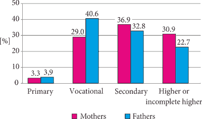
Fig. 1. The level of education of the parents of the examined adolescents aged 18 years old
DDE were found in 16.3% of the examined adolescents, including 2.7% defects classified as fluorosis. The prevalence of DDE and the mean number of the affected teeth have been shown in table 1. The most common type of DDE were demarcated opacities (168/10.4%), followed by diffuse opacities (65/4.0%), hypoplasia (25/1.6%), discolouration (2/0.1%) and a combination of different defects (10/0.6%). Out of 26 noted cases of hypoplasia, dental surface hypoplasia (14/53.8%) and pits (9/34.6%) were the most commonly encountered, whereas grooves (2/7.7%) and missing enamel on the incisal edges (1/3.9%) were less frequent.
Tab. 1. The prevalence of DDE in the population of adolescents aged 18 years old according to the place of residence (urban vs. rural) and sex
| | DDE overall | DDE not classified as fluorosis | Fluorosis |
| Number of affected adolescents % | Mean number of teeth ± SD | Number of affected adolescents | Mean number of affected teeth ± SD | Number of affected adolescents | Mean number of affected teeth ± SD |
| Urban areas | 143/17.9% | 1.32 ± 5.02 | 119/14.9% | 0.91 ± 3.34 | 24/3.0% | 0.41 ± 2.76 |
| Rural areas | 120/14.7% | 1.09 ± 4.73 | 101/12.4% | 0.79 ± 3.34 | 19/2.3% | 0.30 ± 2.29 |
| p | 0.082 | 0.338 | 0.140 | 0.472 | 0.399 | 0.371 |
| Females | 130/15.3% | 1.11 ± 4.82 | 102/12.0% | 0.69 ± 2.88 | 28/3.3% | 0.42 ± 2.70 |
| Males | 133/17.4% | 1.31 ± 4.95 | 118/15.4% | 1.03 ± 3.78 | 15/2.0% | 0.28 ± 2.33 |
| p | 0.264 | 0.410 | 0.047* | 0.041* | 0.095 | 0.271 |
| Total | 263/16.3% | 1.21 ± 4.88 | 220/13.7% | 0.85 ± 3.34 | 43/2.7% | 0.35 ± 2.53 |
*statistical significance
Demarcated opacities were found in 1.8% of all examined teeth, diffuse opacities in 1.2% and hypoplasia – in 0.1%. The defects most commonly involved the enamel of maxillary central incisors and first premolars (fig. 2). Diffuse opacities were more evenly distributed than demarcated opacities and hypoplasia.

Fig. 2. The distribution of DDE according to given teeth in the entire examined population of adolescents aged 18 years old
Powyżej zamieściliśmy fragment artykułu, do którego możesz uzyskać pełny dostęp.
Mam kod dostępu
- Aby uzyskać płatny dostęp do pełnej treści powyższego artykułu albo wszystkich artykułów (w zależności od wybranej opcji), należy wprowadzić kod.
- Wprowadzając kod, akceptują Państwo treść Regulaminu oraz potwierdzają zapoznanie się z nim.
- Aby kupić kod proszę skorzystać z jednej z poniższych opcji.
Opcja #1
29 zł
Wybieram
- dostęp do tego artykułu
- dostęp na 7 dni
uzyskany kod musi być wprowadzony na stronie artykułu, do którego został wykupiony
Opcja #2
69 zł
Wybieram
- dostęp do tego i pozostałych ponad 7000 artykułów
- dostęp na 30 dni
- najpopularniejsza opcja
Opcja #3
129 zł
Wybieram
- dostęp do tego i pozostałych ponad 7000 artykułów
- dostęp na 90 dni
- oszczędzasz 78 zł
Piśmiennictwo
1. Seow WK: Developmental defects of enamel and dentine: challenges for basic science research and clinical management. Aust Dent J 2014; 59 (1 suppl.): 143-154.
2. Wright JT: Normal formation and development defects of the human dentition. Pediatr Clin North Am 2000; 47(5): 975-1000.
3. Vargas-Ferreira F, Ardenghi TM: Developmental enamel defects and their impact on child oral health-related quality of life. Braz Oral Res 2011; 25(6): 531-537.
4. Sujak SL, Abdul Kadir R, Dom TNM: Esthetic perception and psychosocial impact of developmental enamel defects among Malaysian adolescents. J Oral Sci 2004; 46(4): 221-226.
5. Sleiter R, von Arx T: Developmental disorders of permanent teeth after injuries of their primary predecessors. A retrospective study. Schweiz Monatsschr Zahnmed 2002; 112(3): 214-219.
6. Holan G, Topf J, Fuks AB: Effect of root canal infection and treatment of traumatized primary incisors on their permanent successors. Endod Dent Traumatol 1992; 8(1): 12-15.
7. Gisoo FF, Mohseni A: Prevalence study of etiologies of developmental defects of enamel of first permanent molar among six to seven years old children. Curr Res Dent 2010; 1: 19-22.
8. Enache R, Maxim A, Păsăreanu M: Risk factors involved in the development of enamel defects. J Roman Med Dents 2010; 14: 71-74.
9. Robles MJ, Ruiz M, Bravo-Perez M et al.: Prevalence of enamel defects in primary and permanent teeth in a group of schoolchildren from Granada (Spain). Med Oral Patol Oral Cir Bucal 2013; 18(2): 187-193.
10. Olczak-Kowalczyk D, Danko M, Banaś E et al.: Parenteral nutrition in childhood and consequences for dentition and gingivae. Eur J Paediatr Dent 2017; 18(1): 69-76.
11. Krasuska-Sławińska E, Brożyna A, Dembowska-Bagińska B, Olczak-Kowalczyk D: Antineoplastic chemotherapy and congenital tooth abnormalities in children and adolescents. Contemp Oncol (Pozn) 2016; 20(5): 394-401.
12. Kaczmarek U, Potoczek S, Nowak-Malinowska H et al.: Zaburzenia mineralizacji twardych tkanek zębów w wybranych grupach wieku u dzieci polskich. Badania ankietowe u dzieci w wieku 7-8 lat z zaburzeniami i bez zaburzeń mineralizacji. Czas Stomatol 1992; 45(6-7): 309-312.
13. Wozniak K: Developmental abnormalities of mineralization in populations with varying exposure to fluorine compounds. Ann Acad Med Stetin 2000; 46: 305-315.
14. Balmer RC, Laskey D, Mahoney E, Toumba KJ: Prevalence of enamel defects and MIH in non-fluoridated and fluoridated communities. Eur J Paediatr Dent 2005; 6(4): 209-212.
15. Wong HM, McGrath C, Lo EC, King NM: Association between developmental defects of enamel and different concentrations of fluoride in the public water supply. Caries Res 2006; 40(6): 481-486.
16. Ekanayake L, van der Hoek W: Prevalence and distribution of enamel defects and dental caries in a region with different concentrations of fluoride in drinking water in Sri Lanka. Int Dent J 2003; 53(4): 243-248.
17. Janja J, Sovcikova E, Kočan A et al.: Developmental dental defects in children exposed to PCBs in eastern Slovakia. Chemosphere 2007; 67(9): 350-354.
18. Jan J, Vrbic V: Polychlorinated biphenyls cause developmental enamel defects in children. Caries Res 2000; 34(6): 469-473.
19. Ford D, Seow WK, Kazoullis S et al.: A controlled study of risk factors for enamel hypoplasia in the permanent teeth. Pediatr Dent 2009; 31(5): 382-388.
20. Basha S, Mohamed RN, Swamy HS: Prevalence and associated factors to developmental defects of enamel in primary and permanent dentition. Oral Health Dent Manag 2014; 13(3): 588-594.
21. Păsăreanu M, Florea C: Risk factors involvement in enamel dental dysplasia. J De Med Prevent 2001; 9: 13-17.
22. Kaczmarek U, Potoczek S, Malepszy A et al.: Zaburzenia mineralizacji twardych tkanek zębów w wybranych grupach wieku u dzieci polskich. Frekwencja zaburzeń mineralizacji u badanych w poszczególnych podgrupach rejonów o różnej zawartości fluoru w wodzie do picia. Czas Stomatol 1992; 45(4): 210-213.
23. WHO: Oral Health Surveys. Basic Methods. 5th ed. Geneva 2013.
24. Clarkson J, O’Mullane DM: A modified DDE Index for use in epidemiological studies of enamel defects. J Dent Res 1988; 68(3): 445-450.
25. Dean HT: Classification of mottled enamel diagnosis. J Am Dent Assoc 1934; 21: 1421-1426.
26. de Almeida CM, Petersen PE, Andrè SJ, Toscano A: Changing oral health status of 6- and 12-year-old schoolchildren in Portugal. Community Dent Health 2003; 20(4): 211-216.
27. Muratbegovic A, Zukanovic A, Markovic N: Molar-incisor-hypomineralisation impact on developmental defects of enamel prevalence in a low fluoridated area. Eur Arch Paediatr Dent 2008; 9(4): 228-231.
28. Hoffmann RH, de Sousa M da L, Cypriano S: Prevalence of enamel defects and the relationship to dental caries in deciduous and permanent dentition in Indaiatuba, São Paulo, Brazil. Cad Saude Publica 2007; 23(2): 435-444.
29. Chauhan D, Chauhan T: Prevalence of developmental defects of enamel in mixed and permanent dentition of 9 and 12 year old children of Himachal Pradesh, India: A cross sectional study. Int J Health Allied Sci 2013; 2: 185-188.
30. Ravindran R, Saji AM: Prevalence of the developmental defects of the enamel in children aged 12-15 years in Kollam district. J Int Soc Prev Community Dent 2016; 6(1): 28-33.
31. Arrow P: Prevalence of dental enamel defects of the first permanent molars among school children in Western Australia. Aust Dent J 2008; 53(3): 250-259.
32. Montero MJ, Douglass JM, Mathieu GM: Prevalence of dental caries and enamel defects in Connecticut Head Start children. Pediatr Dent 2003; 25(3): 235-239.
33. Mackay TD, Thomson WM: Enamel defects and dental caries among Southland children. N Z Dent J 2005; 101(2): 35-43.
34. Kanagaratnam S, Schluter P, Durward C et al.: Enamel defects and dental caries in 9-year-old children living in fluoridated and nonfluoridated areas of Auckland, New Zealand. Community Dent Oral Epidemiol 2009; 37(3): 250-259.
35. Olczak-Kowalczyk D, Gozdowski D, Kaczmarek U: Wyniki stomatologicznych badań klinicznych młodzieży w wieku 18 lat i ich omówienie. [W:] Olczak-Kowalczyk D, Mielczarek A (red.): Monitorowanie stanu zdrowia jamy ustnej populacji polskiej w latach 2016-2020. Ocena stanu zdrowia jamy ustnej i jego uwarunkowań w populacji polskiej w wieku 3, 18 oraz 35-44 lata w 2017 roku. Dział Redakcji i Wydawnictw Warszawskiego Uniwersytetu Medycznego 2018: 167-232.
36. Wochna-Sobańska M, Szydłowska-Walendowska B, Lubowiedzka-Gontarek B, Proc P: Występowanie fluorozy i próchnicy u 12-letnich dzieci zamieszkałych na terenach z ponadoptymalnym poziomem fluoru w wodzie pitnej. Czas Stomatol 2009; 62(3): 178-183.
37. Opydo-Szymaczek J: Fluoride Exposure from Diet in Infants and Young Children Fed with the Foodstuffs for Particular Nutritional Use. Dent Med Probl 2012; 49(2): 209-215.
38. Opydo-Szymaczek J, Gerreth K: Enamel fluorosis and its association with dental caries in a nonfluoridated community of Wielkopolska, Western Poland. Fluoride 2013; 46(4): 234-238.
39. Vargas-Ferreira F, Salas MMS, Nascimento GG et al.: Association between developmental defects of enamel and dental caries: A systematic review and meta-analysis. J Dent 2015; 43(6): 619-628.
40. Gopalakrishnan P, Vasan RS, Sarma PS et al.: Prevalence of dental fluorosis and associated risk factors in Alappuzha district, Kerala. Natl Med J India 1999; 12(3): 99-103.
41. Nik-Hussein N, Majid ZA, Mutalib KA et al.: Prevalence of developmental defects of enamel among 16-year-old children in Malaysia. Annal Dent Univ Malaya 1999; 6(1): 11-16.


