Piotr Sobiech1, Anna Turska-Szybka1, Dariusz Gozdowski2, *Dorota Olczak-Kowalczyk1
Caries distribution pattern in primary dentition in children in early childhood from the Warsaw agglomeration
Rozmieszczenie prГіchnicy w uzДҷbieniu mlecznym u dzieci w okresie wczesnodzieciДҷcym z aglomeracji warszawskiej
1Department of Pediatric Dentistry, Medical University of Warsaw, Poland
Head of Department: Professor Dorota Olczak-Kowalczyk, PhD, DMD
2Department of Experimental Statistics and Bioinformatics, Warsaw University of Life Science, Poland
Head of Department: Professor Dorota Olczak-Kowalczyk, PhD, DMD
Streszczenie
WstДҷp. Wysoka czДҷstoЕӣДҮ wystДҷpowania i poziom prГіchnicy wczesnego dzieciЕ„stwa (ECC) u dzieci w wieku 3 lat w Polsce wskazujД… na wysokie ryzyko zachorowania na tДҷ chorobДҷ dzieci mЕӮodszych. PrГіchnica najczДҷЕӣciej rozwija siДҷ na powierzchniach okludalnych zДҷbГіw trzonowych. Rozmieszczenie prГіchnicy w uzДҷbieniu mlecznym zmienia siДҷ wraz z wiekiem. U dzieci mЕӮodszych zДҷby sieczne szczДҷki sД… najczДҷЕӣciej dotkniДҷte prГіchnicД…, natomiast u starszych – zДҷby trzonowe. Brak jest aktualnych danych opisujД…cych rozmieszczenie prГіchnicy u dzieci mЕӮodszych w Polsce.
Cel pracy. Ocena rozmieszczenia prГіchnicy z uwzglДҷdnieniem powierzchni w uzДҷbieniu mlecznym u dzieci w 2. i 3. roku Ејycia z aglomeracji warszawskiej.
MateriaЕӮ i metody. W badaniu przekrojowym dzieci w wieku 12-36 miesiДҷcy w ocenie stanu uzДҷbienia uwzglДҷdniano obecnoЕӣДҮ prГіchnicowych zmian nieubytkowych (d), ubytkowych (p), wypeЕӮnieЕ„ (w), brakГіw zДҷbowych spowodowanych prГіchnicД… (u). OkreЕӣlono czДҷstoЕӣДҮ ECC, jej nasilenie (dpuwz i dpuwp). W analizie statystycznej zastosowano test chi-kwadrat do porГіwnania frakcji (udziaЕӮГіw procentowych).
Wyniki. Zbadano 496 dzieci, w tym 262 (52,8%) chЕӮopcГіw. S-ECC odnotowano u 44,8% badanych, dpuwz i dpuwp osiД…gnДҷЕӮy wartoЕӣci 2,62 ± 3,88 i 4,46 ± 8,42. NajczДҷЕӣciej zmianami prГіchnicowymi byЕӮy objДҷte zДҷby sieczne przyЕӣrodkowe szczДҷki (34,2%) oraz pierwsze zДҷby trzonowe obu ЕӮukГіw (21,0%, w tym 23,5% dla szczДҷki i 18,6% dla Ејuchwy). Zmiany prГіchnicowe najczДҷЕӣciej wystДҷpowaЕӮy na powierzchniach wargowych zДҷbГіw siecznych szczДҷki (19,1%) oraz okludalnych zДҷbГіw pierwszych trzonowych (19,4%).
Wnioski. PrГіchnicДҷ zДҷbГіw mlecznych w okresie wczesnodzieciДҷcym charakteryzuje szybkie przechodzenie zmian nieubytkowych w ubytkowe i obejmowanie kolejno wyrzynajД…cych siДҷ zДҷbГіw. Zmianami prГіchnicowymi najczДҷЕӣciej dotkniДҷte sД… powierzchnie wargowe zДҷbГіw siecznych przyЕӣrodkowych szczДҷki oraz okludalne zДҷbГіw pierwszych trzonowych.
Summary
Introduction. The high incidence and level of early childhood caries (ECC) in children aged 3 years in Poland indicate a high risk of developing this disease in younger children.
Caries most often develops on the occlusal surfaces of molars. The distribution of caries in primary dentition changes with age. In younger children, the maxillary incisors are most often affected by caries, while in the older ones – molars. There are no current data describing caries distribution in younger children in Poland.
Aim. Assessment of caries distribution, taking into account the surface in primary dentition in children in the second and third year of life from the Warsaw agglomeration.
Material and methods. This was a cross-sectional study conducted among children aged 12-36 months to assess dental condition for the presence of non-cavitated (d1) and cavitated caries (d2), fillings (f), and missing (m) teeth (t) and surfaces (s) caused by caries. The frequency of ECC and its intensity (d1d2mft and d1d2mfs) were determined. In the statistical analysis, the chi-square test was used to compare the fractions (percentages).
Results. A total of 496 children were examined, including 262 (52.8%) boys. S-ECC was recorded in 44.8% of the respondents, d1d2mft and d1d2mfs reached the values of 2.62 ± 3.88 and 4.46 ± 8.42, respectively. Central maxillary incisors (34.2%) and the first molars of both arches (21.0%, including 23.5% for the maxilla and 18.6% for the mandible) were most commonly affected by carious lesions. Carious lesions were most often found on the labial surfaces of the maxillary incisors (19.1%) and occlusal first molars (19.4%).
Conclusions. Primary teeth caries in the early childhood period is characterized by a rapid transformation of non-cavitated lesions into cavitated ones and subsequent eruption of teeth. Carious lesions most are most often found on the labial surfaces of the central incisors of the maxilla and occlusal surfaces of the first molars.
Introduction
Exposure of a primary tooth to cariogenic factors shortly after its appearance in the oral cavity is associated with a high risk of carious process and disease progression. The enamel of freshly erupted teeth shows a low degree of mineralisation and high porosity.
High susceptibility to bacterial acids and high enamel permeability contribute to the rapid occurrence of signs of caries following exposure to bacterial acids and transformation of non-cavitated into cavitated lesions (1). Poor mineralisation of the thin dentin layer and the straight course of the wide dentinal tubules promote rapid deepening of the cavity and pulpopathy. The period of early childhood, the second year of a child’s life in particular, seems to be of key importance for maintaining healthy primary dentition. A 2-year-old child usually already has erupted incisors, which are followed by first molars, then canines, and, in some children, also second primary molars. At about 31 months of age, a child already has twenty primary teeth. The time of exposure of the teeth to the oral cavity environment at the age of 36 months ranges from 24 to 30 months for medial incisors, from 18 to 30 months for the lateral incisors, from 17 to 27 months for first molars, from 12 to 22 months for canines and 5 to 17 months for second molars (2). Considering the average duration of post-eruptive maturation of enamel of 2-4 years after tooth eruption, children aged 2-3 years should be considered a group of increased risk of caries and its rapid progression. Therefore, the presence of any carious, non-cavitated or cavitated lesion on the smooth surfaces of a child’s teeth before the age of 3 years is classified as severe early childhood caries (S-ECC). This form is diagnosed in the presence of carious lesions on at least 4 surfaces of the teeth in 3-year-olds, 5 surfaces in 4-year--olds, and at least 6 surfaces in 5-year-olds (3-5). S-ECC refers to “atypical”, “progressive”, “acute” or “rampant” pattern of dental caries.
Aim
The aim of the study was to assess caries distribution in primary dentition in children in the early childhood period from the Warsaw agglomeration depending on age.
Material and methods
The research was conducted in 2011-2017 as part of a programme assessing the oral health and the course of first teething. The study was approved by the Bioethics Committee of the Medical University of Warsaw (KB/221/2009). Cooperative children aged > 12-36 months from Warsaw and areas located no more than 20 km from Warsaw were enrolled in the study after obtaining an informed written consent of the parent/legal guardian for their child to participate in the clinical examination. Children aged less than 12 months and over 36 months, those with intellectual disability, chronic diseases, pharmacotherapy likely to affect dental health, and children residing outside the Warsaw agglomeration were excluded from the study.
The clinical examination of oral health was performed in a dentist’s office equipped with a shadowless lamp, using a mirror and a WHO-621 periodontal probe (6). The number of erupted teeth, the presence of carious lesions, filled and missing teeth due to caries were assessed, considering each tooth surface in subsequent quadrants. Carious lesions were assessed according to the criteria of the modified International Caries Detection and Assessment System (ICDAS II) (7). Non-cavitated lesions with ICDAS-II codes 1-2 were diagnosed as carious when found in the perigingival area or on the chewing surface and were denoted as “d1”. Cavitated lesions (ICDAS II codes ≥ 3) were denoted as “d2”. We estimated the number of teeth present in the oral cavity, the number and percentage of teeth with caries (d1d2mft > 0) as well as d1d2mft and d1d2mfs components, where d1t/d1s stands for teeth/tooth surfaces with non-cavitated lesions (ICDA II code 1 and 2), d2t/ d2s – with carious lesions, mt/ms – missing due to caries, and ft/fs – fillings (6, 7).
The distribution of carious lesions was assessed in the entire study group and in four age subgroups: > 12-18 months, > 18-24 months, > 24-30 months, and > 30-36 months.
The study was conducted by three specialists in pediatric dentistry, who were appropriately trained and calibrated. During calibration, each specialist examined the same group of 10 children independently. Cohen’s kappa coefficients ranging from 0.89 to 0.95 were obtained.
In the statistical analysis, the chi-square test was used to compare the fractions (percentages, e.g., of carious teeth) between the two groups. Statistica 13 was used for statistical analyses. The level of significance was < 0.05 for all analyses.
Results
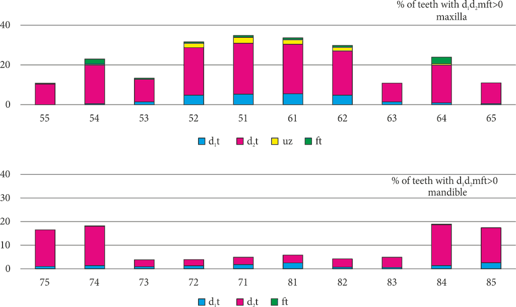
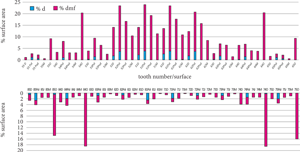
A total of 496 children (mean age 24.16 ± 6.93 months), including 262 (52.8%) boys, participated in the study. The children were divided into age subgroups: 105 (27.2%) in the youngest subgroup, 129 (25.8%) aged >18-24 months, 133 (26.0%) aged > 24-30 months, and 129 (20.1%) children in the oldest subgroup. Table 1 shows the characteristics of the study group, including age, number of assessed and carious teeth and tooth surfaces. Throughout the study group, a statistically significantly higher percentage of teeth and tooth surfaces in the maxilla than in the mandible was affected by caries (tab. 2). There were no differences in the frequency of carious lesions on the left and right side of the dental arches (tab. 3). Carious disease most often involved the maxillary incisors and the first molars of both arches (fig. 1). Central maxillary incisors (34.2%) and first molars of both arches (21.0%, including 23.5% for the maxilla and 18.6% for the mandible) were most commonly involved. Carious lesions (dmf) most often occurred on the labial surfaces of maxillary incisors (19.1%) and occlusal first molars (19.4%) (fig. 2).
Tab. 1. Mean age and number of assessed and carious teeth/tooth surfaces in the entire study group and age subgroups
| Parameters | Age subgroup | Total |
| > 12-18 months | > 18-24 months | > 24-30 months | > 30-36 months |
| Age (months) mean ± SD | 15.52 ± 2.06 | 21.76 ± 1.83 | 27.85 ± 1.77 | 33.75 ± 1.55 | 24.16 ± 6.93 |
| Number of teeth |
| Assessed (100%) | 967 | 1841 | 2325 | 2503 | 7936 |
| Carious n (%) | 75 (7.7%) | 265 (14.4%) | 443 (19.1%) | 522
(20.8%) | 1305 (17.1%) |
| Missing due to caries n (%) | 0
(0.0%) | 15
(0.8%) | 24
(1.0%) | 12
(0.5%) | 51
(0.6%) |
| Number of tooth surfaces |
| Assessed (100%) | 4045 | 7873 | 10090 | 10991 | 32999 |
| Carious n (%) | 114 (2.8%) | 490 (9.2%) | 668
(6.6%) | 938
(8.5%) | 2210 (6.7%) |
Tab. 2. Maxillary and mandibular distribution of caries in primary dentition throughout the study group for teeth and tooth surfaces
| Location | Teeth assessed | Carious teeth | Assessed tooth surfaces | Carious tooth surfaces |
| d1t | d2t | mt | ft | d1s | d2s | ms | fs |
| n = 100% | n/% | n = 100% | n/% |
| Maxilla | 3811 | 112
(2.9) | 727 (19.1) | 49
(1.3) | 43
(1.1) | 16471 | 140
(0.8) | 1331 (8.1) | 201
(1.2) | 59
(0.4) |
| Mandible | 3825 | 54
(1.4) | 285
(7.5) | 2
(0.1) | 33
(0.9) | 16528 | 60
(0.4) | 369
(2.2) | 10
(0.1) | 40
(0.2) |
| p | l | < 0.001* | < 0.001* | < 0.001* | 0.240 | l | < 0.001* | < 0.001* | < 0.001* | 0.053 |
| Total | 7636 | 166
(2.2) | 1012 (13.3) | 51
(0.7) | 76
(1.0) | 32999 | 200
(0.6) | 1700 (5.2) | 211
(0.6) | 99
(0.3) |
*statistically significant, chi-square test
Tab. 3. Maxillary and mandibular distribution of carious lesions in the left and right dental arch in primary dentition throughout the study group
| l | Maxilla | Mandible |
| Location | Teeth assessed | Carious teeth | Teeth assessed | Carious teeth |
| d1s | d2t | mt | ft | d1t | d2t | mt | ft |
| n = 100% | n/% |
| Right arch | 1899 | 54 (2.8) | 372 (19.6) | 26 (1.4) | 21 (1.1) | 1908 | 28 (1.5) | 148 (7.8) | 1 (0.1) | 15 (0.8) |
| Left arch | 1912 | 58 (3.0) | 355 (18.6) | 23 (1.2) | 22 (1.2) | 1917 | 26 (1.4) | 137 (7.1) | 1 (0,1) | 18 (0.9) |
| p | l | 0.713 | 0.616 | 0.586 | 0.772 | l | 0.796 | 0.410 | 0.999 | 0.736 |
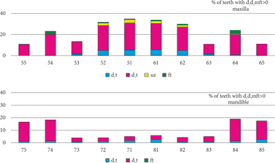
Fig. 1. Caries distribution in individual maxillary and mandibular teeth
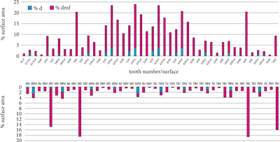
Fig. 2. Caries distribution in individual tooth surfaces in maxillary and mandibular arches throughout the study group
*statistically significant, chi-square test
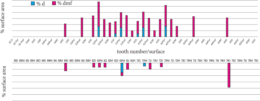
In the youngest age group (> 12-18 months), maxillary teeth were more often involved (58/500; 11.6%) than mandibular dentition (17/467; 3.6%) (fig. 3). No filled teeth were found in any of the children. Maxillary incisors were the most commonly involved group of teeth (13.7%), followed by the maxillary canines (6.4%). First maxillary (5.1%) and mandibular (5.0%) molars, as well as mandibular incisors (3.6%) were less likely to be involved. Carious lesions were most often located on the labial surfaces of lateral maxillary incisors, followed by the labial and, slightly less often, palatal surfaces of medial maxillary teeth and the palatal surfaces of maxillary canines (fig. 3). In the mandible, the occlusal surface of tooth 74 was most frequently affected.

Fig. 3. Caries distribution (d1d2mf) on individual tooth surfaces in maxillary and mandibular arches in children aged > 12-18 months
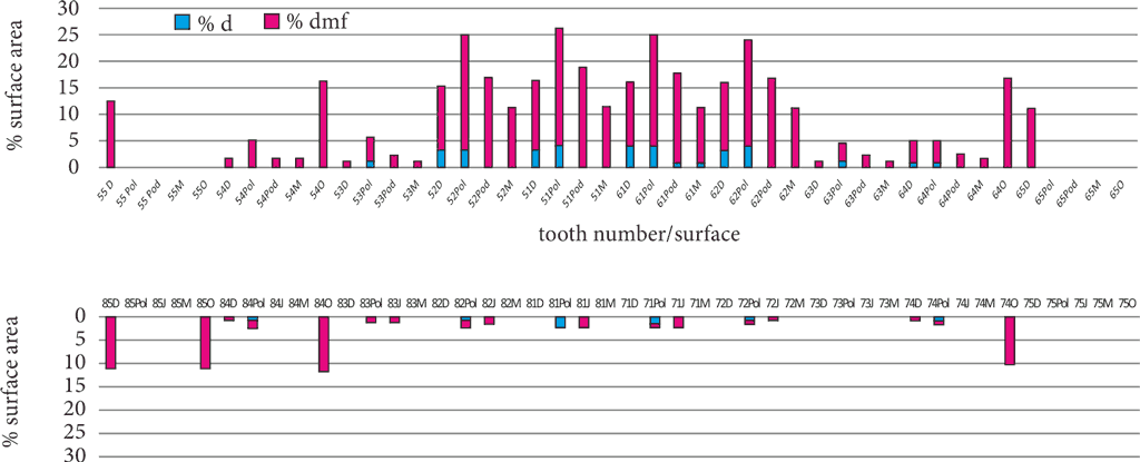
In the group of children > 18-24 months of age, the percentage of carious maxillary teeth (212/923; 23.0%) was also higher than in the mandible (53/918; 5.8%) (fig. 4). Considering the number of teeth with cavitated caries, missing and filled teeth (223), only 16 filled teeth were found. A total of 15 teeth were extracted due to caries. The highest percentage of teeth with d1d2mf > 0 was found in the group of maxillary incisors (32.1%) and maxillary molars (17.4%), followed by mandibular molars (12.5%). Labial surfaces of maxillary incisors and occlusal surfaces of first molars of both arches were the most common surface locations of carious lesions (fig. 4). As for the proximal incisal surfaces, carious lesions were more often found in the distal surfaces. There was no significant difference for the proximal surfaces of first molars.
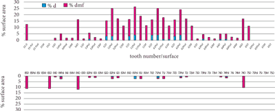
Fig. 4. Caries distribution (d1d2mf > 0) on individual tooth surfaces in dental arches in children aged > 18 to 24 months
Similarly to younger age groups, a higher percentage of maxillary teeth (325/1148; 28.3% vs. 118/1177; 10.0%, respectively) was also involved in children between > 24 and 30 months of age (fig. 5). Considering the number of teeth with cavitated lesions, missing and filled teeth (385), only 23 filled teeth were found. A total of 24 teeth were extracted due to caries. The highest percentage of teeth with d1d2mf > 0 was found in the group of maxillary incisors (42.2%), followed by maxillary molars (20.5%) and mandibular molars (19.5%). Labial surfaces, followed by palatal surfaces were most often involved in maxillary incisors. From 10% to about 20% of proximal surfaces were also affected by caries. In first molars, carious lesions occurred mainly on occlusal surfaces (fig. 5).
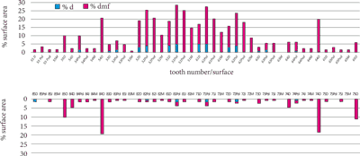
Fig. 5. Caries distribution (d1d2mf > 0) on individual tooth surfaces in maxillary and mandibular arches in children aged > 24 to 30 months
In the oldest age group, the percentage of carious maxillary teeth (336/1240; 27.1%) was almost twice as high as in the mandible (186/1263; 14.7%). Of the teeth with cavitated caries, missing and filled teeth (474), only 37 teeth were filled. A total of 12 teeth were extracted due to caries. A comparison of the percentages of carious teeth in dental groups showed the highest percentages in the maxillary incisors (37%), mandibular molars (28.2%) and maxillary molars (22.8%). Carious lesions were mainly located on the labial and palatal surfaces of maxillary incisors and occlusal surfaces of molars. However, the percentages of proximal surfaces with caries were significantly higher than in the younger age groups (fig. 6). In the group of incisors, the rates of carious lesions on the distal and mesial surfaces were similar. In the group of first molars, carious lesions were more often found on the distal rather than mesial surfaces.
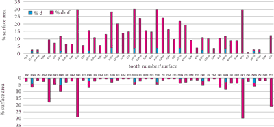
Fig. 6. Caries distribution (d1d2mf > 0) in individual maxillary and mandibular teeth
Discussion
A nationwide study conducted in 2015 among 656 children aged 3 years showed statistically significantly higher severity of caries in the maxilla than in the mandible, which is consistent with our results (8). A total of 13.4% of maxillary teeth and 8.9% of mandibular teeth were affected by caries. The dmft values were 2.67 ± 2.50 and 1.78 ± 1.67. There were no differences in caries frequency between the right and left side of the dental arches. These results are in line with our findings. Similarly, a statistically significant difference was found in the distribution of caries between the maxilla and the mandible in Turkish children (9).
As opposed to children aged 12-36 months from the Warsaw agglomeration, mandibular molars, followed by medial maxillary incisors and maxillary molars were most often affected by caries in 4-year olds (8). The analysis of the distribution of carious lesions in the dental arches in children depending on the age showed a decreasing frequency of caries in the group of maxillary incisors with age and an increasing frequency of caries in molars. It is worth pointing to a similar increase in the frequency of carious lesions in maxillary and mandibular molars.
A study in 153 children aged from 18 to 36 months who attended three randomly selected nurseries in WrocЕӮaw showed that medial maxillary incisors (34%), followed by lateral maxillary incisors (22.4%), and second mandibular molars (19.3%), first mandibular molars (18.9%) and first maxillary molars (17.7%) were most often involved (1).
Canines (0.3%) and mandibular incisors (0.9%) were least affected. Despite the differences in the age of the examined children from the Warsaw agglomeration, the distribution of carious lesions was similar. Maxillary incisors and mandibular molars were at a higher risk of caries throughout the study group. Studies in Turkish children aged 8 to 60 months showed that medial maxillary incisors were also most often involved (9). Similar observations were made by Iranian and Brazilian researchers in children up to 3 years of age (10, 11). Our findings, in line with the results of other authors, show that caries in the anterior maxillary teeth significantly contributes to the overall level of caries in the youngest age groups, with maxillary incisors being the most affected teeth (9-18).
A study in 3-year-olds from three voivodeships of Poland showed that out of 2416 carious surfaces, 771 (31.9%) were tangent, 708 (29.3%) were smooth, and 937 (38.3%) were occlusal surfaces (8). In medial maxillary incisors, caries was more common on the tangent rather than labial surfaces, in the lateral maxillary incisors – on the labial or palatal surfaces, while in molars, lesions on the occlusal surfaces were predominant, which corresponds to the studies in Turkish children (9).
In the medial maxillary incisors, caries was most often present on the medial, followed by labial surface. In the lateral maxillary incisors, carious lesions were most often found on the labial surfaces. In the studies by Doğan et al. (9) in Turkish children and Kumarihamy et al. (15) in Sri Lankan children aged 1-2 years, maxillary incisal caries was most often found on the labial surfaces, followed by medial surfaces, while in a study by Toutouni et al. (13) in children 2-3 years of age – on labial surfaces. Colombian children (mean age 2.8 ± 0.48 years) also most often presented with caries on the labial surfaces of anterior maxillary teeth (14). Iranian researchers found 100% of carious lesions on the smooth surfaces of maxillary incisors in children aged 12-15 months (10). In the oldest age group (26-36 months), lesions in such a location accounted for only 27%. Douglass et al. (19) also noticed that the presence of caries on different incisal surfaces varied with age. Among younger age groups, caries was most common on the labial surfaces of the medial maxillary incisors. In children aged 25-27 months and older, caries predominated on the medial and distal surfaces of medial maxillary incisors.
In our study, children from the Warsaw agglomeration presented mainly with lesions on the labial surfaces, followed by palatal surfaces of incisors, although a constant increase in caries on the proximal surfaces was observed with the age of children, which is in line with the progressive pattern of ECC on the proximal surfaces, more often the distal surfaces of first molars (20, 21).
Contrary to all the studies cited above, Rosemblatt and Zarzar (22) observed the first signs of carious lesions in children under 3 years of age only after the eruption of first molars, starting from the age of 18 months. Changes in the distribution of caries in the dental arches may be associated with changes in the child’s eating habits. Predisposition to maxillary incisal and then first maxillary molar caries in younger age groups may be the result of bottle feeding. This may be due to a build-up of milk film between the upper lip and maxillary incisors in infants fed at night, when their salivation is reduced. On the other hand, the mandibular front teeth are least affected by caries as they are covered with the tongue during suckling, which reduces their exposure to cariogenic factors. The introduction of solid foods requires chewing. In the case of dietary and hygienic errors, the chewing surfaces are exposed to bacterial acids, which, in the case of immaturity of freshly erupted teeth, quickly leads to the formation of cavitated caries.
It is also worth noting that teeth are attacked by caries in accordance with eruption pattern if the risk factors are not controlled when the first signs of caries are detected. Therefore, attention should be paid to the need for dental care already from the first year of life, when it is possible to prevent early lesions or control the already developed disease.
Conclusions
In the early childhood period, caries is more likely to affect the maxillary rather than the mandibular teeth and occurs symmetrically on both sides of the dental arches. Labial surfaces, followed by palatal surfaces of maxillary incisors and occlusal surfaces of first maxillary molars are the most common location of lesions, which are slightly less common in the mandible. As children grow older, proximal surfaces, including the maxillary incisors, become involved.
PiЕӣmiennictwo
1. Kaczmarek U, Grzesiak I: Rozmieszczenie prГіchnicy w zДҷbach mlecznych u wrocЕӮawskich dzieci w wieku 18-36 miesiДҷcy. Dent Med Probl 2006; 43(2): 215-221.
2. Olczak-Kowalczyk D, Boguszewska-Gutenbaum H, Janicha J, Turska-Szybka A: Wybrane zagadnienia zwiД…zane z wyrzynaniem zДҷbГіw mlecznych. Nowa Stomatol 2011; 16(2): 73-76.
3. American Academy of Pediatric Dentistry: Policy on Early Childhood Caries (ECC): Classifications, Consequences, and Preventive Strategies. Pediatr Dent 2016; 38(6): 52-54.
4. Ismail AI, Sohn W: A systematic review of clinical diagnostic criteria of early childhood caries. J Public Health Dent 1999; 59(3): 171-191.
5. Drury TF, Horowitz AM, Ismail AI et al.: Diagnosing and reporting early childhood caries for research purposes. A report of a workshop sponsored by the National Institute of Dental and Craniofacial Research, the Health Resources and Services Administration, and the Health Care Financing Administration. J Public Health Dent 1999; 59(3): 192-197.
6. World Health Organization: Oral Health Surveys – Basic Methods. 5th Edition. World Health Organization, Geneva 2013.
7. International Caries Detection and Assessment System (ICDAS) Coordinating Committee: Rationale and Evidence for the International Caries Detection and Assessment System (ICDAS II) Scotland: Dental Health Services Research Unit 2005. http://www.icdas.org.
8. Olczak-Kowalczyk D, Kaczmarek U, Bachanek T: Monitoring zdrowia jamy ustnej: monitorowanie stanu zdrowia jamy ustnej populacji polskiej w latach 2013-2015: ocena stanu zdrowia jamy ustnej i jego uwarunkowaЕ„ w populacji polskiej w wieku 3, 10 i 15 lat w 2015 roku. Oficyna Wydawnicza WUM, Warszawa 2016: 49-93.
9. Doğan D, DГјlgergil CT, Mutluay AT et al.: Prevalence of caries among preschool-aged children in a central Anatolian population. J Nat Sci Biol Med 2013; 4(2): 325-329.
10. Mohebbi SZ, Virtanen JI, Vahid-Golpayegani M, Vehkalahti MM: Early childhood caries and dental plaque among 1-3-year-olds in Tehran, Iran. J Indian Soc Pedod Prev Dent 2006: 177-181.
11. Santos AP, Soviero VM: Caries prevalence and risk factors among children aged 0 to 36 months. Pesqui Odontol Bras 2002; 16: 203-208.
12. Davies GM, Blinkhorn FA, Duxbury JT: Caries among 3-year-olds in greater Manchester. Br Dent J 2001; 190: 381-384.
13. Toutouni H, Nokhostin MR, Amaechi BT, Zafarmand AH: The Prevalence of Early Childhood Caries among 24 to 36 Months Old Children of Iran: Using the Novel ICDAS-II Method. J Dent (Shiraz) 2015; 16(4): 362-370.
14. Cadavid AS, Lince CM, Jaramillo MC: Dental caries in the primary dentition of a Colombian population according to the ICDAS criteria. Braz Oral Res 2010; 24: 211-216.
15. Kumarihamy SL, Subasinghe LD, Jayasekara P et al.: The prevalence of Early Childhood Caries in 1-2 yrs olds in a semi-urban area of Sri Lanka. BMC Res Notes 2011; 4: 336.
16. Vachirarojpisan T, Shinada K, Kawaguchi Y et al.: Early childhood caries in children aged 6-19 months. Community Dent Oral Epidemiol 2004; 32(2): 133-142.
17. Du M, Bian Z, Guo L et al.: Caries patterns and their relationship to infant feeding and socio-economic status in 2-4-year-old Chinese children. Int Dent J 2000; 50(6): 385-389.
18. Dimitrova MM, Kukleva MP, Kondeva VK: Prevalence of early childhood caries and risk factors in children from 1 to 3 years of age in Plovdiv, Bulgaria. Folia Med (Plovdiv) 2002; 44(1-2): 60-63.
19. Douglass JM, Tinanoff N, Tang JM, Altman DS: Dental caries patterns and oral health behaviors in Arizona infants and toddlers. Community Dent Oral Epidemiol 2001; 29: 14-22.
20. Kawashita Y, Kitamura M, Saito T: Early childhood caries. Int J Dent 2011: 725320.
21. American Dental Association (ADA) Statement on Early Childhood Caries. http://www.ada.org/en/about-the-ada/ada-positions-policies-and statements/statement-on-early-childhood-caries/.
22. Rosemblatt A, Zarzar P: Prevalence of early childhood caries in 12 to 36 month-old children in Recife, Brazil. ASDC J Dent Child 2002; 69: 319-324.