Lidia Pijankowska-Beksa1, Agnieszka Kiryszewska2, Janina Łucja Grzegorczyk2, *Joanna Szczepańska3
The evaluation of the impact of ozone therapy and Fluor Protector fluoride prevention on the number of Streptococcus mutans and Lactobacillus spp., in children dental plaque
Ocena wpływu ozonoterapii i profilaktyki fluorkowej z zastosowaniem preparatu Fluor Protector na liczebność bakterii Streptococcus mutans i Lactobacillus spp. płytki nazębnej u dzieci
1PhD, Department of Paediatric Dentistry, Medical University of Łódź
Head of Department: prof. Joanna Szczepańska, MD, PhD
2Department of Paediatric Dentistry, Medical University of Łódź
Head of Department: prof. Joanna Szczepańska, MD, PhD
3I Department of Microbiology and Laboratory Medical Immunology, Medical University of Łódź
Head of Department: prof. Janina Łucja Grzegorczyk, MD, PhD
Streszczenie
Wstęp. Paciorkowce z rodzaju Streptococcus (głównie Streptococcus mutans oraz Streptococcus sobrinus) uznawane są za najważniejsze bakterie wywołujące próchnicę. Właściwości kwasotwórcze posiadają również występujące w płytce nazębnej G(+) pałeczki z rodzaju Lactobacillus. Powszechnie w profilaktyce próchnicy stosuje się fluorki, które głównie poprzez mechanizmy miejscowe (hamowanie enzymów bakteryjnych oraz wspomaganie procesów remineralizacji) działają przeciwpróchnicowo. W ostatnim czasie wzrosło zainteresowanie ozonem, który uznawany jest za niezawodny środek przeciwbakteryjny.
Cel pracy. Celem pracy była ocena półrocznego wpływu ozonoterapii oraz fluoryzacji kontaktowej na liczebność bakterii próchnicotwórczych płytki nazębnej.
Materiał i metody. Badania mikrobiologiczne płytki nazębnej przeprowadzono u 56 dzieci. Płytkę nazębną pobierano podczas badania wstępnego, przed wykonaniem zabiegów profilaktycznych pierwszych stałych zębów trzonowych, a następnie po 6 miesiącach podczas badania kontrolnego. Przed zabiegiem profilaktycznym przeprowadzano ankietę wśród rodziców, oceniano stan zębów mlecznych i stałych oraz higieny jamy ustnej. Wyniki badań poddano analizie statystycznej z wykorzystaniem testu Manna-Whitneya, testu Wilcoxona oraz współczynnika korelacji rang Spearmana, przy poziomie istotności p ≤ 0,05.
Wyniki. U wszystkich badanych dzieci wyhodowano bakterie S. mutans z płytek nazębnych pobranych podczas pierwszej wizyty, a pałeczki kwasu mlekowego obecne były u ok. 63% dzieci poddawanych ozonoterapii i ok. 67% w grupie lakieru fluorkowego. W badaniu kontrolnym po 6 miesiącach występowanie paciorkowców zmiennych w grupie ozonoterapii obniżyło się do 88% przypadków, a w grupie fluoryzacji kontaktowej do 84,6%. Obecność bakterii Lactobacillus spp. uzyskano u podobnej liczby pacjentów jak w badaniu wstępnym. Nie stwierdzono różnic istotnych statystycznie w liczebności bakterii pomiędzy badaniem wstępnym a badaniem kontrolnym zarówno w grupie osób poddanych ozonoterapii, jak i w grupie preparatu fluorkowego.
Wnioski. Półroczna analiza działania zabiegów ozonoterapii oraz fluoryzacji kontaktowej wykazała, że liczebności bakterii Streptococcus mutans uległy zmniejszeniu w płytce nazębnej u około 1/4 badanych dzieci, chociaż wyniki te nie były istotne statystycznie. Długoterminowy potencjał bakteriobójczy gazowego ozonu i preparatu fluorkowego Fluor Protector kształtują się na podobnym poziomie.
Summary
Introduction. Streptococci (especially Streptococcus mutans and Streptococcus sobrinus) are considered to be the most important bacteria causing caries. G-positive bacilli, such as Lactobacillus present in the dental plaque, also have acidogenic properties. Fluorides are commonly used in the prevention of caries and have anticariogenic effects mainly via local mechanisms (bacterial enzyme inhibition and supporting remineralisation process). The interest in ozone, which is considered a reliable antibacterial agent, has increased recently.
Aim. The aim of the study was to evaluate the long-term impact of ozone and topical fluoridation on the number of cariogenic plaque bacteria.
Material and methods. Microbiological plaque testing was performed in a group of 56 children. Dental plaque was collected during the preliminary examination, prior to preventive treatments of the first permanent molars and after 6 months, during a follow-up evaluation. Before preventive treatment, a survey was conducted among parents, the state of the deciduous and permanent teeth as well as oral hygiene were evaluated. The results were analyzed statistically using the Mann-Whitney test, Wilcoxon test and Spearman rank correlation coefficient, with a significance level of p ≤ 0.05.
Results. Streptococcus mutans were isolated from plaque collected from all children during the first visit. Lactic acid bacteria were present in aprox. 63% of patients treated with ozone and aprox. 67% subjects in the fluoride varnish group. Follow-up after 6 months revealed that the occurrence of S. mutans in the ozone group decreased to 88% of the cases and to 84.6% in the fluoridated group. Lactobacillus spp. were present in similar numbers of patients as in the preliminary test. There were no statistically significant differences in the number of bacteria during first or second evaluation between patients treated with ozone therapy and those undergoing fluoride therapy.
Conclusions. A 6-month assessment of the effectiveness of ozone therapy and topical fluoridation showed that the numbers of plaque Streptococcus mutans decreased in approx. 1/4 of children, however, the results were not statistically significant. Long-term microbicide potential of both ozone gas and Fluor Protector fluoride are at similar level.

Introduction
Caries has affected humans since the dawn of time and still is one of the most common infectious human diseases in different parts of the world. It was considered based on numerous epidemiological and experimental studies in both animals and humans that streptococci (mainly Streptococcus mutans and Streptococcus sobrinus) are the most important bacteria causing caries. This is probably due to their ability to rapidly produce lactic acid from carbohydrates contained in food products, mainly glucose and sucrose (1-4). Gram-positive bacilli from the genus Lactobacillus, which, as opposed to Streptococcus mutans, are not involved in carious process initiation, but in the progression of dental caries, also have acidogenic properties. The presence of mutans streptococci and their ability to produce lactic acid promotes the growth of Lactobacillus. It was shown that Streptococcus mutans are able to independently produce dental plaque, whereas bacteria belonging to the genus Lactobacillus do not have this ability (5, 6). Lactic acid bacteria are characterised by strong acidifying properties. Although common in the oral cavity, they account for only 1% of the oral microflora, though their number increases during active carious processes (7).
Certain criteria must be met in order to classify bacteria as carcinogenic: the ability to produce acids via carbohydrate fermentation, the ability to form extracellular polysaccharides as well as the ability to live in the acidic environment. Production of glucans (dextran and mutan) is also important. Water-insoluble mutan is responsible for bacterial adsorption onto dental surface, followed by bacterial aggregation and co-aggregation. Water-soluble dextran is an energy reserve material in case of dietary carbohydrate deficiency (2, 8).
Most currently used preventive methods aim to eliminate bacteria or inhibit their virulence by neutralising acids produced by the above mentioned cariogenic bacteria in the process of carbohydrate fermentation (9). Fluorine compounds reduce the numbers of S. mutans and Lactobacillus spp. by impairing bacterial cell metabolism as well as inhibiting the production of acids and extra/intracellular polysaccharides. The ability to block enolase, a bacterial enzyme involved in carbohydrate metabolism, is one of the mechanisms of fluoride action. Furthermore, the F- ions strengthen the hard tooth tissue by transforming the hydroxyapatites into more chemically stable fluorapatites (10).
Ozone is another tool against bacteria, both in the aqueous and gaseous phase (11). The structure of bacterial cells renders them susceptible to O3. Due to the lack of cholesterol in bacterial cell membranes, the atomic oxygen formed during ozone decomposition reacts with unsaturated fatty acids, phospholipids and proteins, leading to almost immediate microbial destruction (12, 13). A 10-second application is sufficient for 99% elimination of S. mutans and S. sobrinus (14). The interest in ozone dental treatment has increased recently due to the above mentioned microbiological properties.
Aim
The aim of the study was to evaluate the long-term impact of ozone and in vivo topical fluoridation on the number of plaque Streptococcus mutans and Lactobacillus spp. in children.
Material and methods
Characteristics of the study group
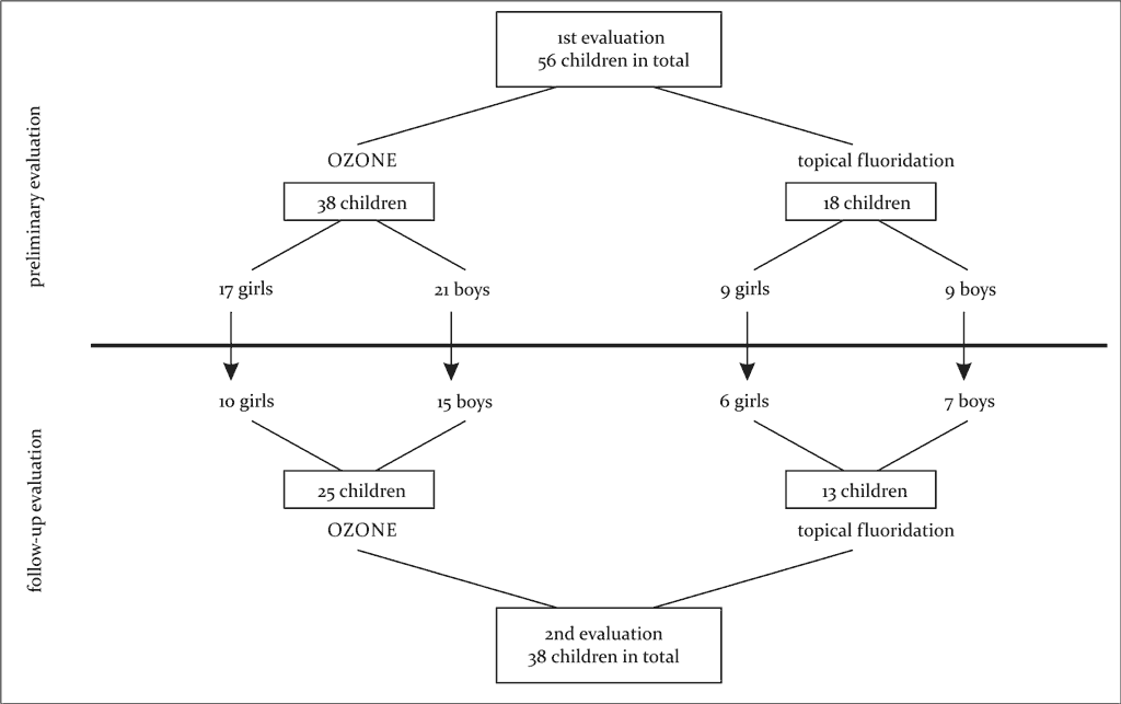
A total of 129 children aged 6-10 years presenting at the Department of Developmental Dentistry, Medical University in Łódź between 2011-2014 were included in the study. After collecting medical history, a questionnaire on health status, health awareness, eating habits and the lifestyle of children was completed by their parents. The children were then classified for two different preventive treatments of the first permanent molars: ozone therapy or topical fluoride treatment using Fluor Protector in accordance with the below description. Microbiological evaluation of the dental plaque was performed in 56 children. The diagram below presents the characteristics of subjects (fig. 1).
Fig. 1. Characteristics of study groups participating in dental plaque microbiological testing
Inclusion criteria: good general health, age 6-10 years.
Exclusion criteria: children with acute or chronic systemic disease, currently receiving antibiotic therapy or a month after antibiotic therapy, children below 6 years or over 10 years old.
Prior to procedures, all children underwent a preliminary examination to determine the state of deciduous and permanent teeth as well as oral hygiene using Green and Vermillion Oral Hygiene Index (OHI). For each patient, dmft, dmfts, DMFT, DMFTS were calculated. DIAGNOdent pen 2190 (Kavo) and standard viaual-tactual scale were used for dental assessment, after cleaning the teeth with a prophylactic toothbrush (with a micromotor). After thorough drying of the teeth, the presence of caries was determined using tip A for an assessment of chewing surfaces. The assessment was performed on the occlusal dental surfaces without fillings or fissure sealant. The tip of the device was brought closer to the tooth in such a way that the radiation beam be set perpendicular to the long tooth axis. The highest value indicated by the device was documented in patient’s records. The values were interpreted by Hibst and Paulus scale, which includes 4 ranges of values:
I (0-13) – no need for preventive treatment,
II (14-20) – the need for intensified preventive treatment performed at home by the patient,
III (21-29) – the need for professional preventive treatment or minimal dental intervention, depending on risk factors for caries,
IV (> 30) – the need for dental intervention as well as professional preventive treatment (15).
Clinical assessment of dental state was performed using a dental mirror and a blunt probe. Dental fissures were considered as affected by carious process if a lesion characterised by hard dental tissue damage was identified. The current DIAGNOdent Scale for dental fissure assessment was modified by the authors. If DIAGNOdent indicated a value above 30 units, yet the clinical assessment classified the lesion as initial or arrested caries (chalky white or brown spots without enamel interruption), the tooth was denoted as requiring checking during the next follow-up visits after 3, 6 and 12 months or more frequently in certain cases. Only teeth with erupted chewing surface, allowing for an assessment of hard dental tissue, were qualified for both procedures. Prevention using Fluor Protector varnish was performed in children with occlusal dental surfaces classified into range I or II according to Hibst and Paulus scale. Ozone therapy was used in healthy teeth as well as teeth with higher DIAGNOdent values, indicating initial caries.
Children as well as their parents received instructions on oral hygiene prior to treatment and during each follow-up visit. Parents were acquainted with the study plan, provided with an explanation and written information on the treatment performed, highlighting the importance of compliance with oral hygiene and proper diet, informed about the importance of these preventive/therapeutic measures and asked to complete a questionnaire. Test results were documented in the clinical assessment record of our own design. The study was approved by the Local Bioethical Committee of the Medical University of Łódź (Resolution No. RNN/63/11/KE).
Sample collection and preparation for analysis
Dental plaque from occlusal molar surfaces as well as vestibular and palatal furrows was sampled for microbiological testing. In total, 94 samples were collected. Full evaluation (preliminary and follow-up 6 months after the first procedure) was performed in 38 patients: 25 children receiving ozone therapy and 13 children receiving Fluor Protector varnishing. Before the study, the hygiene and dietary habits of children were as usual. Dental plaque was sampled during the preliminary examination, prior to prophylactic treatment of the first permanent molars. Then, three months after the first visit, a follow-up visit took place, during which preventive dental treatment was repeated. During a follow-up visit after 6 months, also prior to occlusal surface preventive treatment, dental plaque was collected for a follow-up microbiological testing. Dental plaque was collected using a sterile probe and placed on previously weighed and sterile aluminium foil lining Petri dishes labelled with patient’s individual number. Samples were transferred to the Department of Microbiology & Immunology Laboratory Medicine of the Medical University of Lodz within 10 minutes. OzonyTron (Mymed) emitting ozone via an open system using CA probe (caries) with a ground electrode was used for ozone therapy. Ozone was applied topically. Treatment duration and intensity were determined by the doctor individually for every patient, based on the induction table developed by the manufacturer. Ozone emission lasted from 30 to 120 seconds per tooth surface using medium ozone concentration, and was followed by topical application of Fluor Protector. After dental plaque collection, Fluor Protector varnish was applied using a suitable brush on all enamel surfaces of permanent teeth in patients undergoing topical fluoridation, without departing from the standard management for this type of procedure.
Microbiological analysis of the dental plaque
The delivered plaque samples placed on aluminium foil were weighed to obtain the wet weight of the dental plaque after subtracting the weight of the foil. The material was then transferred to 7 ml of 85% sterile NaCl reduced with cysteine hydrochloride. The suspension of the dental plaque was disrupted for 30 seconds in 100 W ultrasonic disintegrator at a wave amplitude of 5 μm (Measuring & Scientific Equipment, Ltd). The obtained plaque suspension was serially diluted from 100 to 10-3 in sterile 0.85% NaCl solution. The dilutions were inoculated onto TSY20B agar (Becton-Dickinson, USA) and incubated for 48 hrs at 37°C under anaerobic conditions. Undiluted suspension of dental plaque sample was inoculated on M.R.S. Agar (Graso) and also incubated for 48 hrs at 37°C under anaerobic conditions. After this time, colonies grown on TSY20B agar (Becton-Dickinson, USA) were sprayed with 10% mannitol solution and incubated for 3 hrs at 37°C under atmospheric conditions. After incubation with mannitol, the colonies were sprayed with 4% TTC solution (2,3,5-triphenyl tetrazolium chloride) and incubated under the same conditions for 1 hour. The colonies of Streptococcus mutans (pink) grown on TSY20B agar (Becton-Dickinson, USA) and Lactobacillus spp. grown on M.R.S. Agar (Graso) were counted. The number of CFU (Colony Forming Units) per 1 g of wet weight of dental plaque was calculated for each microorganism, taking into account the dilution of a given sample.
Statistical analysis
The obtained results were analysed (for measurable characteristics) statistically. Their minimum and maximum values were shown, the arithmetic means as well as parameters determining the differentiation of the analysed variables: standard deviations, were calculated. For qualitative characteristics, percentages (rates) of the occurrence of their categories were determined. The Mann-Whitney test was used to assess the significance of differences between the mean values in the two groups, while the Wilcoxon test for paired data ranks was used to compare differences in the same group but at different time points (distribution of the characteristics differed from normal distribution). Correlations between qualitative characteristics were assessed using the Spearman’s rank correlation coefficient. The level of significance p ≤ 0.05 was used for all comparisons. Calculations were performed using STATISTICA v. 10 software.
Results
Powyżej zamieściliśmy fragment artykułu, do którego możesz uzyskać pełny dostęp.
Mam kod dostępu
- Aby uzyskać płatny dostęp do pełnej treści powyższego artykułu albo wszystkich artykułów (w zależności od wybranej opcji), należy wprowadzić kod.
- Wprowadzając kod, akceptują Państwo treść Regulaminu oraz potwierdzają zapoznanie się z nim.
- Aby kupić kod proszę skorzystać z jednej z poniższych opcji.
Opcja #1
29 zł
Wybieram
- dostęp do tego artykułu
- dostęp na 7 dni
uzyskany kod musi być wprowadzony na stronie artykułu, do którego został wykupiony
Opcja #2
69 zł
Wybieram
- dostęp do tego i pozostałych ponad 7000 artykułów
- dostęp na 30 dni
- najpopularniejsza opcja
Opcja #3
129 zł
Wybieram
- dostęp do tego i pozostałych ponad 7000 artykułów
- dostęp na 90 dni
- oszczędzasz 78 zł
Piśmiennictwo
1. Al-Mudallal NHA, Al-Jumaily EFA, Muhimen NAA, Al-Shaibany AA: Isolation and identification of Mutans Streptococci bacteria from human dental plaque samples. JNUS 2008; 11: 98-105. 2. Chandrabhan D, Hemlata R, Renu B, Pradeep V: Isolation of dental caries bacteria from dental plaque and effect of tooth pastes on acidogenic bacteria. OJMM 2012; 2: 65-69. 3. Marsh PD, Bradshaw DJ: Dental plaque as a biofilm. J Ind Microbiol 1995; 15: 169-175. 4. Forssen SD, Björklund M, Ouwehand AC: Streptococcus mutans, caries and simulation models. Nutrients 2010; 2: 290-298. 5. Gębska A, Kędzia A, Kochańska B et al.: Effectiveness of incipient caries induction in microaerophilic and/or aerobic conditions using Streptococcus mutans and Lactobacillus acidophilus. In vitro studies. J Stoma 2012; 65(3): 344-358. 6. Marczuk-Kolada G, Jakoniuk P, Łuczaj-Cepowicz E et al.: Liczba bakterii z rodzaju Streptococcus i Lactobacillus w ubytkach próchnicowych przed i po opracowaniu metodą chemo-mechaniczną z użyciem systemu Carisolv. J Stoma 2006; LIX(6): 380-387. 7. Szponar E, Ślebioda Z, Kieliszczyk W et al.: Stan jamy ustnej u młodych dorosłych bez chorób układowych a współwystępowanie Candida, Lactobacillus i Streptococcus. Dent Forum 2009; XXXVII(1): 39-44. 8. Wójtowicz A, Malm A: Mikrobiologiczne podłoże próchnicy w aspekcie jej profilaktyki. Mikrobiol 2009; 65(5): 327-330. 9. Barira I, Shahper NK, Asad UK: Dental caries: From infection to prevention. Med Sci Monit 2007; 13(11): 196-203. 10. Dąbrowska E, Balunowska M, Letko M et al.: Wpływ preparatów fluorkowych i chlorheksydynowych używanych do codziennej higieny na środowisko jamy ustnej. Annales UMCS 2005; LX(73): 333-336. 11. Sadatullah S, Mohamed NH, Razak FA: The antimicrobial effect of 0.1 ppm ozonated water on 24-hour plaque microorganisms in situ. Braz Oral Res 2012; 26(2): 126-131. 12. Gopalakrishnan S, Parthiban S: Ozone – a new revolution in dentistry. J Bio Innov 2012; 1(3): 58-69. 13. Kogut A: Ozonoterapia w praktyce stomatologicznej. Mag Stomatol 2007; 9: 112-118. 14. Stübinger S, Sader R, Filippi A: The use of ozone in dentistry and maxillofacial surgery: A review. Quintessence Int 2006; 37: 353-359. 15. Skomro P: Współczesna diagnostyka laserowa w ocenie zaburzeń mineralizacji na powierzchniach gładkich zębów u pacjentów po leczeniu stałym aparatem ortodontycznym. Implantoprotetyka 2008; IX(4): 47-49. 16. Kaczmarek U: Aspekt bakteryjny próchnicy zębów mlecznych. Dent Med Probl 2004; 41(3): 509-514. 17. Strużycka I, Rucińska K, Radziejewska M: Wybrane metody oceny występowania bakterii z gatunku Streptococcus mutans w środowisku jamy ustnej. Nowa Stomatol 2001; 2: 11-14. 18. Manowiec J, Lisiecka K, Suszczewicz A: Wpływ programu profilaktycznego realizowanego u dzieci przedszkolnych na liczbę Streptococcus mutans i Lactobacillus w ślinie. Dent Med Probl 2003; 40(2): 281-286. 19. Proc P, Filipińska-Skąpska R, Wochna-Sobańska M: Is a bacterial factor crucial in caries development of the youngest children? Nowa Stomatol 2004; 2: 51-55. 20. Hauser-Gerspach I, Pfäffli-Savtchenko V, Dähnhardt JE et al.: Comparison of the immediate effects of gaseous ozone and chlorhexidine gel on bacteria in cavited carious lesion in children in vivo. Clin Oral Invest 2009; 13: 287-291. 21. Baysan A, Beighton D: Assesment of the ozone-mediated killing of bacteria in infected dentine associated with non-cavitated occlusal carious lesions. Caries Res 2007; 41: 337-341. 22. Knight GM, McIntyre JM, Craig GG et al.: The inability of Streptococcus mutans and Lactobacillus acidophilus to form a biofilm in vitro on dentine pretreated with ozone. Aust Dent J 2008; 53: 349-353. 23. Klepacz J, Łęski M: Możliwości wykorzystania ozonu w endodoncji. Dent Med Probl 2008; 45(2): 194-198. 24. Schneider HG: Skuteczność zależy od tego, czy uda nam się pokonać bariery dyfuzji. TPS 2006; 4: 50-51. 25. Holmes J: Clinical reversal of root caries using ozone, double-blind, randomised, controlled 18-month trial. Gerodontology 2003; 20: 106-114. 26. Płuciennik-Stronias M, Zarzycka B, Bołtacz-Rzepkowska E: Wpływ fluoryzacji kontaktowej szkliwa na wzrost bakterii płytki nazębnej. Med Dośw Mikrobiol 2013; 65: 129-132. 27. Zaura-Arite E, ten Cate JM: Effects of fluoride- and chlorhexidine-containing varnishes on plaque composition and on demineralization of dentinal grooves in situ. Eur J Oral Sci 2000; 108: 154-161.