*Karolina Raczkowska-Łabuda, Elżbieta Niemczyk-Cieślak, Lidia Zawadzka-Głos
Congenital tumor of the oral cavity – epulis neonatorum. Case report
Wrodzony guz jamy ustnej – nadziąślak noworodkowy. Opis przypadku
Department of Pediatric Otolaryngology, Medical University of Warsaw, Poland
Head of Department: Associate Professor Lidia Zawadzka-Głos, MD, PhD
Streszczenie
Noworodek z wrodzonym nadziąślakiem może stanowić zaskakujący widok zarówno dla rodziców, jak i medyków zaangażowanych w opiekę nad maleństwem. Te łagodne guzy noworodków mogą być wyjątkowo duże, zajmując znaczną część jamy ustnej i stwarzając ryzyko niedrożności dróg oddechowych oraz znacząco utrudniać karmienie. Diagnostyka prenatalna pozostaje trudna ze względu na późny rozwój guza i niespecyficzne wyniki badań (USG/MRI). Leczeniem z wyboru pozostaje chirurgiczne wycięcie guza. W naszym doniesieniu prezentujemy opis dwóch przypadków noworodków zoperowanych w Klinice Otorynolaryngologii Dziecięcej WUM na przełomie 2020/2021 roku. W obu przypadkach diagnozę postawiono po urodzeniu, a badanie histopatologiczne potwierdziło rozpoznanie wrodzonego guza ziarnistokomórkowego (CGCT). Narośla wycięto w znieczuleniu ogólnym, z dobrym wynikiem pooperacyjnym, co pozwoliło na doustne karmienie w drugiej dobie po zabiegu.
Summary
A newborn baby with congenital epulis can be a startling sight for both parents and medics involved in the care of a neonate. These benign neonatal tumours can be exceptionally big, occupying a significant portion of the oral cavity and posing a risk of airway obstruction and significantly hindering feeding. Prenatal diagnosis remains difficult due to late tumour development and non-specific findings (ultrasound/MRI). The treatment of choice remains surgical excision of the tumour. In our report, we present a description of two cases of neonates operated at the Department of Pediatric Otorhinolaryngology of the Medical University of Warsaw, at the turn of 2020/2021. In both cases the diagnosis was made at birth and histopathological examination confirmed the diagnosis of congenital granular cell tumour (CGCT). The growths were excised under general anaesthesia with a good postoperative outcome, which allowed oral feeding on the second postoperative day.
Introduction
Congenital Granular Cell Tumour of the newborn (CGCT), also known as congenital epulis or Neuman’s tumour, is a rare gingival outgrowth of the anterior alveolar ridges of maxilla and mandible. Originally described in 1871 by Neuman. Congenital epulis is a benign lesion with no tendency to metastasise (1). It is observed three times more often in the canine or incisor region of the maxilla than in the mandibular alveolus (2). The ratio of girls to boys is 10(9):1 (2, 3). It typically grows unidirectionally reaching a size of a few millimetres to a few centimetres. The 2-directional growth – towards the posterior pharyngeal wall, results in restriction of airway patency, leading to airway failure. CGCT impairs neonatal feeding, hence we classify the indications for surgical intervention as expedited. Due to the slow growth of the tumour, prenatal diagnosis is possible only in the third trimester of pregnancy and is limited to imaging examinations (ultrasound, MRI). Imaging studies allow assessment of the degree of tissue differentiation and exclusion of possible birth defect syndrome (2, 3).
We present a description of two cases of CGCT, female newborns, hospitalized at the Department of Neonatology, of the Medical University of Warsaw, operated at the turn of 2020/2021 by surgeons of the Department of Pediatric Otorhinolaryngology, WUM.
Methodology
This case report presents the diagnostic process, treatment and 4-month follow-up of two female patients (operated on the 4th and 6th day of life) with postoperatively confirmed granulosa cell tumour in histopathological examination.
Results
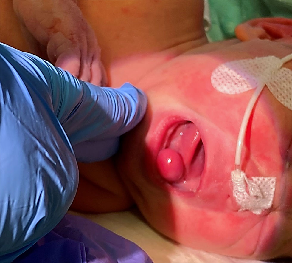
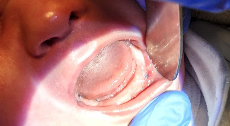
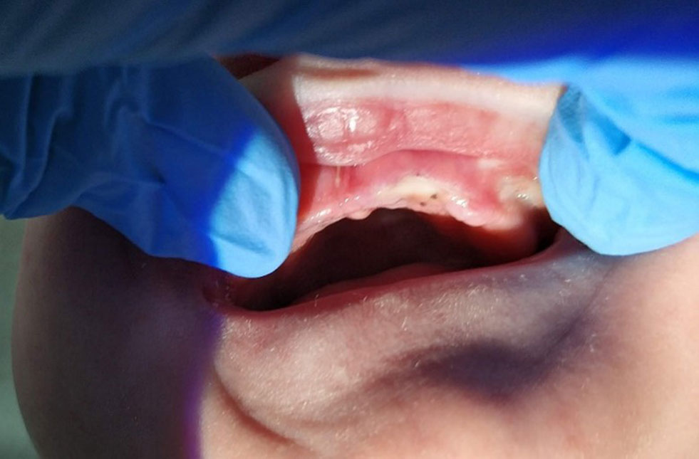
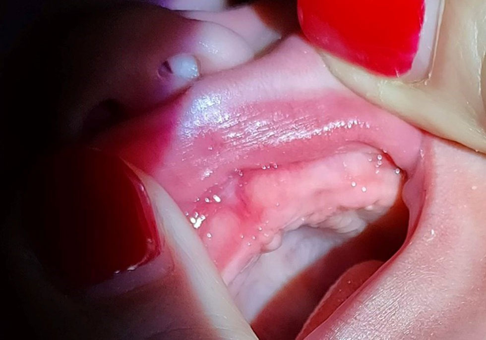
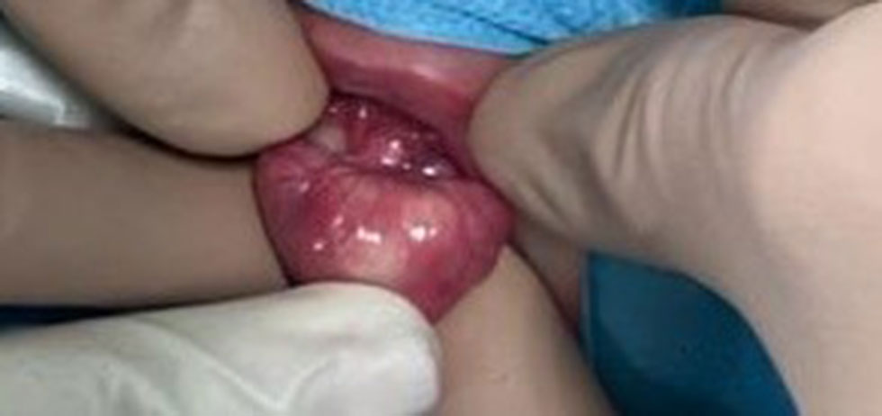
In 2020/2021 (December-February), 2 patients with Neuman’s tumour were operated on the Department of Pediatric Otorhinolaryngology, WUM. In both cases they were female newborns. The first girl (Sz.Z.) (fig. 1) underwent surgery on the 4th day of life; the patient with G1P1 was born at 39 EGA with 7-8-9-10 Apgar points, weight 3610 g, pregnancy complicated by maternal genital tract infection – GBS (+). The second patient (S.K.) (fig. 2) was operated on in the 6th day of life. The girl with G1P1, born at 41 EGA with 9-10-10-10 Apgar points, weight: 3640 g; course of pregnancy uncomplicated. In the case of Sz.Z. intraoral examination revealed an elastic, smooth-surface, pedunculated mass measuring 1.5 x 1.0 x 0.5 (cm3) in the region of alveolar 72-74 (mandibule, left side). In the case of second girl (S.K.) a 1.0 x 1.2 x 0.5 (cm3) tumour was located in the region of the future maxillary canine, also on the left side. In both cases, a single lesion was found covered with smooth, unaltered gingival mucosa. Both tumours prevented normal closure of the mouth and interfered with breastfeeding. Simple surgical excision under general anaesthesia was indicated as the method of treatment. In patient Sz.Z., a mucosal flap was dissected and approximated with dissolvable sutures., in patient S.K. a simple excision was proceded (fig. 3). The removed growths were subjected to histopathological examination obtaining the result: congenital granular cell tumor/congenital epulis neonatorum. The uncomplicated postoperative period allowed breastfeeding of both girls on the second day after the surgery. Newborns were followed up for 5 days postoperatively and discharged home. The follow-up visits confirmed proper healing with the correction of the primary alveolar cavity (figs. 4-7). Follow-up examinations were performed 1 week, 1 month and 3 months after surgery. In both patients, the alveolar processes are covered with mucosa of regular morphology.
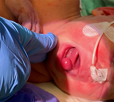
Fig. 1. Congenital epulis of the mandible. Patient Sz.Z.

Fig. 2. Congenital epulis of the maxilla (typical location). Patient S.K.
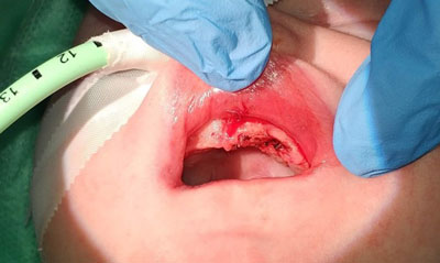
Fig. 3. Patient S.K. – status after simple tumour excision

Fig. 4. Patient Sz.Z. – one week after tumour removal with dissection of the mucosal flap. Remnants of a dissolvable suture can be seen

Fig. 5. Patient S.K. – one week after removal of Neuman’s tumour
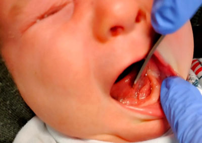
Fig. 6. Patient Sz.Z. – one month after surgery

Fig. 7. Patient S.K. – one month after surgery
Discussion
There are a multitude of terms in the literature of congenital epulis (CE) – congenital granular cell tumour; congenital granular cell myoblastoma; congenital granular cell fibroblastoma, or Neuman’s tumour (1-3). Regardless of its name, it is a rare, benign tumour of undetermined aetiology (1-14). Growths (CE) are a heterogeneous group of tumours derived from undifferentiated mesenchymal cells, fibroblasts, myoblasts, histiocytes, Schwann cells, or odontogenic epithelium (3-6). Some authors raise the issue of hormonal (estrogen) dependence of the tumour, due to predominance of the female sex (2, 3, 6). The ratio of girls to boys is on average 9:1 (8-10:1) (2, 3, 7).
In general, congenital epulis is an isolated pathology, without association with known congenital syndromes. Nevertheless, in single reports before 2000, we can find reports of Neuman’s tumour co-occurrence with polydactyly (8), goiter (9), X chromosome trisomy (8), polyposis (10, 11), mandibular hypoplasia (11), or neurofibromatosis (12) – which is not confirmed in our two observations.
Clinically, a congenital epulis of neonatorum is a single, hard tumour with a smooth surface, more rarely multiple lobes, pedunculated or with a wide base, pink rarely red and nontender on palpation (fig. 8). Its size varies from a few millimetres to about 10 cm at its widest diameter (13). The dimensions of our patients’ tumours can be described as average, they were 1.5 x 1.0 x 0.5 (cm3) and 1.0 x 1.2 x 0.5 (cm3) respectively. The typical location of congenital granular cell tumour (CGCT) is the alveolar process of the maxilla, lateral to the midline, in the area of the canines and incisors (2, 14) (fig. 2). The analogous location on the mandible is involved three times less frequently (2) (fig. 1) – as in the case of patient Sz.Z. Multiple lesions or maxillary and mandibular locations have been described in 5-16% of cases (15) of Neuman’s tumors.

Fig. 8. Morphology of congenital epulis
Out of more than 200 cases of epulis neonatorum described in the English literature, spontaneous regression has been documented in only eight cases (4), hence the recommendations in the literature to adopt a wait-and-see and non-operative stance in selected cases of congenital granulosa cell tumors in which no feeding or respiratory disorders are found. Both of our patients required feeding through a gastric probe before surgical removal of the lesion (fig. 1), which was the basis for qualification for the expedited procedure.
The gold standard management for CGCT is complete surgical excision under general (16) or local (17) anaesthesia within hours (3) to several days (16, 18) after birth. The literature contains isolated case reports of excision of congenital epulis using carbon dioxide laser (18) and erbium-chromium-yttrium-scandium-galus-garnet (Er, Cr: YSGG) laser (19). No recurrence of CE has been reported after surgical excision, even if the removal was incomplete, and the dentition in the area of the lesion usually remains intact (20, 21). It is worth noting that in the first patient from our Clinic, a mucosal flap was dissected at the site of the resected tumour and approximated with dissolvable sutures. In the second patient simple excision was used, leaving the wound to heal by granulation. In both cases, healing was normal, without complications, with a very good aesthetic result (fig. 5, 6). By the time of publication, no tumour recurrence had been observed. A single case of a hypoplastic deciduous left incisor, premolar and maxillary first molar in the region in which a 2.5 cm CGCT was surgically removed 11 days after birth was described (22). During the surgical procedure, the mucoperiosteal flaps were elevated, which, according to the authors, may have impaired the development of these three deciduous teeth. Bearing in mind the above report, both our patients were referred to the Children’s Dental Clinic for dental check-up during the period of eruption of deciduous teeth.
Conclusions
Congenital epulis of the newborn is a benign tumour with no reported history of metastasis (1-23). Due to its location and size, it may cause mechanical obstruction of the oral cavity leading to feeding difficulties, cyanosis, dyspnoea, and in the perinatal/postnatal period may be the cause of death due to asphyxia (2, 3). Very small lesions are asymptomatic and sometimes regress spontaneously (4). Large lesions disrupting feeding and breathing require expedited surgical intervention, i.e. simple surgical excision under local or general anaesthesia (16, 17). Surgical excision is curative and no recurrence has been reported in long-term postoperative follow-up (16-19). Excision of the tumour is a safe procedure (23), and allows rapid implementation of breastfeeding. CGCT do not correlate with congenital syndromes, so patients do not require genetic counselling (4). Differential diagnosis includes teratomas, odontogenic tumours, neurodermal tumours, hemangiomas, rhabdomyomas, fibromas, myoblastomas, or dermoid cysts (1-3, 6). Postoperative histopathological examination is the gold standard for statement of a definitive diagnosis (2, 3, 13, 16).
Piśmiennictwo
1. McGuire T, Gomes P, Frelich M, Sandor K: Congenital Epulis: A Surprise in the Neonate. J Can Dent Assoc 2006; 72: 747-750.
2. Sowa A, Borszewska-Kornacka M, Małdyg J, Dębska M: Congenital epulis of a newborn twin – a case report. Ginekologia Polska 2013; 8: 733-736.
3. Kumar RM, Bavle RM, Umashankar DN, Sharma R: Congenital epulis of the newborn. J Oral Maxillofac Pathol 2015; 19(3): 407.
4. Ritwik P, Brannon RB, Musselman RJ: Spontaneous regression of congenital epulis: a case report and review of the literature. J Med Case Reports 2010; 4: 331.
5. Khattala K, Rami M, Elmadi A et al.: A case report of a huge congenital granular cell epulis operated under local anesthesia. Pan Afr Med J 2012; 12: 105.
6. Bosanquet D, Roblin G: Congenital Epulis: A Case Report and Estimation of Incidence. International Journal of Otolaryngology 2009; 2009: Article ID 508780.
7. Jain N, Sinha P, Singh L: Large Congenital Epulis in a Newborn: Diagnosis and Management. Ear, Nose & Throat Journal 2020; 99(7): NP79-NP81.
8. Kim ES, Gross TL: Prenatal ultrasound detection of a congenital epulis in a triple X female fetus: a case report. Prenat Diagn 1999; 19(8): 774-776.
9. Sarihan H, Gedík Y, Mocan H et al.: Congenital epulis. Case report. Scand J Plast Reconstr Surg Hand Surg 1995; 29(1): 77-79.
10. Ophir D, Marshak G: Congenital granular cell epulis causing polyhydramnios. Head Neck Surg 1985; 7(5): 427-430.
11. Koch BL, Myer C, Egelhoff JC: Congenital epulis. AJNR Am J Neuroradiol 1997; 18(4): 739-741.
12. Hoyme HE, Musgrave SD, Browne AF, Clemmons JJ: Congenital oral tumor associated with neurofibromatosis detected by prenatal ultrasound. Clin Pediatr (Phila) 1987; 26(7): 372-374.
13. Song WS, Kim JW, Kim YG, Ryu DM: A case report of congenital epulis in the fetus. J Oral Maxillofac Surg 2005; 63(1): 135-137.
14. Neville BW, Damm DD, Allen CM, Bouquot JE: Oral and maxillofacial pathology. Saunders/Elsevier, St. Louis 2009: 537-539.
15. Charrier JB, Droullè P, Vignaud JM et al.: Obstructive congenital gingival granular cell tumor. Ann Otol Rhinol Laryngol 2003; 112(4): 388-391.
16. Silva GC, Vieira TC, Vieira JC et al.: Congenital granular cell tumor (congenital epulis): a lesion of multidisciplinary interest. Med Oral Patol Oral Cir Bucal 2007; 12(6): E428-430.
17. Merrett SJ, Crawford PJ: Congenital epulis of the newborn: a case report. Int J Paediatr Dent 2003; 13(2): 127-129.
18. Lapid O, Shaco-Levy R, Krieger Y et al.: Congenital epulis. Pediatrics 2001; 107(2): E22-10.1542/peds.107.2.e22.
19. Olivi G, Constacurta M, Maturo P, Docimo R: Removal of a fibrous epulis with Er, Cr: YSGG laser: case report. Eur J Paediatri Dent 2007; 8(3): 149-152.
20. Welbury RR: Congenital epulis of the newborn. Br J Oral Surg 1980; 18(3): 238-243.
21. Marakoglu I, Gursoy UK, Marakoglu K: Congenital epulis: report of a case. ASDC J Dent Child 2002; 69(2): 191-192.
22. Sunderland R, Sunderland EP, Smith CJ: Hypoplasia following congenital epulis. Br Dent J 1984; 157(10): 353.
23. Cheong SP, Fahy CJ, Craigie MJ: Anesthesia for excision of an intraoral mass in a neonate: use of a laryngeal mask during removal of congenital epulis. Anaesthesia and Intensive Care 2008; 36(1): 116-118.