*Anna Szufnara1, Sylwia Majewska-Beśka2, Agata Szczesio-Włodarczyk3, Joanna Nowak3, Joanna Szczepańska2
Shear bond strength of Equia Forte with enamel and dentin of permanent immature teeth – in vitro study
Ocena wytrzymałości połączenia materiału Equia Forte z powierzchnią szkliwa i zębiny zębów stałych z niezakończonym rozwojem – badania in vitro
1Doctoral studies, Department of Paediatric Dentistry, Medical University of Łódź
Head of Department: Professor Joanna Szczepańska, MD, PhD
2Department of Paediatric Dentistry, Medical University of Łódź
Head of Department: Professor Joanna Szczepańska, MD, PhD
3University Material Research Laboratory, Medical University of Łódź
Head of Laboratory: Professor Jerzy Sokołowski, MD, PhDi
Streszczenie
Wstęp. Rozwój nowych materiałów szkło-jonomerowych pozwala leczyć głębokie ubytki próchnicowe w sposób minimalnie inwazyjny. Zastosowanie w praktyce znajdują również bioaktywne materiały, które poprzez swoje właściwości remineralizujące regenerują tkanki zęba. Przykład stanowi bioaktywne szkło – preparat Sylc.
Cel pracy. Celem pracy była ocena wytrzymałości połączenia szkło-jonomeru Equia Forte z powierzchnią szkliwa i zębiny zębów stałych z niezakończonym rozwojem. Analizie został poddany również wpływ piaskowania preparatem Sylc oraz starzenia termicznego na jakość adhezji materiału względem tkanek zęba.
Materiał i metody. Badania przeprowadzono na zębach stałych niedojrzałych. Materiał szkło-jonomerowy do wypełnień stanowiła Equia Forte (GC), a bioaktywne szkło – preparat Sylc (GlaxoSmithKline). Wyróżniono 8 grup badanych, uwzględniając: typ tkanki zęba – szkliwo/zębina, przygotowanie powierzchni ubytku – piaskowanie/brak piaskowania, szoki termiczne w postaci 500 termocykli w przedziale temperatur 5-55°C. Otrzymane wyniki zostały ukazane jako średnia i odchylenie standardowe. Oceniano normalność rozkładu danych, jednorodność wariacji, istotność różnic między dwiema niezależnymi grupami zmiennych dyskretnych lub zmiennych odbiegających od normalności. Określano również prawdopodobieństwo ujawnionych różnic między danymi wynikami, przez czysty przypadek.
Wyniki. Wartości średnich naprężeń dla poszczególnych tkanek wynosiły odpowiednio dla szkliwa i zębiny: 7,57 MPa ± 2,72 i 6,74 MPa ± 1,63, natomiast po wypiaskowaniu powierzchni szkliwa i zębiny preparatem Sylc: 7,53 MPa ± 1,88 i 5,74 MPa ± 2,03. Wypiaskowanie preparatem Sylc powierzchni zębiny zęba stałego z niezakończonym rozwojem istotnie statystycznie osłabiło wytrzymałość połączenia materiału szkło-jonomerowego Equia Forte z tą tkanką, w warunkach wytworzonych poprzez zadanie 500 termocykli (6,91 MPa ± 1,94 vs 4,34 MPa ± 2,00, P2-str. bootstrap < 0,004).
Wnioski. W badaniach doświadczalnych piaskowanie ubytku preparatem Sylc osłabia wytrzymałość połączenia materiału Equia Forte względem szkliwa i zębiny zębów stałych z niezakończonym rozwojem. Szoki termiczne wpływają na osłabienie adhezji materiału do powierzchni badanych tkanek.
Summary
Introduction. Development of new glass-ionomer materials allows to treat deep caries lesions in a minimally invasive manner. Bioactive materials, which regenerate tooth tissue through their remineralising properties, are also used in practice. The Sylc preparation is an example of bioactive glass.
Aim. The aim of this study was to evaluate the shear bond strength of glass-ionomer Equia Forte with enamel and dentine surfaces of permanent immature teeth. The effect of sandblasting with Sylc as well as thermal ageing on the quality of material adhesion in relation to tooth tissues was also analysed.
Material and methods. The study was conducted on immature permanent teeth. Equia Forte (GC) was used as glass-ionomer filling material whereas the Sylc (GlaxoSmithKline) preparation served as bioactive glass. The subjects of the study were classified into eight study groups according to: the type of tooth tissue – enamel/dentin, cavity surface preparation: sandblasting/no sandblasting, thermal shocks including 500 thermocycles at a temperature ranging 5-55°C. The obtained results are shown as mean and standard deviation. The normality of data distribution, homogeneity of variations, significance of differences between two independent groups of discrete variables or variables deviating from normality were assessed. The probability of revealed differences between the given results was also determined by pure chance.
Results. The mean values of shear bond stresses for individual tissues were 7.57 MPa ± 2.72 for enamel and 6.74 MPa ± 1.63 for dentin. After sandblasting the enamel and dentin surfaces with the Sylc preparation, the values were 7.53 MPa ± 1.88 and 5.74 MPa ± 2.03, respectively. Sanding with Sylc of the dentin surface of a permanent tooth with incomplete development statistically significantly weakened the strength of the bond of the Equia Forte glass ionomer material with this tissue under the conditions created by applying 500 thermocycles (6.91 MPa ± 1.94 vs. 4.34 MPa ± 2.00, P2-str. bootstrap < 0.004).
Conclusions. The experimental study shows that sandblasting with the Sylc preparation weakens the shear bond strength of the Equia Forte material against enamel and dentin of permanent immature teeth. Thermal shocks weaken adhesion of the material to the surface of the studied tissues.

Introduction
Advances in materials science have made glass ionomer materials a popular filling in both permanent and deciduous dentition. In paediatric dentistry, it is extremely important to choose appropriate material that will enable to fill the cavity, allow to preserve as much hard tooth tissue as possible and provide remineralisation. Glass ionomers have an ability to release fluoride ions over a longer period of time after application. Hence, they can be used in treatment of deep caries lesions. When treating a child, it is extremely important to introduce therapeutic procedures that will reduce treatment duration and the number of necessary medical appointments. The ability of glass ionomers to bond in humid environments, increasingly quicker polymerisation process and simple application procedure make them material of choice in cases of limited patient cooperation (1-3).
The material used for filling cavities in permanent immature teeth should be characterised by good adhesion to tooth tissue, which will enable it to stay in the oral cavity for a long time without having to be replaced. The potential of bioactive materials which are characterized with remineralisation abilities should be used in tooth tissue regeneration. An increase in the environmental pH value has a beneficial effect so it is recommended in treatment of deep caries lesions (4).
A group of bioactive materials includes bioactive glasses which permanently bond to bone tissue. Their invention is attributed to Hench, who in 1969 produced glass which is marked with number 45S5 (SiO2 [45%], P2O5 [6%], CaO [24.5%], Na2O [24.5%]). The name refers to the chemical composition – SiO2 accounts for 45% of weight, while the molar ratio of Ca:P is 5 (5). In dentistry, bioactivity translates into the material’s ability to form a hydroxyapatite (HA) layer on the enamel and dentin surface. The Sylc powder from the Bioglass group is an example of such a preparation. In contact with demineralised tooth surface, this preparation induces formation of HA and B-tricalcium phosphate (B-TCP) compounds, which leads to tooth remineralisation. Application of bioactive glass on the surface of dentine results in alkalisation of its surface. However, the alkalisation is weaker than that observed after application of the MTA (mineral trioxide aggregate) material (Medcem, Weinfelden, Switzerland) or calcium hydroxide (PULPODENT Corporation, Oakland, CA) (4). This feature is directly reflected in good antimicrobial properties of Bioglass acting against Streptococcus mutans and Porphyromonas gingivalis (6).
The effect of bio-glass sandblasting of enamel and dentine of permanent immature teeth on the shear bond strength of glass-ionomer filling with tooth tissue is not reported in professional literature. Researchers analysed the effect of sandblasting dentin surface with bioactive glass on the adhesion quality of composite cement. An analysis conducted by Carvalho showed that this procedure does not weaken the bond between the filling and tooth tissue (7). However, studies conducted by Ballal et al. (4) indicate that alkalisation of the environment induced by application of bio-glass has a negative effect on shear bond strength of both etching and self-etching composite materials.
Aim
The aim of the study was to evaluate shear bond strength of Equia Forte glass-ionomer with enamel and dentine surfaces of permanent immature teeth. Effects of sandblasting with the Sylc powder and thermal ageing on the quality of adhesion to tooth tissue were also analysed.
Material and methods
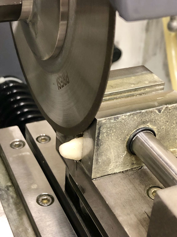
The study was approved by the Bioethics Committee of the Medical University of Lodz (number RNN/154/19/KE as of 12 March 2019). 132 samples of permanent immature molars (first, second and third), removed for surgical, orthodontic or inflammatory reasons, were used in the study. The extracted teeth were stored in 2% sodium azide solution at room temperature until the experiment. To prepare the teeth for the study, their roots were cut off, whereas the crown was cut mesio-distally into two parts using Presi-Mecatome T 201 precision cutting machine (Presi, France), equipped with a diamond-cutting disc (fig. 1). The teeth were placed in PVC rings (fig. 2). The teeth were placed in PVC rings of the following size: height – 2 cm and internal diameter – 18 mm. The inside of the ring was filled with acrylic (fig. 2, 3). In order to obtain a smooth and parallel structure, tooth surfaces were wet ground at a speed of 320 rpm with SiC paper (grit 180, 240, 400, 600) with the use of grinding-polishing machine Presi Minitech 233 (Presi, France) (fig. 4). The tooth samples were randomly divided into 8 study groups (tab. 1):
1. tooth,
2. tooth + sandblasting,
3. dentine + thermocycler,
4. dentine + sandblasting + thermocycler,
5. enamel,
6. enamel + sandblasting,
7. enamel + thermocycler,
8. enamel + sandblasting + thermocycler.

Fig. 1. Presi-Mecatome T 201 diamond-cutting disc
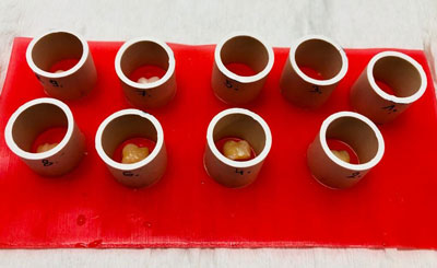
Fig. 2. Sample preparation
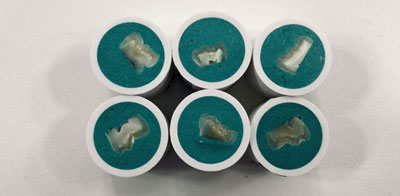
Fig. 3. Prepared dentine samples
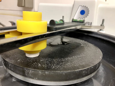
Fig. 4. Presi Minitech 233 polishing-grinding machine
Tab. 1. Mean stress values in the shear test for enamel and dentine
| Lp./No | Grupa badana | n | Średnia
Mean (MPa) | SD | Max (MPa) | Min (MPa) |
| 1 | Zębina
Dentine | 27 | 6,74 | 1,63 | 10,00 | 3,55 |
| 2 | Zębina + piaskowanie
Dentine + sandblasting | 29 | 5,74 | 2,03 | 9,34 | 1,42 |
| 3 | Zębina + termocykler
Dentine + thermocycler | 11 | 6,91 | 1,94 | 7,79 | 5,25 |
| 4 | Zębina + piaskowanie + termocykler
Dentine + sandblasting + thermocycler | 11 | 4,34 | 2,0 | 7,63 | 0,29 |
| 5 | Szkliwo
Enamel | 15 | 7,57 | 2,72 | 9,53 | 5,90 |
| 6 | Szkliwo + piaskowanie
Enamel + sandblasting | 15 | 7,53 | 1,88 | 10,20 | 4,24 |
| 7 | Szkliwo + termocykler
Enamel + thermocycler | 12 | 6,38 | 3,3 | 7,90 | 3,50 |
| 8 | Szkliwo + piaskowanie + termocykler
Enamel + sandblasting + thermocycler | 12 | 5,27 | 1,77 | 7,38 | 2,73 |
SD – odchylenie standardowe
Powyżej zamieściliśmy fragment artykułu, do którego możesz uzyskać pełny dostęp.
Mam kod dostępu
- Aby uzyskać płatny dostęp do pełnej treści powyższego artykułu albo wszystkich artykułów (w zależności od wybranej opcji), należy wprowadzić kod.
- Wprowadzając kod, akceptują Państwo treść Regulaminu oraz potwierdzają zapoznanie się z nim.
- Aby kupić kod proszę skorzystać z jednej z poniższych opcji.
Opcja #1
29 zł
Wybieram
- dostęp do tego artykułu
- dostęp na 7 dni
uzyskany kod musi być wprowadzony na stronie artykułu, do którego został wykupiony
Opcja #2
69 zł
Wybieram
- dostęp do tego i pozostałych ponad 7000 artykułów
- dostęp na 30 dni
- najpopularniejsza opcja
Opcja #3
129 zł
Wybieram
- dostęp do tego i pozostałych ponad 7000 artykułów
- dostęp na 90 dni
- oszczędzasz 78 zł
Piśmiennictwo
1. Santamaría RM, Abudrya MH, Gül G et al.: How to Intervene in the Caries Process: Dentin Caries in Primary Teeth. Caries Res 2020; 54(4): 306-323.
2. Iaculli F, Salucci A, Di Giorgio G et al.: Bond strength of self-adhesive flowable composites and glass ionomer cements to primary teeth: A systematic review and meta-analysis of in vitro studies. Materials (Basel) 2021; 14(21). Epub ahead of print.
3. Peric T, Markovic E, Markovic D et al.: Meta-analysis of in-vitro bonding of glass-ionomer restorative materials to primary teeth. Materials (Basel) 2021; 14(14): 1-11.
4. Ballal NV, Gandhi P, Kashyap NN: Influence of particulate alkaline biomaterial remnants in dentin on the adhesion of two resin-based bonding systems. Microsc Res Tech 2021; 84(5): 1036-1041.
5. Mocquot C, Attik N, Pradelle-Plasse N et al.: Bioactivity assessment of bioactive glasses for dental applications: A critical review. Dent Mater 2020; 36(9): 1116-1143.
6. Palza Cordero H, Castro Cid R, Diaz Dosque M et al.: Li-doped bioglass® 45S5 for potential treatment of prevalent oral diseases. J Dent 2021; 105. Epub ahead of print.
7. Carvalho EM, Lima DM, Carvalho CN et al.: Effect of airborne-particle abrasion on dentin with experimental niobophosphate bioactive glass on the microtensile bond strength of resin cements. J Prosthodont Res 2015; 59(2): 129-135.
8. Szczesio-Wlodarczyk A, Sokolowski J, Kleczewska J et al.: Ageing of dental composites based on methacrylate resins – A critical review of the causes and method of assessment. Polymers (Basel) 2020; 12(4): 1-18.
9. Karadas M, Atıcı MG: Bond strength and adaptation of pulp capping materials to dentin. Microsc Res Tech 2020; 83(5): 514-522.
10. François P, Greenwall-Cohen J, Goff S Le et al.: Shear bond strength and interfacial analysis of high-viscosity glass ionomer cement bonded to dentin with protocols including silver diammine fluoride. J Oral Sci 2020; 62(4): 444-448.
11. Meral E, Baseren N: Shear bond strength and microleakage of novel glass-ionomer cements: An In vitro Study. Niger J Clin Pract 2019; 22(4): 566-572.
12. Sheikh Hasani Y, Paryab M, Saffarpour A et al.: The Effect of Disinfection with Chlorhexidine on the Shear Bond Strength of Equia Resin-Modified Glass Ionomer Cement to Dentin in Permanent Teeth after Two Thermocycling Protocols. J Dent (Shiraz, Iran) 2017; 18(4): 265-271.
13. Cebe MA, Polat S, Cebe F et al.: Bonding performance of two newly developed self-adhering materials between zirconium and dentin. Niger J Clin Pract 2015; 18(2): 221-226.
14. Latta MA, Tsujimoto A, Takamizawa T et al.: Enamel and dentin bond durability of self-adhesive restorative materials. J Adhes Dent 2020; 22(1): 99-105.
15. Latta MA, Radniecki SM: Bond strength of self-adhesive restorative materials affected by smear layer thickness but not dentin desiccation. J Adhes Dent 2020; 22(1): 79-84.