© Borgis - New Medicine 1/2014, s. 24-25
*Konrad Wroński1, 2
Caput medusae in alcoholic liver disease – case report
1Faculty of Medicine, Department of Oncology, University of Warmia and Mazury, Olsztyn, Poland
Head of Department: prof. Sergiusz Nawrocki, MD, PhD
2Department of Surgical Oncology, Hospital Ministry of Internal Affairs with Warmia and Mazury Oncology Centre, Olsztyn, Poland
Head of Department: Andrzej Lachowski, MD
Summary
Caput medusae is the symptom of portal hypertension due to cirrhosis of liver. This symptom is encountered in medical practice less and less due to earlier diagnostic and effective treatment of portal hypertension. In this article the author present a case of a man who reported to the hospital because of huge ascites due to cirrhosis of liver. After draining ascitic fluid the symptom of caput medusae was observed.
INTRODUCTION
Caput medusae is the symptom of portal hypertension due to cirrhosis of liver (1-3). This symptom is encountered in medical practice less and less due to earlier diagnostic and effective treatment of portal hypertension (1, 4, 5).
CASE REPORT
A 53-year-old man, Caucasian race, was admitted to the hospital because of the huge ascites and general destruction. He had history of alcohol consumption about 80-120 gram/day for 20 years and he was heavy smoker – 20 cigarette/day for 20 years. The patient had suffered from cirrhosis of the liver for 5 years. Serology for viral hepatitis B and C were negative. During palpable examination, drew attention to the huge ascites (fig. 1). A umbilical revealed the presence of varicose veins (fig. 2). After drainage 2 liters of ascetic fluid were seen superficial epigastric veins radiating from a umbilical large venous varix (fig. 3).
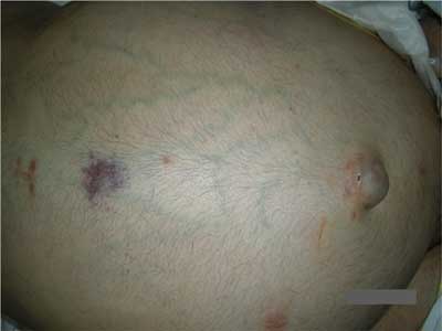
Fig. 1. Dilated superficial epigastric veins radiating from a umbilical large venous varix.
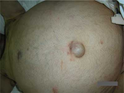
Fig. 2. Umbilical large venous varix.
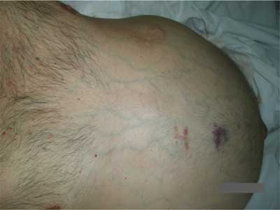
Fig. 3. Dilated superficial epigastric veins and bruising caused by thrombocytopenia.
The patient serum total bilirubin was 4.0 mg/dL, platelets – 11.000/mm3, white blood cells – 4.300/mm3, gamma glutamyltranspeptidase – 735 U/L, asparate aminotransferase – 40 U/L, alanine amnotranserase – 18 U/L.
The Doppler ultrasound was performed. The superficial epigastric veins were enlarge during this examination. Upper gastrointestinal endoscopy showed grade 3 esophageal varices but with no signs of recent hemorrhage.
The patient had drainage of the ascites. Within a few days the patient had evacuated 10 liters of ascitic fluid. Ascitic fluid had straw-color.
DISCUSSION
Medical dictionaries assign caput medusae the French pathologist Jean Cruveilhier (1791-1874) and the German pathologist Paul Clemens Von Baumgarten (1791-1873). In Stedman’s Medical Dictionary caput medusae is defined as „Cruveilhier’s sign; varicose veins radiating from umbilicus, seen in the Cruveilhier-Baumgarten syndrome” (6). The Cruveilhier-Baumgarten syndrome is „cirrhosis of the liver with patent umbilical or periumbilical veins and varicose periumbilical veins” (6). The Cruveilhier-Baumgarten marmur is „the venous hum heard over collateral veins, connecting the portal and caval venous systems, on the abdominal wall” (6).
Caput medusae is one of the fundamental symptom of portal hypertension due to cirrhosis of the liver (1, 7, 8). Blood from the portal venous system is shunted through the umbilical veins into the abdominal wall veins (1).
There have been reported a few cases of bleeding from umbilical varices (7-9). In the case described in Bahner and Holland article (7) the patient exsanguinated, in the other case (8) embolization of the umbilical vein was successful performed, and in the third case (9) transjugular intrahepatic portosystemic shunt (TIPS) was deployed.
Piśmiennictwo
1. Hari Kumar K, Rastogi SK: Caput medusae in alcoholic liver disease. Niger J Clin Pract 2011; 14: 508-509. 2. Singh NK, Cheema U, Khalil A: Caput medusa. BMJ Case Report 2010; 10.1136/bcr.03.2010.2795. 3. Bahner DR, Holland RW. Exsanguinating hemorrhage from a caput medusae: cutaneous variceal bleeding. J Emerg Med 1992; 10: 19-23. 4. Fitzgerald JB, Chalmers N, Abbott G et al.: The Use of TIPS tocontrol bleeding caput medusae. Br J Radiol 1998; 71: 558-560. 5. Douglas JG: Umbilical haemorrhage – an unusual complication of cirrhosis. Postgrad Med J. 1981; 57: 461-462. 6. Stedman’s medical dictionary. Baltimore: Williams and Wilkins, 1976. 7. Bahner DR Jr, Holland RE 3rd: Exsanguinating hemorrhage from caput medusa: cutaneous cariceal bleeding. J Emerg Med 1992; 10: 19-23. 8. Bell R, Thompson JF, Waugh RC et al.: Control of life-threatening bleeding from caput medusae by umbilical vein embolisation. Lancet 1989; 1: 736. 9. Sze DY, Magsamen KE, McClenathan JH et al.: Portal hypertensive hemorrhage from a left gastroepiploic vein caput medusa in an adhesed umbilical hernia. J Vasc Interv Radiol 2005; 16: 281-285.
