© Borgis - Postępy Nauk Medycznych 6/2016, s. 414-418
*Teresa Jackowska1, 2, Małgorzata Nieścior2, Monika Grzelczyk-Wielgórska2
Invasive Streptococcus pyogenes infections – cases review**
Inwazyjne zakażenia Streptococcus pyogenes – opisy przypadków
1Department of Pediatrics, Centre of Postgraduate Medical Education, Warsaw
Head of Department: prof. Teresa Jackowska, MD, PhD
2Department of Pediatrics, Father Jerzy Popiełuszko “Bielański” Hospital, Independent Public Health Care Institution in Warsaw
Head of Department: prof. Teresa Jackowska, MD, PhD
Streszczenie
Streptococcus pyogenes jest patogenem, który może wywoływać zakażenia o różnorodnym przebiegu, zarówno ze względu na ciężkość zakażenia, jak i obraz kliniczny. Jest czynnikiem etiologicznym zakażeń dróg oddechowych (gardła, ucha środkowego, zatok obocznych nosa, zapalenia płuc) oraz skóry i tkanek miękkich, mogących prowadzić do martwiczego zapalenia powięzi, a także zapalenia stawów, zapalenia opon mózgowo-rdzeniowych. Celem pracy było przedstawienie pięciu przypadków z inwazyjnym zakażeniem paciorkowcowym, leczonych w oddziale pediatrycznym.
Dzieci w wieku od 3-10 lat były hospitalizowane w latach 2010-2014 w Klinicznym Oddziale Pediatrycznym w Warszawie z powodu zakażenia inwazyjnego o etiologii Streptococcus pyogenes. Każdy z pacjentów prezentował całkowicie odmienny obraz kliniczny zakażenia.
U pacjentów potwierdzono obecność paciorkowca gr. A według Lancefielda (GAS) w lokalizacji fizjologicznie jałowej. Początkowo stosowano leczenie empiryczne, które zmieniono na antybiotykoterapię celowaną po otrzymaniu antybiogramu.
Obraz kliniczny paciorkowcowych zakażeń inwazyjnych jest różnorodny. Szybkie postawienie właściwego rozpoznania oraz celowana antybiotykoterapia gwarantują powodzenie leczenia.
Summary
Streptococcus pyogenes is a pathogen causing infections of various course regarding both the severity, as well as clinical picture. It is the etiological factor of the upper respiratory tracts infections (pharyngitis, otitis media, sinusitis, pneumonia), as well as skin and soft tissues infections, which may lead to necrotising fasciitis, arthritis and meningitis. The aim of this study was to present 5 cases of invasive streptococcus infections, treated in paediatric department
Paediatric patients (aged 3 to 10 years) were hospitalised (from 2010 to 2014) at the Warsaw Clinical Paediatric Department due to invasive infection of Streptococcus pyogenes etiology. Each of the patients displayed markedly different clinical picture of infection.
Patients were confirmed to have streptococcus infection of the Group A (according to Lancefield grouping) in physiologically sterile location. Empiric therapy was conducted initially and switched to antibiotic therapy after receiving an antibiogram.
The clinical picture of invasive streptococcal infections is diverse. A quick diagnosis and aimed antibiotic therapy ensure successful treatment.

Introduction
Streptococcus pyogenes (Group A streptococcus according to Lancefield grouping, GAS) is one of the most frequent infecting pathogen in children, occurring in the form of asymptomatic carriage, localised disease, organ disease and sepsis (1, 2). It may be the cause of different infections, such as pharyngitis and tonsillitis, lymphadenitis, scarlet fever, localized abscess (middle ear, sinuses, lungs, pleura, the meninges, appendages, joints and bones) as well as necrotising fasciitis, sepsis and toxic shock syndrome (1, 3). A group A streptococcal infection may result in complications, such as rheumatic fever with heart involvement and glomerulonephritis (1, 3). The incidence of invasive disease caused by Streptococcus pyogenes (S. pyogenes) in the world is estimated to be equal to 663 thousands cases a year (including 163 thousands deaths) (2). Polish National Institute of Public Health epidemiological reports from the last 3 years indicate constant increase in group A streptococcal invasive infections. In the year 2011 there was 3455 infections per year at incidence rate of 9.0/100 thousands; in the year 2012 there was 4286 infections per year at incidence rate of 11.1/100 thousands; in the year 2013 there was 5294 infections per year at incidence rate of 13.7/100 thousands; in the year 2014 there was 5597 infections per year at incidence rate of 14.54/100 thousands (4). This is probably associated with increasing virulence of the streptococcus and the presence of the M protein which blocks the complement system and inhibits phagocytosis (1, 3). Invasive infections are most prevalent in immunodeficient patients and patients with chronic diseases, although they occur also in immunocompetent patients (3).
Cases overview
Between 2010 and 2014, five paediatric patients (three females and two males) were hospitalised at the Clinical Paediatrics Department of Bielański Hospital in Warsaw with an invasive disease caused by group A streptococcus. The youngest patient was 3 years and 3 months old, and the oldest patient was 10 years old. Four patients were hospitalized from January to March, and one female patient with the diagnosis of soft tissue inflammation of the shin was hospitalized in July. All the paediatric patients were in a good condition, without immune disorder history (tab. 1).
Tab. 1. Patients’ characteristics
| | Girl
(JK)
Patient 1 | Girl
(ZW)
Patient 2 | Girl
(AW)
Patient 3 | Boy
(AW)
Patient 4 | Boy
(MP)
Patient 5 |
| Age (years) | 3 3/12 | 3 6/12 | 7 3/12 | 8 4/12 | 10 |
| Diagnosis | Pneumonia and pleuritis | Soft tissue inflammation | Left temporal lobe abscess in the course of acute middle ear inflammation | Acute sinusitis.
Pneumonia and pleuritis | Right shoulder inflammation in the course of GAS sepsis.
Acute sinusitis. Scarlet fever |
| S. pyogenes cultivated, group A | Pleural fluid | Wound smear | Abscess culture | Blood culture | Blood culture, throat smear |
| Hospitalization duration (days) | 24 | 24 | (16 + 28)
44 | 21 | 5 and further treatment in department of surgery in other hospital |
| Initial treatment | Cefotaxime, clindamycin | Ceftriaxone | Ceftriaxone | Cefotaxime,
clindamycin | Cefotaxime, clindamycin |
| Targeted therapy | Crystalline penicillin, clindamycin | Crystalline penicillin, clindamycin | Ceftriaxone,
clindamycin, meropenem | Crystalline penicillin, clindamycin,
linezolid | Crystalline penicillin, clindamycin |
Female patient 1
Female patient (JK) aged 3 years and 3 months was admitted to the paediatric department with dyspnoea (SpO2 86%), dry cough and fever lasting for 3 days, with the temperature up to 39°C. Pneumonia was diagnosed with pleural reaction. Blood culture test result was proper. There was a need for pleural cavity drainage and 750 mL of fluid was extracted. The fluid culture showed an increase in S. pyogenes number. During hospitalization general condition improved (fig. 1 and 2).
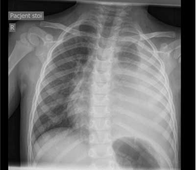
Fig. 1. Patient 1 (JK). Almost total shading of the left lung with mediastinum shift to the right
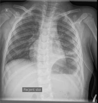
Fig. 2. Patient 1 (JK). 8th day of the treatment [ndash] clinical picture improvement
Female patient 2
Female patient (ZW) aged 3 years and 3 months was admitted to the paediatric department with the right shin swelling, fever up to 40°C and diarrhoea, which occurred twenty four hours before admission. Soft tissue inflammation of the shin and prerenal azotemia was diagnosed. The starting point of the infection was the wound caused by a mosquito bite. Although third generation cephalosporin was included in the treatment, the general condition of the girl deteriorated. Acute phase proteins concentration increased (CRP from 136.7 mg/l to 426 mg/l), as well as white blood cells (WBC from 17.1 thous./μl to 22.9 thous./μl), with insignificant decrease of procalcitonin concentration (PCT from 76.5 ng/ml to 58.6 ng/ml). During the first three days of the treatment the female patient still had a fever and her urea level was raised (72.4 ng/ml). Physical examination revealed meningism symptoms (neck stiffness, Brudzinski’s symptom). To rule out meningitis a lumbar puncture procedure was performed with the correct result. Several ultrasound examinations of the shin and surgical consultation was performed to rule out necrotising fasciitis. There were no indications for surgical intervention. After receiving the wound culture (S. pyogenes) result, the treatment was switched to crystalline penicillin with clindamycin. The general condition of the girl started to improve as fever as well as oedema and infiltration of the right shin subsided. After 4 days of antibiogram compliant treatment (penicilin, clindamycin susceptibility), the CRP and PCT values decreased considerably (51.6 mg/l and 1.2 ng/ml) accordingly).
Female patient 3
Powyżej zamieściliśmy fragment artykułu, do którego możesz uzyskać pełny dostęp.
Mam kod dostępu
- Aby uzyskać płatny dostęp do pełnej treści powyższego artykułu albo wszystkich artykułów (w zależności od wybranej opcji), należy wprowadzić kod.
- Wprowadzając kod, akceptują Państwo treść Regulaminu oraz potwierdzają zapoznanie się z nim.
- Aby kupić kod proszę skorzystać z jednej z poniższych opcji.
Opcja #1
29 zł
Wybieram
- dostęp do tego artykułu
- dostęp na 7 dni
uzyskany kod musi być wprowadzony na stronie artykułu, do którego został wykupiony
Opcja #2
69 zł
Wybieram
- dostęp do tego i pozostałych ponad 7000 artykułów
- dostęp na 30 dni
- najpopularniejsza opcja
Opcja #3
129 zł
Wybieram
- dostęp do tego i pozostałych ponad 7000 artykułów
- dostęp na 90 dni
- oszczędzasz 78 zł
Piśmiennictwo
1. Rutkowski Z: Choroby zakaźne i pasożytnicze u dzieci. PZWL, Warszawa 2001: 14-24.
2. Carapetis JR, Steer AC, Mulholland EK, Weber M: The global burden of group A streptococcal diseases. Lancet Infect Dis 2005; 5: 685-694.
3. Imohl M, Reinert R, Ocklenburg C, Linden M: Epidemiology of invasive Streptococcus pyogenes disease in Germany during 2003-2007. FEMS Immunol Med Microbiol 2010; 58: 389-396.
4. Narodowy Instytut Zdrowia Publicznego [ndash] Państwowy Zakład Higieny. Zakład Epidemiologii Główny Inspektorat Sanitarny. Departament Zapobiegania oraz Zwalczania Zakażeń i Chorób Zakaźnych u Ludzi; http://www.pzh.gov.pl/oldpage/epimeld/index_p.html.
5. Paul SP, Jerwood S: Group A streptococcal septicemia, meningitis and cerebral abscess: case report and literature review. The Turkish Journal of Pediatrics 2012; 54: 180-183.
6. Martin J, Murchan S, O’Flanagan D, Fitzpatrick F: Invasive group A streptococcal disease in Ireland 2004-2010. Euro Surveill 2011; 16(41): pii=19988; http://www.eurosurveillance.org/images/dynamic/EE/V16N41/art19988.pdf.
7. Filleron A, Jeziorski E, Michon E-L et al.: Current insight in invasive group A streptococcal infection in pediatrics. Eur J Pediatr 2012; 171: 1589-1598.
8. Committee on Infectious Diseases, American Academy of Pediatrics. Severe invasive group A streptococcal infections: A subject review. Pediatrics 1998; 101(1): 136-140.
9. Miodek M, Gawrysiak M, Kucharski P, Niedzielski J: Rozległa ropowica ściany klatki piersiowej jako powikłanie wiatrówki u 13-letniego chłopca [ndash] opis przypadku. Przegl Ped 2012; 42(1): 31-33.
10. Dale JB, Penfound TA, Chiang EY, Walton WJ: New 30-valent M-protein based vaccine evoked cross-opsonic antibodies against non-vaccine serotypes of group A streptococci. Vaccine 2011; 29: 8175-8178.
11. Szczypa K, Sadowy E, Izdebski R et al.: Group A streptococci from invasive disease episodes in Poland are remarkably divergent at the molecular level. J Cinic Microbiol 2006; 44(11): 3975-3979.
12. Stevens DL, Gibbons AE, Bergstrom R, Winn V: The Eagle effect revisited: efficacy of clindamycin, erthromycin, and penicillin in the treatment of streptococcal myositis. J Infect Dis 1988; 158: 23-28.


