Rohan Anna1, *Nyc Małgorzata2, Rogóż Anna3, Fugiel Jarosław4
Changes in plantar pressure distribution after long-distance running
Ocena zmian rozkładu nacisku stóp pod wpływem biegu długodystansowego
1Department of Human Anatomy, Wroclaw Medical University, Poland
Head of Department: Professor Bohdan Gworys, MD, PhD
2Faculty of Technology and Natural Science, Karkonoska State College, Jelenia Góra, Poland
Dean of Faculty: Wioletta Palczewska, MD, PhD
3Vertex, Rehabilitation Unit, Wroclaw, Poland
4Department of Biostructure, University School of Physical Education, Wroclaw, Poland
Head of Department: Professor Teresa Sławińska-Ochla, MD, PhD
Streszczenie
Bieg długodystansowy to ogromny wysiłek dla organizmu. W jego trakcie dochodzi między innymi do znacznych obciążeń układu ruchu, a w szczególności stóp, które ze względu na swoje dystalne położenie i bezpośredni kontakt z podłożem podlegają znacznym naciskom. Długotrwały wysiłek powoduje zmęczenie tkanek miękkich stabilizujących sklepienie podeszwowe stopy, co z kolei wpływa na rozkład ciężaru ciała na poszczególne strefy w obrębie stopy. Długotrwała praca mięśni i więzadeł wywołuje przeciążenie pewnych pól oraz odciążenie innych.
Celem pracy była analiza i ocena tych zmian zachodzących pod wpływem biegu długodystansowego w obrębie poszczególnych stref w stopie.
Badaniom poddano 36 mężczyzn, którzy byli uczestnikami VIII Półmaratonu Ślężańskiego.
Badanie przeprowadzono w dwóch etapach: przed i po biegu.
Ocena stóp została wykonana metodą pedobarometryczną. Pomiarów dokonano za pomocą Platformy E.P.S./R1. Na podstawie badania przeprowadzonego przed biegiem i analizy dotyczącej obciążeń stóp w pozycji stojącej zaobserwowano, że w badanej grupie biegaczy w większym stopniu obciążana jest stopa lewa. Przed biegiem masa ciała w pozycji stojącej nie była rozłożona równomiernie na obie stopy. U większości biegaczy bardziej obciążona była lewa kończyna, która częściej pełni funkcję podporową, co może wynikać z asymetrii funkcjonalnej. Wyniki badań wykazały, że obraz zmian po biegu był różny w stopie prawej i lewej. Rozkład obciążeń w stopie prawej nie zmienił się istotnie, natomiast w stopie lewej nastąpił wzrost obciążenia przodostopia i tyłostopia oraz zmalało obciążenie śródstopia. Zmiana ta wpłynęła na typologię stopy; po biegu zwiększyła się liczba biegaczy, u których sklepienie podłużne tej stopy uległo uniesieniu.
Summary
Long-distance running is a huge effort for the organism. During the run, a significant load is put on the movement system, especially on feet, which, due to their distal location and direct contact with the ground, are subject to significant pressure. Prolonged exercise causes fatigue of the soft tissues that stabilize the arches of the foot, which, in turn, influences the distribution of the weight between the foot zones. Prolonged activity of muscles and tendons causes an overload in certain fields and lowers the load in other zones.
The aim of this paper was to analyze and assess the changes of pressure in the individual foot zones after long-distance running.
The study involved 36 men who participated in the 8th Ślęża Half Marathon and was conducted in two stages: the measurements were taken before and after the race.
The analysis was conducted with pedobarometry with the Platform E.P.S./R1 apparatus. It was observed that more weight was put on the left foot. Before the race, the load was not evenly distributed between the feet in a standing position. In the majority of the runners, left foot was more loaded. Left foot has a supportive role, which may be caused by a functional asymmetry. The results indicated that the changes in plantar pressure varied between feet. Plantar pressure distribution did not significantly change in the right foot, while in the left foot, the pressure increased on the forefoot and hindfoot, and decreased on the midfoot. The change influenced foot typology – after the run, the number of runners with high-arched feet increased.
Introduction
Running is one of the most natural exercise for human, and from ancient times, it has been one of the Olympic sports. Its popularity has been constantly growing, as evidenced by the growing number of joggers, as well as the number of runners participating in mass events. Running owes its popularity to its simplicity, as it requires no special skills, training space nor equipment (1, 2). This type of exercise has a positive influence on the general health status, inducing several positive changes in the organism, which may protect against the diseases of civilization (3).
Properly dosed exercise induces the lowering of the blood pressure and heart rate, as well as improves metabolism and immune system function. Jogging also has a huge impact on the psyche: in runners, a high-spirited state, known as “runner’s high” is observed after the prolonged and intense exercise (4, 5). It has also been proven that certain substances released to the bloodstream during runnin cause the so-called conditioned pain modulation, i.e. reduce the pain sensation (6).
However, it must be noted that long-distance running is a huge effort for the body. During the run, a significant load is put on the movement system, especially on the feet, which, due to their distal location and direct contact with the ground, are subject to significant pressure. Prolonged exercise causes fatigue of the soft tissues that stabilize the arches of the foot, which, in turn, influences the plantar pressure distribution. Prolonged activity of muscles and tendons causes an overload in certain foot zones and lowers the load in other. The influence of the long-distance running on the plantar pressure has been studied repeatedly (7-12).
Aim
The aim of the study was to assess the changes in plantar pressure after long-distance running. Changes in plantar pressure, as well as in the pressure distribution between individual foot zones, were assessed. The results from before and after the race were compared.
The study sought to answer the following research questions:
– Will the half marathon influence the load distribution between the right foot and the left foot?
– Will long-distance run influence the load distribution between forefoot, hindfoot and midfoot of the left foot and the right foot?
– Will the long-distance run directly influence the foot type in runners?
– Will there be any differences in the load distribution in individual foot zones of the right foot and left foot before and after the long-distance run?
Material and methods
The study was conducted, with the consent of the Organizer, during the 8th Ślęża Half Marathon, which took place on the 21th March 2015 in Sobótka. The study included 36 male volunteers participating in the race. All of the participants agreed to take part in the study according to the Declaration of Helsinki. The mean age of the participants was 35.3 ± 10.4 years.
The study was conducted in two stages: before and after the race.
The pedobarometrical analysis was conducted with the Platform E.P.S./R1 apparatus, measuring 700 x 500 x 5 mm, equipped with 2304 sensors distributed on the active surface.
The runner was to step on the pedobarometrical platform so that the feet were situated on two sides of a vertical line drawn on a mat. Then, the subject was asked to perform three slow steps in place in order to set the feet at ease on the mat. Subsequently, the participant stood erect with upper limbs along the body, looking straight ahead. At the signal, he remained motionless for 30 seconds. During the time, the measurements were taken.
In this paper, the following measurements were analyzed:
1. The load on the right foot and the left foot (expressed in %).
2. The load on the forefoot, midfoot and hindfoot (expressed in %). Based on the percentage of the load on the midfoot, the person’s foot type was automatically determined by the program (tab. 1).
Tab. 1. Foot type based on the load distribution
| Pressure on midfoot [%] | Foot type |
| 0-7 | very high foot arch |
| 7-14 | high foot arch |
| 14-21 | slightly high arch |
| 21-28 | normal foot |
| 28-35 | sligthly flat foot |
| 35-42 | flat foot |
| 42 and more | very flat foot |
3. Mean pressure on individual anatomical foot zones (expressed in kPa). In the Biomech Studio software, the results were analyzed after having been divided to ten zones: hallux, toes 2 to 5, head of the 1st metatarsal bone, head of the 2nd metatarsal bone, head of the 3rd metatarsal bone, head of the 4th metatarsal bone, head of the 5th metatarsal bone, midfoot, medial heel, lateral heel.
For every parameter analyzed, following statistical characteristics were calculated: mean (x) and standard deviation (SD). The normal distribution of the variables was assessed with Shapiro-Wilk test. Results with p < 0.05 were considered statistically siginificant.
Results
Before the race, the load was not evenly distributed between the feet in a standing position. In the majority of the runners, left foot was more loaded. The mean load put on the left foot before the race was 55.5%, and the mean load on the right foot – 44.5%. After the race, a change in the load distribution was observed. The load on the left foot decreased to 53.6%, and the load on the right foot increased by 1.9% to 46.4%. The differences of the load of the feet before and after the run were not statistically significant. In spite of the transfer of the part of the load from the left foot to the right foot, higher load was still put on the left limb, and the differences were statistically significant (tab. 2).
Tab. 2. Weight distribution between the right foot and the left foot before and after the half-marathon race and differences between feet
| Foot | Time of the measurement | x | SD | Z | p |
| Right [%] | before the race | 44.5 | 5.8 | 3.25 | 0.03* |
| after the race | 46.4 | 6.4 |
| Left [%] | before the race | 55.5 | 5.7 | 5.73 | 0.05* |
| after the race | 53.6 | 6.4 |
*p-values of statistical significance p < 0.05
Furthermore, the weight distribution between forefoot, midfoot and hindfoot was assessed. Data were analyzed separately for each anatomical part of the right foot and left foot, comparing the measurements taken before and after the race.
Mean pressure on left forefoot was 43.0% before the race, and 45.2% after the race, and the difference was statistically significant (tab. 3). Mean pressure on right forefoot was 43.0% before the race, and 44.4% after the race, but the difference was not statistically significant (tab. 3). Mean pressure on forefoot was similar for both feet, but more pressure was put on the left forefoot after the race when compared with the right forefoot.
Tab. 3. Load on forefoot, midfoot and hindfoot of the right foot and left foot before and after the long-distance race
| Foot | Time of measurement | x | SD | Z | p |
| Right | Forefoot | before the race | 43.01 | 5.75 | 1.00 | 0.32 |
| after the race | 44.02 | 4.99 |
| Midfoot | before the race | 21.76 | 7.95 | 1.37 | 0.17 |
| after the race | 20.69 | 7.38 |
| Hindfoot | before the race | 35.23 | 4.61 | 0.41 | 0.68 |
| after the race | 35.29 | 4.64 |
| Left | Forefoot | before the race | 43.02 | 5.0 | 3.07 | 0.002* |
| after the race | 45.22 | 5.4 |
| Midfoot | before the race | 20.12 | 6.56 | 3.29 | 0.001* |
| po biegu | 16.12 | 8.55 |
| Hindfoot | before the race | 36.86 | 2.70 | 2.72 | 0.006* |
| after the race | 38.68 | 4.05 |
*p-values of statistical significance p < 0.05
The analysis of the pressure on left midfoot indicated that mean pressure decreased by 6%. Before the race the pressure was 22.1%, and after the race – 16.1%. The difference was statistically significant (tab. 3). Mean pressure on left midfoot was 21.8% before the race, and 21.0% after the race. The difference was not statistically significant (tab. 3). As with the forefoot, mean pressure was similar for both feet before the race, and after the race, the pressure on the right foot increased.
The mean pressure on left hindfoot was 36.9% before the race and 38.9% after the race. The difference was statistically significant (tab. 3). Mean pressure on right foot was 35.2% and did not change after the race – 35.3%. The pressure on hindfoot was higher for left foot, and the difference increased after the race.
The analysis of the results indicated that in the case of the left foot, the pressure on the forefoot and hindfoot increased, and the pressure on the midfoot decreased. In the case of the right foot, the differences in pressure before and after the race were not statistically significant. Therefore, it has been concluded that the typology of the left foot had changed – the longitudinal arch had raised.
In the subsequent part of the analysis, the changes in pressure in 10 anatomical zones of the foot were observed. It was observed that in the right foot, the pressure on the head of the 1st metatarsal bone had decreased (tab. 4). In the left foot, changes in the pressure were observed in the midfoot and in the medial and lateral heel. In each of these zones, the pressure decreased after the race. Mean pressure on the midfoot decreased by 1.61 kPa (11.9%), on the medial heel – by 3.34 kPa (8.6%), and on the lateral heel – by 3.61 kPa (9.9%) (tab. 5).
Tab. 4. Changes in load distribution in the right foot
| Foot zone | Before the race [kPa] | After the race [kPa] | Difference |
| x | SD | x | SD | Z | p |
| Big toe | 10.06 | 7.64 | 8.17 | 7.12 | 1.24 | 0.21 |
| Toes 2 to 5 | 4.44 | 4.27 | 4.39 | 4.60 | 0.2746 | 0.78 |
| Head of the 1st metatarsal bone | 15.44 | 4.74 | 13.61 | 4.26 | 2.0616 | 0.04* |
| Head of the 2nd metatarsal bone | 26.17 | 6.44 | 27.17 | 7.43 | 0.5947 | 0.55 |
| Head of the 3rd metatarsal bone | 29.33 | 7.50 | 31.94 | 6.58 | 1.3186 | 0.19 |
| Head of the 4th metatarsal bone | 29.00 | 9.32 | 30.44 | 7.16 | 0.6097 | 0.54 |
| Head of the 5th metatarsal bone | 24.56 | 10.58 | 25.33 | 10.30 | 0.1293 | 0.90 |
| Midfoot | 14.94 | 4.56 | 13.56 | 4.76 | 1.3065 | 0.19 |
| Medial heel | 25.33 | 6.63 | 26.94 | 5.23 | 0.879 | 0.38 |
| Lateral heel | 26.39 | 6.82 | 26.89 | 5.02 | 0.5917 | 0.55 |
*p-values of statistical significance p < 0.05
Tab. 5. Changes in load distribution in the left foot
| Foot zone | Before the race [kPa] | After the race [kPa] | Differences |
| x | SD | x | SD | Z | p |
| Big toe | 10.33 | 8.11 | 10.06 | 7.23 | 0.53 | 0.59 |
| Toes II to V | 3.56 | 3.57 | 3.67 | 4.01 | 0.04 | 0.97 |
| Head of the I metatarsal bone | 15.00 | 5.29 | 15.06 | 4.92 | 0.28 | 0.98 |
| Head of the II metatarsal bone | 25.89 | 5.20 | 25.89 | 7.79 | 0.26 | 0.80 |
| Head of the III metatarsal bone | 27.83 | 5.35 | 28.78 | 8.63 | 0.49 | 0.62 |
| Head of the IV metatarsal bone | 25.67 | 9.75 | 26.17 | 10.55 | 0.15 | 0.88 |
| Head of the V metatarsal bone | 19.89 | 9.76 | 19.89 | 8.57 | 0.12 | 0.91 |
| Midfoot | 13.56 | 5.86 | 11.95 | 5.60 | 2.41 | 0.02* |
| Medial heel | 38.67 | 6.83 | 35.33 | 7.27 | 2.58 | 0.01* |
| Lateral heel | 36.33 | 5.47 | 32.72 | 4.81 | 2.96 | 0.00* |
*p-values of statistical significance p < 0.05
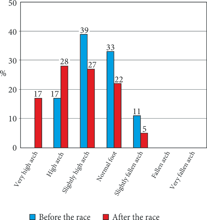
The change of the type of arching of the feet confirmed the observations of pressure change. The change of the type of arching was assessed based on the load on the midfoot before and after the race. It was found that after the race, the percentage of persons with high and very high arch was higher that before the race (fig. 1).
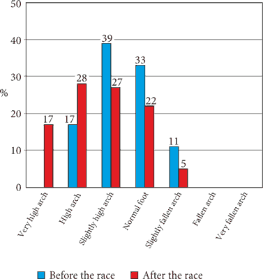
Fig. 1. Typology of feet before and after the race
Discussion
During the long-distance run, there is a significant load put on feet. This leads to the passive and active overload of the motor system, which manifests by a change in the foot support pattern, which can result in the lowering of the foot arch. In a study by Kuraś (13) on athletes, a decrease in the Clarke angle has been observed. Similarly, Stawczyk (14) reported lowering of the foot arch of the launching leg in high jump athletes and long jump athletes when compared to the opposite leg of the athletes. This was explained by a greater load on the launching foot. However, this observation was not confirmed by Gradek et al. (15). Greater load on feet was also observed in obese and overweight persons (16-18). The described increased load during locomotion may affect the lowering of the feet arch and therefore, lead to a decrease in their fitness (19).
Our research revealed that the load on anatomical structures during the race results in different weight distribution on the feet after the race. The change in load was also manifested in their arching. Before the race, body mass in a standing position was not distributed evenly between the two feet. In most of the runners, the left foot was more loaded than the right one. The left foot is more likely to have a supportive function, which may be due to a functional asymmetry. It is assumed that the adoption of the stance posture and the functional privilege of the right upper extremity provide the basis for preferring the left lower extremity for supportive functions. The extremity is also more frequent to be used as a launching extremity at the jumps, as it performs supportive-launching functions (20). After a long-distance race, in which the movements are symmetrical, part of the load has been transferred to the right foot, although a difference in the load between the feet remained.
After analyzing the load of the forefoot, midfoot and hindfoot, it was found that the character of changes differed between the right foot and the left foot. The distribution of weight in the right foot did not change significantly, while in the left foot, an increase in the load in the forefoot and hindfoot, as well as a decrease in the load on the midfoot, were observed. The change influenced the foot typology; after the race, a higher number of runners had a high or a very high arch of the foot. This complies with the views of Skarżyński (21), who has stated that long-distance runners land on their heel during the run, rolling over on their foot and only then launching, making the foot arch less loaded. Classical technique, in which a runner lands on the midfoot, is only used by the best marathoners (22). The study subjects did not belong to this group of athletes, therefore, they may have increased their load on left hindfoot after the race, as the left side was more loaded in them during the race. At the same time, a significant increase in pressure on the left median and lateral heel has been observed in the analysis of the load on 10 foot zones. In a study concerning foot load after a 60-minute race, Escamilla-Martinez et al. (9) have also observed changes in foot pressure – changes leading a tendency for pronation and a pressure increase of the medial heel and II metatarsal head. A tendency for pronation has also been noted by Cowley and Marsden in runners after a half-marathon (7). However, Masłoń and Golec (23) have reported that an increase in pressure on lateral metatarsal heads in some of the participants. The authors explain this phenomenon by the adaptation to the training, resulting from the change in proportion of the support phase to the swing phase in the course of the race.
It is believed that physical activity is associated with a correct foot arching (18, 19, 24-28). However, too much stress may be detrimental to foot structure and functioning.
Conclusions
1. Long-distance run affects the weight distribution between the feet. In our participants, the left foot was more loaded, but the differences in weight distribution were reduced after the race.
2. In both feet after the long-distance run, pressure distribution pattern has changed. In the left foot, pressure is increased on forefoot and midfoot, and decreased on midfoot, resulting in a higher arch. In the right foot, the changes in pressure distribution before and after the race have not been relevant.
3. After the long-distance run, the load on feet changes, and the typology of the feet changes accordingly.
Piśmiennictwo
1. Nowak PF: Poziom zaangażowania polskich biegaczy w ich sportową pasję. In: Siwiński W, Pluta B (ed.): Teoria i metodyka rekreacji ruchowej w świetle aktualnych badań. Bogucki Wydawnictwo Naukowe, Poznań 2012: 290-301.
2. Nowak PF, Supiński J: Uczestnictwo w biegach maratońskich, a zdrowotność polskich biegaczy. Rozprawy Naukowe Akademii Wychowania Fizycznego we Wrocławiu 2014; 45: 41-47.
3. Maciantowicz J: Ruch fizyczny o charakterze wytrzymałościowym (bieg) zapobiega starzeniu się, leczy z patologicznych chorób społecznych. Polish J Sport Med 2003; 19(4): 133-138.
4. Galdino G, Romero TR, Silva JF et al.: The endocannabinoid system mediates aerobic exercise-induced antinociception in rats. Neuropharmacology 2014; 77: 313-324.
5. Raichlen DA, Foster AD, Gerdeman GL et al: Wired to run: exercise-induced endocannabinoid signaling in humans and cursorial mammals with implications for the ‘runner’s high’. J Exp Biol 2012; 215: 1331-1336.
6. Masataka U, Wanseok L, Courtney AM, Shelby CH: Influence of Moderate Intensity Physical Activity Levels and Gender on Conditioned Pain Modulation. J Sport Sci 2016; 34.5: 467-476.
7. Cowley E, Marsden J: The effects of prolonged running on foot posture: a repeated measures study of half marathon runners using the foot posture index and navicular height. J Foot Ankle Res 2013; 6(1): 20.
8. Deleu PA, Matricau G, Leemruse T: Impact of 90 minutes running exercise on plantar loading of the forefoot: a prospective study on symptom-free athletes. J Foot Ankle Res 2008; 1(1): O18.
9. Escamilla-Martinez E, Martinez-Nova A, Gómez-Martin B et al.: The Effect of Moderate Running on Foot Posture Index and Plantar Pressure Distribution in Male Recreational Runners. J Am Podiat Med Assn 2013; 103(2): 121-125.
10. Griffin NL, Richmond BG: Cross-sectional geometry of the human forefoot. Bone 2005; 37(2): 253-260.
11. Karagounis P, Prionas G, Armenis E et al.: The impact of the Spartathlon ultramarathon race on athletes’ plantar pressure pattems. J Foot Ankle Res 2009; 2(4): 173-178.
12. Nagel A, Fernholz F, Kibele C, Rosenbaum D: Long distance running increases plantar pressures beneath the metatarsal heads: a barefoot walking investigation of 200 marathon runners. Gait Post 2008; 27: 152-155.
13. Kuraś Z: Czynnościowe badania stopy u średnio i długodystansowców. AZS Warszawa. Kultura Fizyczna 1958; 8: 567-574.
14. Stawczyk Z: Skoczność dosiężna a wysklepienie stopy. Roczniki Naukowe WSWF Poznań 1965; 10: 229-240.
15. Gradek J, Mleczko M, Bora P: Wysklepienie stóp młodych lekkoatletek. JPES 2004; 6-7: 11-14.
16. Ślężyński J, Rottermund J: Cechy plantograficzne stóp kobiet w średnim i starszym wieku w zależności od charakteru pracy oraz czynników środowiskowych i osobniczych. JPES 1999; 4: 41-68.
17. Furgał W, Adamczyk A: Ukształtowanie sklepienia stopy u dzieci w zależności od wskaźnika masy ciała. Polish J Sport Med 2009; 25(3): 189-199.
18. Trocińska A: Charakterystyka wybranych parametrów budowy stóp kobiet i mężczyzn uprawiających karate. In: Marecki B (ed.): Sport i turystyka we współczesnym stylu życia. AWF. Poznań 2009: 89-95.
19. Lichota M, Plandowska M, Mil P: Wysklepienie stóp zawodników wybranych dyscyplin sportowych. Pol J Sport Tourism 2013; 20: 135-146.
20. Malinowski A: Auksologia: rozwój osobniczy człowieka w ujęciu biomedycznym. Uniwersytet Zielonogórski, Zielona Góra 2004.
21. Skarżyński J: Biegiem przez życie. Mega Sport, Szczecin 2012.
22. Thurgood G, Sepstead G, Stankiewicz C: Bieganie od rekreacji do maratonu. Solis, Warszawa 2014.
23. Masłoń A, Golec E: Ocena wpływu biegów rekreacyjnych na wzorzec obciążenia przodostopia w fazie odbicia. Med Rehabil 2013; 17(3): 13-22.
24. Abshire D, Metzler B: Bieganie naturalne. Buk Rower. Warszawa 2013.
25. Furgał W, Adamczyk A: Ukształtowanie sklepienia stopy u dzieci w zależności od poziomu aktywności fizycznej. Med Sport 2008; 24, 5(6) 311-317.
26. Lizis P, Puszczałowska-Lizis E: Charakterystyka zmian podeszwowej powierzchni stóp oraz związek z wybranymi cechami budowy ciała koszykarzy I ligi polskiej. Physiother 2006; 14(1): 43-52.
27. Trzcińska D, Tabor P, Olszewska E: Plantograficzna analiza wybranych parametrów budowy stóp młodych siatkarzy. JPES 2008; 52(4): 207-212.
28. Zieliński JR, Ilnicka L: Stopy ciężarowców w świetle badań ciągłych. JPES 1992; 35(3): 43-52.