Magdalena Frankowska1, Jolanta Biszewska1, Jarosław Łuczaj2, *Emilia Duchnowska1, Bożena Kosztyła-Hojna1, Jerzy Robert Ładny3, Maciej Zdrojkowski1
Facial nerve palsy in acute otitis media – case study
Porażenie nerwu twarzowego w przebiegu ostrego zapalenia ucha środkowego – studium przypadku
1Department of Clinical Phonoaudiology and Speech Therapy, Medical University of Bialystok, Poland
2Department of Otolaryngology, Medical University of Bialystok, Poland
3Department of Emergency Medicine, Medical University of Bialystok, Poland
Streszczenie
Dzięki narządowi słuchu i równowagi człowiek zachowuje orientację przestrzenną, posiada zdolność lokalizacji poszczególnych dźwięków oraz ich dyskryminacji. Bez poprawnie funkcjonującego narządu słuchu niemożliwe byłoby wykształcenie mowy, która jest środkiem komunikacji i interakcji społecznych. Dlatego ważne jest obserwowanie stanu zdrowia poszczególnych zmysłów, w tym słuchu, a w razie niepokojących sygnałów lub wypadków będących przyczyną ich uszkodzenia zgłoszenie się do specjalisty. Umożliwi to podjęcie wczesnej diagnostyki oraz interwencji.
Dźwięk odbierany jest przez człowieka za pośrednictwem przewodzeniowej oraz odbiorczej części aparatu słuchu. Ważne jest, by przy pogorszeniu słuchu stwierdzić, której części dotyczy problem, ponieważ postępowanie w przypadku niedosłuchu odbiorczego i przewodzeniowego jest inne.
Dzięki wnikliwej diagnostyce można ustalić miejsce uszkodzenia słuchu. Podstawowy element diagnostyki stanowi wywiad. Następnie przeprowadza się pełne badanie laryngologiczne. Kolejnym krokiem jest przebadanie pacjenta za pomocą odpowiednich testów subiektywnych (próby stroikowe, audiometria, badania akumetryczne) oraz obiektywnych (OAE, tympanometria, potencjały wywołane z pnia mózgu). Badania te wskazują na lokalizację oraz stopień uszkodzenia słuchu oraz odpowiedni sposób leczenia indywidualnego pacjenta.
Ostre zapalenie ucha środkowego to przypadłość dotykająca głównie dzieci, jednak zdarza się też u osób dorosłych – częściej mężczyzn. Wywołane jest zazwyczaj infekcjami wirusowymi i bakteryjnymi górnych dróg oddechowych. Charakterystyczne są: nagłe pojawienie się dolegliwości, wysięk w jamie bębenkowej oraz ból i stan zapalny ucha. Ostre zapalenie ucha środkowego może doprowadzić do powikłań, takich jak: porażenie nerwu twarzowego, tympanoskleroza, zapalenie błędnika, zapalenie wyrostka sutkowego czy ropień mózgu.
Summary
The hearing and balance organ maintains human spatial orientation as well as the ability to localize and discriminate individual sounds. Without properly functioning hearing organ, it would be impossible to develop speech, which is a mean of communication and social interaction. Therefore, it is important to observe the condition of particular senses, including hearing, and in case of disturbing symptoms or accidents causing damage, contact a specialist. This will enable early diagnosis and intervention.
Sound is received by human organism through the conductive and receiving parts of the hearing organ. It is important to determine which part of hearing organ is impaired, since the procedure for receiving and conductive hearing loss is different.
Precise diagnosis may determine the location of hearing damage. The basic element of diagnostics is case history. Further, a complex laryngological examination is conducted. The next step is the examination of the patient with appropriate subjective tests (Weber tests, distraction test, whispered voice test) and objective tests (OAE, tympanometry, brainstem auditory evoked potentials). These tests indicate the location and extent of hearing damage and also suggest the appropriate treatment for the individual patient.
Acute otitis media is a condition that mainly affects children, but it also occurs in adults – more often men. It is usually caused by viral and bacterial infections of the upper respiratory tract. Characteristic is the sudden onset of discomfort, exudation in the tympanic cavity, along with ear pain and inflammation. Acute otitis media may lead to complications such as facial nerve palsy, tympanosclerosis, labyrinthitis, mastoiditis, and brain abscess.

Introduction
The basic system that receives and processes the external impulses is the hearing organ. It consists of three basic parts: the ear, in which the outer, middle and inner part is distinguished; auditory-vestibular nerve and hearing centers in the central nervous system (CNS) (1, 2).
The facial nerve is divided into two parts:
• locomotor – provides facial muscles and stapedius muscle,
• sensory-secretory – an intermediate nerve supplying the tear gland and salivary glands.
Both fragments have their origin in medulla oblongata and run through the internal auditory canal, and further through the facial nerve canal on the temporal bone. Outline of the course of the facial nerve is presented in figure 1.
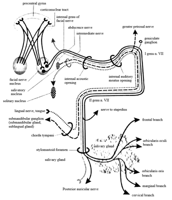
Fig. 1. The facial nerve course (3)
One of the most common children diseases is acute otitis media, the incidence of which decreases with the child’s age. However, the disease also affects adults, much more often men. Acute otitis media (AOM) may be determined by genetic factors, family episodes and anomalies in body’s anatomy (3-6).
Peripheral paralysis is the most common type of facial nerve palsy. It concerns the motor fibers of the facial muscles of the half of the face. As a result of such paralysis, the face loses its symmetry (7).
Facial nerve inflammation may occur as a result of viral infection, otitis media – in acute otitis media, the infection may spread to the facial nerve that runs on the medial tympanic cavity causing paralysis, while in chronic inflammation, the development of cholesteatoma or bone distortion may occur, ear polyps or granulation tissue – of mastoid process, parotid gland, and then as a result of the spread of inflammation to the nerve trunk. Paralysis may also occur as a result of stroke or intracranial tumors (7, 8).
The aim of the study is to analyze the diagnostic and therapeutic process in a patient with facial nerve palsy in the course of acute otitis media based on the analysis of the patient’s medical records and literature on the subject.
Case report
40-year-old patient reported to the Department of Otolaryngology, University Hospital in Bialystok because of left ear pain and impairment of facial muscles on the left side lasting for 3 days. The patient was not chronically ill and had no allergies. In case history, 7 days before otitis, the patient reported an upper respiratory infection with rhinitis.
Description of laryngological examination on admission
Nose: Vestibule free. The mucous membrane and turbinate without changes.
Mouth and throat: No change.
Neck: No change in palpation.
Face: Peripheral paresis of the nerve VII on the left side.
Ears: White, purulent, pulsating secretion from the left ear. LE eardrum retracted.
Weber test: Sound lateralizes to LE.
Rinne test: RE positive, LE negative.
Whispered voice test: RE: 6 m whisper, LE: 0 m whisper, 2 m speech.
CT scan of craniofacial area without contrast: The features of intracranial hemorrhage or pathological foci of hypodense in the brain perceptible by CT scan have not been demonstrated. Symmetrical ventricular system. Middle brain structures not displaced. Soft tissue masses/condensed contents mostly fill the mastoid cells on the left side of the temporal bone and the mastoid antrum and completely fill the tympanic cavity with obliteration of the ossicles. Thickening of the soft tissues of external left auditory canal. Segmental thinning of bone plates of the left pyramid of the temporal bone. Outer and middle ear structures on the right side regular. Inner ear structures on both sides without any noticeable changes. Thickening of the maxillary sinus mucosa.
Thoracic Rtg: Diaphragm and costophrenic angles. Lung fields without fresh focal lesions. Vascular hila. Mediastinal contours in Rtg regular.
Tests analysis before the surgery
While interpreting test results, it has been stated that the level of hearing is better in the right ear. The left ear has impaired conductive hearing.
While analyzing the results of tuning fork tests, left ear conductive hearing loss has been confirmed. After the Weber test, the sound lateralized to the left ear, and in the result of Rinne test was positive for the right ear and negative for the left ear.
Whispered voice test showed that the patient heard the whisper with his right ear at a distance of 6 meters, while with his left ear he did not hear the whisper.
On 19/03/2018 left ear paracentesis in local anesthesia has been performed, purulent contents evacuated, material for bacteriological examination taken. After the surgery general condition of the patient was good. The next day, the incision has been widened, and the ear cleared of purulent contents.
After the surgery, patient stayed in control isolation, peripheral puncture and analgesics, antibiotics, steroids have been administered, as well as dry dressing has been applied to the ear.
Growth of Streptococcus pyogenes has been obtained from the ear culture, patient received an antibiotic consistent with the antibiogram.
Powyżej zamieściliśmy fragment artykułu, do którego możesz uzyskać pełny dostęp.
Mam kod dostępu
- Aby uzyskać płatny dostęp do pełnej treści powyższego artykułu albo wszystkich artykułów (w zależności od wybranej opcji), należy wprowadzić kod.
- Wprowadzając kod, akceptują Państwo treść Regulaminu oraz potwierdzają zapoznanie się z nim.
- Aby kupić kod proszę skorzystać z jednej z poniższych opcji.
Opcja #1
29 zł
Wybieram
- dostęp do tego artykułu
- dostęp na 7 dni
uzyskany kod musi być wprowadzony na stronie artykułu, do którego został wykupiony
Opcja #2
69 zł
Wybieram
- dostęp do tego i pozostałych ponad 7000 artykułów
- dostęp na 30 dni
- najpopularniejsza opcja
Opcja #3
129 zł
Wybieram
- dostęp do tego i pozostałych ponad 7000 artykułów
- dostęp na 90 dni
- oszczędzasz 78 zł
Piśmiennictwo
1. Latkowski JB: Otolaryngologia. Podręcznik dla studentów i specjalizujących się lekarzy. PZWL Wydawnictwo Lekarskie, Warszawa 2017.
2. Byczkowska-Lipińska L: Mechanizmy biologiczne jako systemy przetwarzania i transmisji danych. Automatyka 2010; 14(3/1): 403-409.
3. Kuczkowski J: Metody badań i rehabilitacji w otorynolaryngologii. Harmonia Universalis (Grupa Wydawnicza Harmonia), Gdańsk 2018.
4. Janczewski G, Latkowski JB, Olszewski J, Kosiek K: Algorytmy diagnostyki i postępowania w otolaryngologii. Podręcznik dla lekarzy rodzinnych i pozostałych lekarzy podstawowej opieki zdrowotnej. Termedia Wydawnictwa Medyczne, Poznań 2010.
5. Niemczyk K, Jurkiewicz D, Składzień J et al.: Otolaryngologia kliniczna. Tom 2. Medipage, Warszawa 2015.
6. Kuczkowski J: Aktualne problemy w rozpoznawaniu i leczeniu ostrego i wysiękowego zapalenia ucha środkowego. Forum Medycyny Rodzinnej 2011; 5(4): 287-294.
7. Walowska J: Propozycja rehabilitacji porażonego nerwu twarzowego. Rehabilitacja w Praktyce 2014; 1(6): 27-28.
8. Szefler J, Głowacka P, Patalong-Ogiewa M: Kinesiology taping jako metoda wspierająca terapię ośrodkowego uszkodzenia nerwu VII. Annales Academiae Medicae Silesiensis 2012; 66(1): 73-76.
9. Radzikowski A: Ostre zapalenie ucha środkowego. Lekarz Rodzinny 2011; 3(163): 292-305.
10. Brydak LB: Grypa chorobą rodziny. Family Medicine & Primary Care Review 2011; 13(2): 281-286.
11. Arnold W, Ganzer U (red. nauk. I wyd. pol. Kręcicki T): Checklist Otolaryngologia. MedPharm Polska, Wrocław 2014.
12. Hassman-Poznańska E: Ostre zapalenie ucha środkowego. Polski Przegląd Otorynolaryngologiczny 2012; 3(1): 210-214.
13. Drożdżyńska M, Sobieraj-Garbiak I, Chlasta A, Jastrzębska M: Toksyna botulinowa i jej zastosowanie w medycynie. Diagnostyka Laboratoryjna 2015; 51(2): 139-146.
14. Morales RC: Ustno-twarzowa terapia regulacyjna. Wydawnictwo Fundacja Promyk Słońca, Wrocław 2009.
15. Jaraczewska E: Kinesiotaping i jego zastosowanie w programach rehabilitacyjnych – opis przypadków. Rehabilitacja w Praktyce 2009; 4: 23-27.

