Maciej Pilch, Anna Tuszyńska, Karolina Raczkowska-Łabuda, *Lidia Zawadzka-Głos
A paediatric patient with nasal septal perforation – symptoms, diagnosis and treatment options
Pacjent pediatryczny z perforacją przegrody nosa – objawy, diagnostyka, możliwości leczenia
Department of Paediatric Otolaryngology, Medical University of Warsaw, Poland
Head of Department: Associate Professor Lidia Zawadzka-Głos, MD, PhD
Streszczenie
Wstęp. Przegroda nosa jest anatomiczną strukturą, która nie tylko oddziela od siebie obie jamy nosa, ale jest elementem podporowym dla nosa zewnętrznego i wpływa na fizjologię nosa. Defekt w budowie przegrody nosa – skrzywienia i ubytki – wpływają na strumień przepływu powietrza przez jamy nosa. Wystąpienie perforacji przegrody nosa jest jednym z czynników, które powodują upośledzenie drożności nosa na drodze zmian wtórnych. W zależności od zgłaszanych dolegliwości i zaawansowania zmian w obrębie przegrody nosa istnieją różne możliwości leczenia. Wybór sposobu leczenia jest dobierany indywidualnie dla każdego pacjenta.
Cel pracy. Celem tej pracy jest omówienie problemów, z jakimi zgłasza się pacjent, metod diagnostycznych oraz przedstawienie możliwości leczenia dzieci z perforacją przegrody nosa. Analizie poddano pacjentów Kliniki Otolaryngologii Dziecięcej Warszawskiego Uniwersytetu Medycznego.
Materiał i metody. Praca przedstawia przypadki 18 pacjentów w wieku 3-17 lat przyjętych w Poradni Laryngologicznej w Klinice Otolaryngologii Dziecięcej Warszawskiego Uniwersytetu Medycznego.
Wyniki. W latach 2011-2017 do Kliniki zgłosiło się 18 pacjentów w wieku 3-17 lat, średnia wieku to 12,83 roku, SD – 4,34. Najczęściej perforacja przegrody nosa występowała jako powikłanie po zabiegu septoplastyki (4) oraz po przebytym urazie nosa. Najczęstsze objawy, z jakimi zgłaszali się pacjenci, to krwawienia z nosa oraz strupienie. U około 1/3 pacjentów perforacji przegrody nosa towarzyszyło zniekształcenie nosa zewnętrznego.
Wnioski. W literaturze naukowej jest niewiele doniesień na temat pacjentów pediatrycznych z perforacją przegrody nosa. Wszystkie uzyskane informacje zostały przeanalizowane na podstawie doniesień dotyczących pacjentów dorosłych i pediatrycznych. Występujące objawy miały różne nasilenie w zależności od etiologii.
Summary
Introduction. Nasal septum is an anatomical structure which not only separates both nasal cavities, but is also a supporting element for the external nose, and has an impact on its physiology. Structural abnormalities of the nasal septum, such as deviations and defects, affect the airflow in the nasal cavities. Nasal septal perforation is one of the factors that impair nasal patency due to secondary pathological changes. There are various treatment options, depending on the symptoms reported and the stage of septal lesions. Treatment strategy is selected individually for each patient.
Aim. The aim of this paper was to discuss the symptoms, diagnostic methods and treatment options in children with nasal septal perforation. Patients of the Department of Paediatric Otolaryngology, Medical University of Warsaw, were included in the analysis.
Material and methods. We present cases of 18 patients aged between 3 and 17 years, admitted to the Outpatient ENT Clinic at the Department of Paediatric Otolaryngology, Medical University of Warsaw.
Results. A total of 18 patients aged between 3 and 17 years (mean age 12.83 years, SD = 4.34) reported to our clinic. In most cases, nasal septal perforation developed as a complication of septoplasty (4) or nasal trauma. Nasal bleeding and crusting were the most commonly reported symptoms. Perforation was accompanied by external nasal deformity in about 1/3 of patients.
Conclusions. Only few reports on paediatric patients with nasal septal perforation may be found in literature. All data obtained was analysed based on reports describing adult and paediatric patients. The severity of symptoms varied depending on the aetiology.
Introduction
Nasal septum is an anatomical structure composed of membranous, cartilaginous and osseous elements. Soft tissues dominate in a child’s nose. However, during the craniofacial development, nasal bones elongate and the surface area of the vomer and the ethmoid bone increases. The nasal vasculature is very abundant. The nasal septum is supplied by the septal branches: anterior and posterior ethmoidal branches, as well as by the septal branches arising from the palatosphenoidal artery, which is the largest artery supplying the nasal septum. These vessels form a multilayer arterial network of the nasal septal mucosa (1).
The external nose is a very complex anatomical structure. The nasal root is located superiorly on the border with the glabella. Below lies the nasal dorsum, which is made up of bony parts, i.e. nasal bones supported by the vertical plate of the ethmoid and the vomer, as well as cartilaginous parts, i.e. the lateral nasal cartilages, which connect to the trapezoid cartilage on the nasal dorsum. The lower portion of the external nose consists of the nasal apex, nasal wings, columella and anterior nostrils. The main cartilage that builds this part of the nose is the greater alar cartilage, which gives rise to lateral crura located within the nasal wings; and the medial crura connect to the nasal septum, forming the columella and providing shape, support and projection to the nasal apex. The cartilage of the nasal septum also plays an important role in the structure of the external nose. It is the main support structure for the nasal apex and the cartilaginous dorsum (2).
Nasal septal perforation is very rare in children. The mechanism underlying this type of damage involves ischaemia of the mucoperichondrial-periosteal flaps. If both sides are equally affected for a sufficiently long time, this may lead to irreversible changes in the nasal mucosa, triggering transformations that cause damage followed by defects in the cartilage or, less often, both cartilage and bone of the nasal septum.
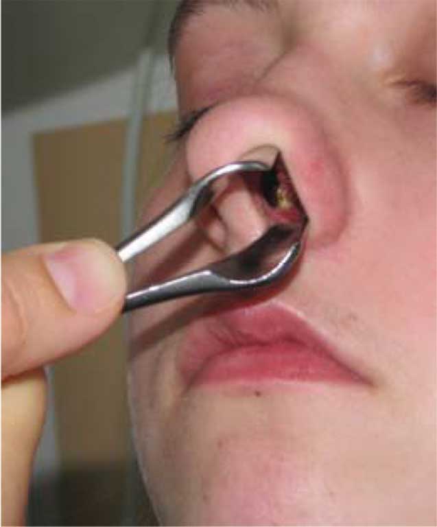
Symptoms that may be seen in these patients include e.g. itching in the nasal cavity, crusting, recurrent nasal bleeding, nasal obstruction, wheezing, and secretions moving down the back of the throat. In younger children, some of the symptoms may be identified only by parents or during a thorough ENT examination (fig. 1).
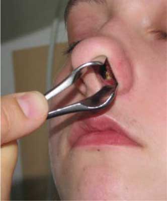
Fig. 1. Nasal septal perforation in an otolaryngological examination
A thorough medical history is important for accurate diagnosis. Data on patient’s habits, such as nose picking, abuse of decongestants, use of irritants or nasal drugs, should be collected. It is also worth inquiring about past nasal injuries, nasal bone fractures, minor nasal surgical procedures and operations, episodes of anterior nasal packing, as well as potential foreign body impaction in the nasal cavity.
Proper development and growth of the nasal septum not only determine the structure and appearance of the external nose, but they also play a role in the development of adjacent structures, such as the maxilla and the premaxilla. They are also important for maintaining facial proportions and aesthetics.
Aim
The aim of this paper was to present cases of patients aged up to 18 years, who reported to our Clinic in the Department of Paediatric Otolaryngology, Medical University of Warsaw due to nasal septal perforation for further diagnosis and treatment. We compared the causes of nasal septal defects, as well as analysed the symptoms presented by these patients.
Material and methods
We analysed medical records of patients admitted to the Department of Paediatric Otolaryngology, Medical University of Warsaw, with the diagnosis of nasal septal perforation. These were records from an ENT examination in our Clinic and qualification for surgical treatment.
Results
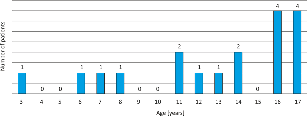
A total of 18 patients aged 3 to 17 years (mean age 12.83 years, SD = 4.34) were reported to our ENT clinic in the Department of Paediatric Otolaryngology, Medical University of Warsaw in the years 2011-2017 (fig. 2). There were 12 boys and 6 girls (fig. 3).

Fig. 2. The number of patients in different age groups
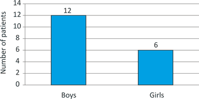
Fig. 3. The number of patients in gender groups
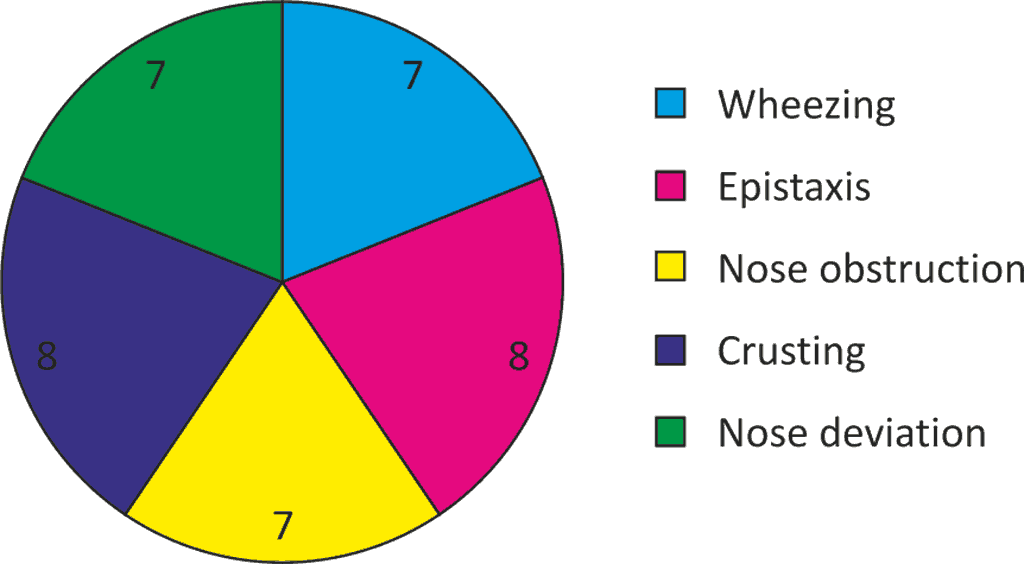
The patients most often reported nasal bleeding (n = 8) and crusting (n = 8), followed by nasal obstruction (n = 7) and nasal wheezing (n = 7). Two patients reported no symptoms. Altered shape of the external nose was an important aspect assessed during otolaryngological examination to select appropriate treatment modality (n = 7) (fig. 4).

Fig. 4. Symptoms
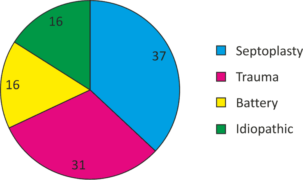
The most common causes of nasal septal perforation in the study group included iatrogenic septal perforation as a complication after septoplasty (37%) and nasal injuries (31%). Other, less common, though equally important, causes of nasal septal defects included foreign body (a battery) impaction (17%) and spontaneous nasal septal perforation, which occurred without any apparent reason (17%) (fig. 5).

Fig. 5. Causes of nasal septal perforation
Discussion
Very few reports on nasal septal perforation in paediatric patients may be found in the literature. We therefore based our analysis and conclusions mostly on scientific reports on adult patients, which were extrapolated to patients aged up to 18 years. The analysis of the possible causes underlying nasal septal perforation in our patients showed that these were mostly iatrogenic complications (68%), complications after septoplasty (37%) or after nasal injury (31%). Both these situations involve the use of anterior nasal packing, which exerts compression on the nasal septal mucosa. Under these circumstances, patient’s gender or age do not play an important role. However, when considering a history of injury in relation to gender, nasal septal perforation was more common in boys (5:1) than in girls. Septoplasty or nasal trauma may be complicated by septal haematoma, and, consequently, bacterial infection in the form of septal abscess. This type of complication can lead to perforation (4).
Batteries are the most dangerous foreign bodies found in the nasal cavity. It just takes a short time for the current flowing through the septal structures to cause damage, i.e. necrosis of the mucosa and deeper tissues. The electrolyte in the battery, which causes mucosal and vascular damage through chemical burns, also contributes to this process. Patients with a foreign body in the nasal cavity included in our analysis were aged between 3 and 8 years, and batteries accounted for 100% of these cases. It is therefore necessary to pay particular attention to the possibility of foreign body impaction in the nasal cavity in the youngest patients, and, in the case of an impacted battery, remove the object immediately to prevent further mucosal damage (3).
Each patient should receive appropriate treatment depending on the aetiology of nasal septal perforation. Patients with perforation of an unknown aetiology should be referred to a rheumatology outpatient clinic for extended diagnosis to exclude systemic diseases (lupus, sarcoidosis, granulomatosis with vasculitis) (1). Patients should be also informed about the need to use nasal moisturisers or oils and, in the case of bacterial infection, local or (in extremely rare cases) systemic antimicrobial therapy (1, 5, 6).
Conclusions
The symptoms reported by our patients were mild-to-moderate and improved after conservative treatment. However, due to insufficient patient’s satisfaction, a decision was made to include patient-tailored surgical treatment. Rhinoplasty is indicated in each patient with external nose deformity to restore the supporting structure and proper external nasal shape. Surgical nasal septal reconstruction is of particular importance in the youngest patients. Appropriate and well-tailored surgical treatment is the best method to allow for the proper growth of the midface, which depends on the development of nasal septum (3).
Piśmiennictwo
1. Kridel RWH, Foda HMT: Prevention, Management and Repair of Nasal Septal Perforation. [In:] Papel I (ed.): Facial Plastic and Reconstructive Surgery. 3rd ed. Thieme, New York 2008: 663-666.
2. Aleksandrowicz R, Ciszek B: Anatomia kliniczna głowy i szyi. Wyd. I, 3 dodruk. PZWL, Warszawa 2018.
3. Gryczyńska D (red.): Otolaryngologia dziecięca. alfa-medica press, Bielsko-Biała 2007.
4. Dąbrowska-Bień J, Skarżyński P: Complications in septoplasty based on a large group of 5639 patients. Eur Arch Otorhinolaryngol 2018; 275(7): 1789-1794.
5. Mullaces M: Management of nasal septal perforation using silicone nasal septal button. Acta Otorhinolaryngol Ital 2006; 26(4): 216-218.
6. Papay FA, Eliachar I, Risica R, Carroll M: Large Septal Perforation Repair Using Inferior Turbinate Sliding Advancement Flap. Am J Rhinol 1989; 3(4): 185-189.