Radosław Cylke1, Magdalena Kwapisz1, Agata Ostaszewska1, *Małgorzata Kołodziejczak1, 2
Reconstruction of anal sphincter muscles in post-traumatic sphincter damage with fecal incontinence – case report
Rekonstrukcja mięśni zwieraczy odbytu w pourazowym uszkodzeniu zwieraczy z nietrzymaniem stolca – opis przypadku
1Department of General Surgery and Transplantology, University Clinical Center, Medical University of Warsaw, Infant Jesus Clinical Hospital, Warsaw
2Warsaw Proctology Center, St. Elisabeth Hospital, Mokotów Medical Center, Warsaw
Streszczenie
Pozapołożnicze urazy okolicy odbytu należą do rzadkości. Przyczyną izolowanych uszkodzeń są najczęściej wypadki podczas pracy przy maszynach gospodarczych lub urazy typu wbicia na pal zadane przez ostre przedmioty, a także wypadki komunikacyjne. Autorzy przedstawiają rzadki przypadek młodego mężczyzny, który doznał przed 8 laty rozległego urazu zmiażdżeniowego miednicy, z oderwaniem odbytnicy oraz rozległymi urazami okolicy ud i pośladków z odwarstwieniem płata skórno-tłuszczowego w przebiegu wypadku komunikacyjnego. W ośrodku chirurgii dziecięcej wykonano wówczas stabilizację złamań oraz wytworzono kolostomię, którą zamknięto w 15. roku życia pacjenta. Z powodu utrzymujących się objawów nietrzymania stolca mężczyznę zakwalifikowano do późnej rekonstrukcji zwieraczy. Przed operacją wykonano szczegółową diagnostykę obrazową. Po 6 miesiącach od zabiegu pacjent deklarował znaczną poprawę w zakresie jakości życia oraz trzymania stolca (3/20 punktów w skali oceny zaawansowania nietrzymania stolca według Wexnera). Po całkowitym zagojeniu rany pacjentowi zalecono elektrostymulację zwieraczy. Autorzy konkludują, że nie ma jednoznacznych wytycznych dotyczących postępowania u pacjentów z urazami odbytnicy i odbytu. Leczenie powinno być zindywidualizowane dla każdego przypadku. W sytuacji zaopatrywania uszkodzenia aparatu zwieraczowego w trybie ostrym kluczowa jest weryfikacja, czy uraz nie obejmuje również narządów wewnątrzotrzewnowych, gdyż determinuje to dalsze postępowanie i ewentualną możliwość wykonania pierwotnej definitywnej operacji rekonstrukcyjnej. Dla pomyślnej funkcjonalności zwieraczy istotne jest także pooperacyjne wdrożenie ćwiczeń wzmacniających mięśnie, terapii „biofeedback” i elektrostymulacji.
Summary
Extra-obstetric injuries to the anal area are rare. The cause of isolated damage is most often accidents when working with utility machines or impalement injuries caused by sharp objects, as well as transport accidents. The authors present a rare case of a young man who, 8 years ago, suffered an extensive pelvic fracture injury, with rectal detachment, and extensive injuries to the thigh and buttock area with skin-fat lobe detachment in the course of a transport accident. At the pediatric surgery center, fracture stabilization was performed and colostomy was created, which was closed when patient completed 15 years of age. Due to persistent symptoms of fecal incontinence, the man was qualified for a late reconstruction of sphincters. Prior to the operation, detailed imaging diagnostics were performed. 6 months after the procedure, the patient declared significant improvements in quality of life and fecal retention (3/20 points on the Wexner Faecal Incontinence Quality of Life Scale). After complete healing of the wound, the patient was prescribed sphincter electrostimulation. The authors conclude that there are no clear guidelines for the management of patients with rectal and anal injuries. Treatment should be individualized on a case-by-case basis. In the situation of supplying the acute damage to the sphincter apparatus, it is crucial to verify that the injury does not also include the intraperitoneal organs, as this determines further proceedings and a possibility of primary definitive reconstructive surgery. For the successful functionality of sphincters, it is also important to implement muscle-strengthening exercises, “biofeedback” therapy and electrostimulation.

Introduction
Extra-obstetric injuries to the anal area are rare. The cause of isolated damage is most often accidents when working on utility machines or impalement injuries caused by sharp objects, as well as transport accidents. They may also be the result of sexual practices or offences and iatrogenic damage during endoscopic, radiological or surgical procedures. For the most part, however, they are accompanied by other injuries to the skeletal system and to pelvic organs, abdomen or even lower extremities. Diagnosis and treatment in each case therefore requires an individual approach. Based on the extent of the damage found, it is necessary to choose a treatment strategy that will ensure an effective supply of the primary injury and avoid complications in the form of fecal incontinence. The authors present a rare case of a young man who suffered a pelvic and perineal injury with damage to the anal sphincter apparatus as a result of a transport accident.
Case report
A nineteen-year-old man, a high school student with post-traumatic anal sphincter muscle damage, was admitted for elective reconstructive surgery to the Department of General Surgery and Transplantation Hospital of the Infant Jesus Clinical Hospital.
8 years before, the patient suffered extensive pelvic fracture injury, with rectal detachment and extensive injuries to the thighs and buttocks area with skin-fat lobe detachment in the course of a transport accident (cyclist in collision with a truck). At the pediatric surgery center, fracture stabilization was performed and colostomy was created. Subsequently, the approximate area of the thighs and buttocks with tissue defect was provided with a body fat transplant into the sacral region. At the age of 15, a colostomy was closed in the patient and the continuity of the gastrointestinal tract was restored. In the physical proctological examination, extensive trauma scars were found in the xlack of sphincter muscle systolic function (fig. 1). Because of persistent symptoms of gas and fecal incontinence (16/20 points on the Wexner Faecal Incontinence Quality of Life Scale, tab. 1) he was qualified for corrective surgery.
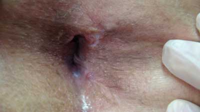
Fig. 1. Anus image before sphincter reconstructive surgery
Tab. 1. Pre-operative assessment of the Wexner Fecal Incontinence Quality of Life Scale
| Incontinence type | Frequency |
| | Never | Rarely
(< 1 once a week) | Sometimes
(≥ 1 once a week) | Often
(< 1 once a day) | Always
(≥ 1 once a day) |
| Solid stool | 0 | 1 | 2 | 3 | 4 |
| Loose stool | 0 | 1 | 2 | 3 | 4 |
| Gas | 0 | 1 | 2 | 3 | 4 |
| Necessity to wear a diaper | 0 | 1 | 2 | 3 | 4 |
| Lifestyle change | 0 | 1 | 2 | 3 | 4 |
Preoperatively, transrectal and transperineal ultrasound examination showed the loss of the internal sphincter continuity on 1/2 of the posterior circuit in its mid- and distal parts and the loss of the external sphincter continuity on the posterior and posterior-left circuits in the distal part. Systolic function in the dynamic study was not shown.
Before surgical intervention, the large intestine was typically prepared. The patient received antibiotic prophylaxis – cefazoline 1 g i.v. During the operation, a scar on the posterior anal periphery was cut immobilizing the stumps of the external sphincter muscle. Stumps were prepared and partially mobilized, stating a defect of approximately 40% of the muscle periphery. Then the muscle stumps were stitched with absorbable sutures, and above, a partial closure of the anoderm was performed. At the perimeter, the wound was left open for drainage. The perioperative course was uncomplicated. In the postoperative period without increased temperature, slight pain complaints (treatment used: paracetamol 4 x 1 g, metamisole 4 x 1 g, ketoprofen 2 x 100 mg). Due to the failure to exclude the functioning of the operated gastrointestinal tract (the patient was reconstituted digestive continuity 4 years earlier), it was decided to use prolonged antibiotic therapy and strict diet up to the 4th day after surgery. After completion of the healing process, the patient was prescribed daily electrostimulation of pelvic floor muscles and anal sphincters using a rectal electrode.
Powyżej zamieściliśmy fragment artykułu, do którego możesz uzyskać pełny dostęp.
Mam kod dostępu
- Aby uzyskać płatny dostęp do pełnej treści powyższego artykułu albo wszystkich artykułów (w zależności od wybranej opcji), należy wprowadzić kod.
- Wprowadzając kod, akceptują Państwo treść Regulaminu oraz potwierdzają zapoznanie się z nim.
- Aby kupić kod proszę skorzystać z jednej z poniższych opcji.
Opcja #1
29 zł
Wybieram
- dostęp do tego artykułu
- dostęp na 7 dni
uzyskany kod musi być wprowadzony na stronie artykułu, do którego został wykupiony
Opcja #2
69 zł
Wybieram
- dostęp do tego i pozostałych ponad 7000 artykułów
- dostęp na 30 dni
- najpopularniejsza opcja
Opcja #3
129 zł
Wybieram
- dostęp do tego i pozostałych ponad 7000 artykułów
- dostęp na 90 dni
- oszczędzasz 78 zł
Piśmiennictwo
1. Dudding TC, Vaizey CJ, Kamm MA: Obstetric anal sphincter injury: incidence, risk factors, and management. Ann Surg 2008; 247(2): 224-237.
2. Herzig DO: Care of the patient with anorectal trauma. Clin Colon Rectal Surg 2012; 25(4): 210-213.
3. Jeganathan AN, Cannon JW, Bleier JIS: Anal and Perineal Injuries. Clin Colon Rectal Surg 2018; 31(1): 24-29.
4. Kołodziejczak M, Kosim A, Sudoł-Szopińska I: Przyczyny, diagnostyka i leczenie poporodowych uszkodzeń zwieraczy odbytu. Ginekologia po Dyplomie 2013; 15(2): 59-65.
5. Sultan A, Fernando R: Risk factors and management of obstetric perineal injury. Obstetrics, Gynaecology & Reproductive Medicine 2007; 17(8): 238-243.
6. Kołodziejczak M, Ciesielski P: Atlas technik operacyjnych w proktologii. Borgis, Warszawa 2019.
7. Buppasiri P, Lumbiganon P, Thinkhamrop J et al.: Antibiotic prophylaxis for third- and fourth-degree perineal tear during vaginal birth. Cochrane Database Syst Rev 2014; (10): CD005125.
8. Mazur-Bialy AI, Kolomanska-Bogucka D, Oplawski M et al.: Physiotherapy for Prevention and Treatment of Fecal Incontinence in Women-Systematic Review of Methods. J Clin Med 2020; 9(10).


