© Borgis - New Medicine 4/2014, s. 139-141
*Konrad Wroński1,2
Marjolin’s ulcer of the thigh after burn injury – case report and review of literature
1Department of Oncology, Faculty of Medicine, University of Warmia and Mazury, Olsztyn, Poland
Head of Department: prof. Sergiusz Nawrocki, MD, PhD
2Department of Surgical Oncology, Hospital Ministry of Internal Affairs with Warmia and Mazury Oncology Centre, Olsztyn, Poland
Head of Department: Andrzej Lachowski, MD
Summary
Marjolin’s ulcer is defined as a tumor which is arising from a chronic wound, scar or chronic inflammation. It is a rare chronic cutaneous ulcer which is often malignancy. The majority of malignancy in Marjolin’s ucers are squamous cell carcinomas. Rarely described malignancies in Marjolin’s ulcers are basal cell carcinoma, melanoma, adenocarcinoma, liposarcoma, fibrosarcoma and osteogenic sarcoma.
The author of this article present a case of a 70-year-old Caucasian man who was admitted to the hospital because of Marjolin’s ulcer. The author performed a literature review on Marjolin’s ulcer diagnosis and treatment.
INTRODUCTION
Marjolin’s ulcer is defined as a tumor which is arising from a chronic wound, scar or chronic inflammation (1, 2). It is a rare chronic cutaneous ulcer which is often malignancy (1, 2). The majority of malignancy in Marjolin’s ucers are squamous cell carcinomas. Rarely described malignancies in Marjolin’s ulcers are basal cell carcinoma, melanoma, adenocarcinoma, liposarcoma, fibrosarcoma and osteogenic sarcoma (3).
CASE REPORT
A 70-year-old Caucasian man was referred to the Department of Surgical Oncology Hospital Ministry of Internal Affairs with Warmia and Mazury Oncology Centre in Olsztyn due to Marjolin’s ulcer of the right tight (fig. 1). In an interview with the patient, he had the burn injury 15 years ago and from 3 years he observed tumor which was arising from this chronic wound. Marjolin’s ulcer in histopathologically examination was: squamous cell carcinoma G2. CT scan showed no infiltration carcinoma cells on important important nerve or vascular structures or bone. The patient had no any other symptoms, there was no history of weight loss and loss of appetite. The patient was treated chronically for hypertension and benign prostatic hyperplasia (BPH). He had no surgeries and there was no history of carcinoma in patient family. Blood test and other routine hematological examinations and biochemical tests were within normal limits.
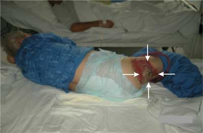
Figure 1. Clinical photograph of Marjolin’s ulcer on the right tight from burn injury.
The chronic wound after burn injury had diameter 20 centimeters. In the central part of this wound was located Marjolin’s ulcer diameter 11 centimeters (fig. 2). Regional lymph nodes in palpable examination were not enlarged.
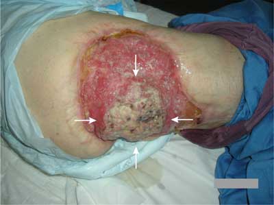
Figure 2. The chronic wound on the right tight had diameter 20 centimeters and in the central part of chronic wound was located Marjolin’s ulcer diameter 11 centimeters.
The patient was admitted to the Department of Surgical Oncology to wide local excision of Marjolin’s ulcer. The patient was informed about the possibility of treating Marjolin’s ulcer and possible complications associated with surgery. The patient refused surgery and decided that he wanted to undergo radiotherapy treatment. The patient was transferred for further treatment to the Department of Radiotherapy Hospital Ministry of Internal Affairs with Warmia and Mazury Oncology Centre in Olsztyn.
DISCUSSION
French surgeon Jean-Nicholas Marjolin first time described ulceration in a burn scar in 1828 (4). Dupuytren first time noted malignancy in „Marjolin’s ulcer” (5). DaCosta first time described forming malignant tumors inside „Marjolin’s ulcer” (6).
The mechanism of malignant transformation of Marjolin’s ulcer is still unclear (7). Some theories consider that the toxin, chronic irritation and inflammation, ultraviolet rays and environmental interaction affected the malignant transformation (7).
The majority of Marjolin’s ulcers are observed on the extremities in 60%, on the head and face in 30% and on the trunk in 10% of cases (5). The reason for such anatomical site predilection is still not understood. There are also described cases of Marjolin’s ulcers in the genitalia and the breast skin after burn (8, 9).
There are described two variants of Marjolin’s ulcer. An acute form in which malignant transformation is observed within 12 months from the date of the injury or the inflammation process. The chronic form is more frequent and malignancy developed slowly with average latency period of 36 years (10, 11). The interval period from injury/scar to malignant transformation is typically from 25 to 40 years (10). In studies from western countries malignant transformation in men are observed three times more frequently than in women (11).
Marjolin’s ulcers presents histopathologically in 75% to 96% of cases as squamous cell carcinoma (12). Rarely described malignancies in Marjolin’s ulcers are basal cell carcinoma, melanoma, adenocarcinoma, liposarcoma, fibrosarcoma and osteogenic sarcoma (3).
Most of Majrolin’s ulcers are mistaken for an infected wounds, scars or ulcerations. Symptoms such as: foul-smelling, painful with exudates and bloody drainage, non-healing ulcers with a rich network of blood vessels suggest malignant transformation (13).
Malignant cells presented in Marjolin’s ulcers are aggressive and have great tendency to metastasize than other types of skin cancers. Metastasis to the regional lymph nodes, brain, liver, lung or kidney are reported in different articles (14, 15).
The treatment of Marjolin’s ulcers should be multidisciplinary it means that it should be treated in oncological centers. Treatment include surgery, chemotherapy and radiotherapy (16). Surgical treatment of Marjolin’s ulcers include wide local excision, block dissection of the regional lymph nodes and amputation in advanced tumors (13, 16). Wide local excision together with skin grafting or primarily delayed is preferred in treatment of Marjolin’s ulcers. Surgical margin in wide local excision should be at least 20 mm (13, 16).
The prognosis in Marjolin’s ulcers depends on various factors. The most important factors are: age, size, grade and stage of tumor, metastases and presence of local recurrence. Long-term follow-up is recommended in all cases. Most series indicate that the incidence of recurrence after surgery is in the range of 20% to 50% (3). Prognosis for patients with Marjolin’s ulcer range from 65% to 75% for 3 year survival time. For patients with Marjolin’s ulcer with metastasis 3 year survival time is from 35% to 50% (17).
Prevention is better than cure and it is important that all chronic scars, wounds and ulcers should have multiple biopsies to avoid missing malignant ulcers (7, 18).
CONCLUSIONS
1. Marjolin’s ulcer is defined as a tumor which is arising from a chronic wound, scar or chronic inflammation.
2. The majority of malignancy in Marjolin’s ucers are squamous cell carcinomas.
3. Most of Majrolin’s ulcers are mistaken for an infected wounds, scars or ulcerations.
4. The treatment of Marjolin’s ulcers should be multidisciplinary it means that it should be treated in oncological centers.
5. All chronic scars, wounds and ulcers should have multiple biopsies to avoid missing malignant ulcers.
Piśmiennictwo
1. Agale SV, Kulkarni DR, Valand AG et al.: Marjolin’s Ulcer – a diagnostic dilemma. JAPI 2009; 57: 593-594. 2. Suhag V, Sunita, Singh S, Nimbrian VK et al.: Marjolin’s ulcer developing in electric burns: a rare case report. Pak J Med Sci 2005; 21(3): 375-376. 3. Hill BB, Sloan DA, Lee EY et al.: Marjolin’s ulcer of the foot caused by nonburn trauma. South Med J 1996; 89(7): 707-710. 4. Marjolin NJ: Ulcere. Dictionnaire de medicine 1828; 21: 31. 5. Treves N, Pack GT: The development of cancer in burn scars. Surg Gynecol Obstet 1930; 51: 749-782. 6. DaCosta JC: Carcinomatous changes in an area of chronic ulceration, or Marjolin’s ulcer. Ann Surg 1903; 37: 495-502. 7. Nthumba PM: Marjolin’s ulcers: theories, prognostic factorsand their peculiarities in spina bifida patients. World J Surg Oncol 2010; 8: 108. 8. Chintamani M, Shankar M, Singhal V et al.: Squamous cell carcinoma developing in the scar of Fournier’s gangrene – case report. BMC Cancer 2004; 274(1): 16. 9. Calikapan GT, Akan M, Karaca M et al.: Marjolin ulcer of the scalp: report of 5 cases and review of the literature. J Craniofac Surg 2008; 19: 1020-1025. 10. Wysocki WM, Komorowski A, Wojewoda T et al.: Two different cases of Marjolin’s ulcer and recommendations for practice. TOSJ 2010; 2: 83-85. 11. Davalos BA, Cortes-Flores AO, Bandera-Delago A: Malignant neoplasm in burn scar: Marjolin’s ulcer: report of two cases and review of the literature. Cir Cir 2008; 76: 329-331. 12. Ozek C, Celik N, Bilkay U et al.: Marjolin’s ulcer of the scalp: report of 5 cases and review of the literature. J Burn Care Rehabil 2001; 22: 65-69. 13. Malheiro E, Pinto A, Choupina M: Marjolin’s ulcer of the scalp: case report and literature review. Ann Burns Fire Disasters 2001; 14: 39-41. 14. Tutela RR Jr, Granick M, Benevenia J: Marjolin’s ulcer arising in pressure ulcer. Adv Skin Wound Care 2004; 17: 462-467. 15. Kowal-Vern A, Criswell BK: Burn scar neoplasms: a literature review and statistical analysis. Burns 2005; 31: 403-413. 16. Ochenduszkiewicz U, Matkowski R, Szynglarewicz B et al.: Marjolin’s ulcer: malignant neoplasm arising in scars. Rep Pract Oncol Radiother 2006; 11(3): 135-138. 17. Lack W, McKinley T.: Marjolin’s ulcer: incidental diagnosis of squamous cell carcinoma on hemipelvectomy for recalcitrant pelvic osteomyelitis. IOWA Orthop J 2010; 30: 174-176. 18. Nthumba PM.: Marjolin’s ulcer in sub-Sahara Africa. World J Surg 2010; 34: 2272-2277.
