© Borgis - New Medicine 4/2014, s. 115-117
Karolina Raczkowska-Łabuda, Lechosław P. Chmielik, *Lidia Zawadzka-Głos
Macroscopic evaluation of intraoperative changes in children with chronic sinusitis
Department of Pediatric Otolaryngology, Medical University of Warsaw, Poland
Head of Department: Lidia Zawadzka-Głos, MD, PhD
Summary
Introduction. The chronic sinusitis issue within the pediatric population is quite popular in the literature. The vide variety of information on microbiology, diagnosis or treatment are opposite to the relatively short experience of endoscopic surgery in pediatrics. This article is the FESS experience summary of Department of Pediatric Otolaryngology, Medical University of Warsaw.
Aim. Endeavour to intraoperative macroscopic evaluation of changes in children with CRSs. Analysis of the type and incidence of pathological changes observed macroscopically during FESS was undertaken as well as an parallel attempt to correlate the level of inflammation markers with severity of disease.
Material and methods. Retrospective analysis of case records of 44 patients of the Department of Pediatric Otolaryngology, Medical University of Warsaw, hospitalized in the year 2013. During that time 37 patients with a diagnosis of chronic maxillary sinusitis required surgical intervention. 33 children (average-aged 10.4 years) were qualified to functional endoscopic surgery. The youngest patient was 4 years old and the oldest 18. 11 patients presented the polypoid lesions of sinuses or nasals. At 9 confirmed the coexistence of sinus polyps with nasal polyps.
Results. 26 patients had oedematous-inflammatory lesions and 11 polypoid. Among the group of listed above 11 children, 9 had either sinuses or nasal polyps. A common deviation in the results of morphology in children with CRSs is higher level of monocytes. Inflammation markers within the population of FESS qualified children were low.
Conclusions. 1. Oedematous-inflammatory lesions are frequently intraoperatively identified. 2. The CRSs without polyps is the common type of chronic sinusitis in children. 3 Nasal polyps often coexist with sinus polyps. 4 Markers of inflammation in CRSs are low.
INTRODUCTION
The chronic sinusitis issue within the paediatric population is quite popular in the literature. The wide variety of informationes on microbiology, diagnosis or treatment are opposite to the relatively short experience of endoscopic surgery in pediatrics. Endeavour to intraoperative macroscopic evaluation of changes in children with CRSs. Analysis of the type and incidence of pathological changes observed macroscopically during FESS was undertaken as well as an parallel attempt to correlate the level of inflammation markers with severity of disease.
MATERIAL AND METHODS
Retrospective analysis of case records of 44 patients of the Department of Pediatric Otolaryngology, Medical University of Warsaw, hospitalized in the year 2013. During that time 37 patients with a diagnosis of chronic maxillary sinusitis required surgical intervention. 33 children (avg-aged 10.4 years) were qualified to functional endoscopic surgery. The youngest patient was 4 years old and the oldest 18. 11 patients presented the polypoid lesions of sinuses or nasals. At 9 confirmed the coexistence of sinus polyps with nasal polyps. There was one choanal polyp and onefold change of maxillary sinus. Furthermore analyzed the results of a patient’s blood test (morphology, CRP and ESR) searching for statistically significant deviations. Samples were taken not later than 7 days prior to surgery.
RESULTS
26 patients had oedematous-inflammatory lesions (fig. 1) and 11 polypoid (fig. 2, 3).
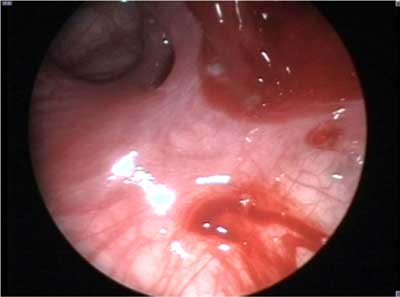
Fig. 1. Oedematous-inflammatory lesions nearby natural ostium of maxillary sinus.
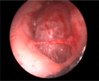
Fig. 2. Nasal polyp.
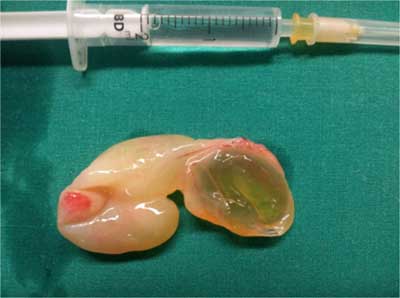
Fig. 3. Choanal polyp.
Among the group of listed above 11 children, 9 had either sinuses or nasal polyps. 30% children with polyps were diagnosed with cystic fibrosis. They were the youngest in the group of patients (4yrs, 4yrs, 8yr). A common deviation in the results of morphology in children with CRSs is higher level of monocytes. Inflammation markers within the population of FESS qualified children were low.
DISCUSSION
Chronic rhinosinusitis (CRS) in children is very challenging to diagnose and treat because the symptoms depend on age (1-3). According to the European Consensus on Rhinosinusitis (EP3OS) (4) CRS is defined by 2 or more symptoms lasting beyond 12 weeks. The symptoms are: facial pain/pressure, nasal blockage or nasal discharge (anterior/posterior), reduction or loss of smell. Other authors agreed that CRS in children is characterized by symptoms persisting beyond 8 weeks (5). Chronic illness have a negative impact on a child’s quality of life. It is connected with complications such as chronic antibiotic therapy, school absences, poor sleep patterns, impaired school performance, or irritability (6).
During normal childhood development the ethmoid and maxillary sinuses are usually present at birth. Most commonly, the sphenoid sinuses are pneumatized by age 5 years, and the frontal sinuses appear by age 7 years but are not completely developed until adolescence. Thus, children are predisposed to sinus infection at an early age. In young children, the most common sinuses involved are the ethmoid and maxillary sinuses (7).
Nasal polyps in the paediatric population may develop due to chronic inflammation of the mucous membrane caused by allergic rhinitis, chronic sinusitis, asthma, or aspirin allergy. Polyps, if present, should prompt an evaluation for cystic fibrosis, an allergy, GERD and drug’s intolerance syndroms (7, 8). Medical therapy should always be prior to surgical one and it continues to be prolonged courses of antibiotics. The vast majority of patients requires longterm conservative treatment. Staged surgical intervention with initial adenoidectomy followed by partial or anterior ethmoidectomy should be considered only when medical therapy failed (9, 10).
Functional endoscopic sinus surgery (FESS) was elaborated by Messerklinger and Wigand in Europe and performed via the ostiomeatal complex (10, 11). FESS has become the standard surgical treatment for chronic sinusitis. The external approaches are adjunct and used in tumor or foreign body management (12).
Conscious of the international guidelines, in the Department of Pediatric Otolaryngology Medical University of Warsaw, we perform endoscopic surgery after or parallel with adenoidectomy secondary to conservative treatment. Subjected to analysis intraoperatively evaluated changes of mucous membrane in children with a diagnosis of CRS.
CONCLUSIONS
1. Oedematous-inflammatory lesions are frequently intraoperatively identified.
2. The CRSs without polyps is the most common type of chronic sinusitis in children.
3. Nasal polyps often coexist with sinus polyps.
4. Markers of inflammation in CRSs are low.
Piśmiennictwo
1. Wu A, Shapiro N, Bhattacharyya N: Chronic Rhinosinusitis in Children: What are the Treatment Options? Immunol Allergy Clin N Am 2009; 29: 705-717. 2. Hamilos D: Chronic rhinosinusitis: Epidemiology and medical management. J Allergy Clin Immunol 2011; 128 (4): 693-707. 3. Poole MD: Pediatric endoscopic sinus surgery: the conservative view. Ear Nose Throat J 1994; 73: 221-27. 4. Fokkens W, Lund V, Mullol J: On behalf of the European Position Paper on Rhinosinusitis and Nasal Polyps group. 2007. Rhinol 2007; 20(Suppl): 1-136. 5. Smart B: Pediatric Rhinosinusitis and Its Relationship to Asthma and Allergic Rhinitis. Pediatric Asthma, Allergy & Immunology 2005; 18 (2): 88-98. 6. Kay DJ, Rosenfeld RM: Quality of life for children with persistent sinonasal symptoms. Otolaryngol Head Neck Surg 2003; 128(1): 17-26. 7. Barghouth G, Prior J, Lepori D et al.: Paranasal sinuses in children: size evaluation of maxillary, sphenoid, and frontal sinuses by magnetic resonance imaging and proposal of volume index percentile curves. Eur Radiol 2002; 12(6): 1451-1458. 8. Ariji Y, Kuroki T, Moriguchi S et al.: Age changes in the volume of the human maxillary sinus: a study using computed tomography. BIR 1994; 23(3). 9. Spaeth J, Krügelstein U, Schlöndorff G: The paranasal sinuses in CT-imaging: Development from birth to age 25. Int J Pediatr Otorhinolaryngol 1997; 1(14): 25-40. 10. Ramadan HH (Ed.): Medical Treatment of Pediatric Sinusitis Clinical Presentation. MedScape, 2014. 11. Chmielik M (Ed.): Otolaryngologia dziecięca. Akademia Medyczna w Warszawie 2004; (4): 30-37. 12. Hebert RL, Bent JP: Meta-analysis of outcomes of pediatric functional endoscopic sinus surgery. Laryngoscope 1998; 108: 796-99. 13. Wigand ME, Steiner W, Jaumann MP: Endonasal sinus surgery with endoscopical control: from radical operation to rehabilitation of the mucosa. Endoscopy 1978; 10(4): 255-60. 14. Messerklinger W: Endoscopy of the nose. Urban & Schwarzenberg, Baltimore 1978. 15. Patel A, Meyers AD (Ed.): Surgical Treatment of Chronic Maxillary Sinusitis Surgical Overview. MedScape, 2013.
