Agnieszka Kozubska1, *Joanna Szczepańska2
The effectiveness of formocresol amputation in deciduous molars – a retrospective study**
Skuteczność amputacji formokrezolowej w leczeniu miazgi zębów trzonowych mlecznych – badanie retrospektywne**
1Doctoral Studies, Department of Paediatric Dentistry, Medical University of Łódź
2Department of Paediatric Dentistry, Medical University of Łódź
Head of Department: Professor Joanna Szczepańska, MD, PhD
Streszczenie
Wstęp. Metodą leczenia próchnicowego obnażenia miazgi oraz nieodwracalnego częściowego zapalenia miazgi jest zabieg amputacji. W tym celu był dotychczas stosowany głównie formokrezol, który stanowił „złoty standard” leczenia chorób miazgi zębów mlecznych.
Cel pracy. Określenie skuteczności stosowania formokrezolu w zabiegu amputacji w zębach mlecznych.
Materiał i metody. Analizie retrospektywnej poddano dokumentację medyczną pacjentów urodzonych w 2006-2007 roku, u których wykonano zabiegi amputacji formokrezolowej w latach 2010-2012. Uzyskano zgodę Komisji Bioetycznej przy Uniwersytecie Medycznym w Łodzi o numerze RNN/137/18/KE oraz pisemne zgody pacjentów/ich rodziców/opiekunów prawnych.
Wyniki. W badaniu przeanalizowano zabiegi w 102 zębach mlecznych u 64 dzieci, w tym w 49 zębach pierwszych i drugich trzonowych górnych i 53 dolnych. W 78 przypadkach doszło do próchnicowego obnażenia miazgi, w pozostałych 24 stwierdzono nieodwracalne częściowe zapalenie miazgi bez widocznego odsłonięcia miazgi. U 37 osób wykonano po jednym zabiegu pulpotomii, u 19 pacjentów konieczne było zastosowanie leczenia amputacyjnego w 2 zębach, u 6 osób w 3 zębach, u 1 pacjenta w 4 zębach oraz u 1 pacjenta w 5 zębach trzonowych mlecznych. Zabieg pulpotomii został powtórzony w wyniku utraty wypełnienia szkło-jonomerowego w 19 przypadkach. Ogółem usunięto 63 zęby, w tym 25 o czasie, a 38 przedwcześnie. Podczas ostatniego przeprowadzonego badania na przełomie 2016/2017 roku 31 zębów spośród 102 było obecnych w jamie ustnej. Stwierdzono brak 8 zębów przy jednoczesnym braku stałego następcy. Analiza 102 przypadków wykazała średnio dwuipółletni okres utrzymania zębów w jamie ustnej po amputacji formokrezolowej.
Wnioski. Analiza retrospektywna zastosowania formokrezolu w zabiegu amputacji w zębach mlecznych wykazała skuteczność kliniczną tego preparatu, co pozwalało na utrzymanie w jamie ustnej zęba mlecznego dotkniętego stanem zapalnym miazgi przez okres od 2 do prawie 3 lat po zabiegu.
Summary
Introduction. The treatment of carious denudation of pulp and irreversible partial pulp inflammation is pulpotomy. In this method formocresol was used as the “gold standard”.
Aim. Assessment of the effectiveness of the use of formocresol in pulpotomy in deciduous teeth.
Material and methods. Formocresol amputation performed in 2010-2012 in children born in 2006-2007 were analysed. The consent of the Bioethical Committee at the Medical University of Łódź as well as written consent from all the subject/parents of all the subjects/legal guardians of all the subjects were obtained.
Results. In this research, the amputation in 102 deciduous teeth, including 49 first and second upper molars, and 53 lower molars, performed on 64 children were analysed. In 102 amputation cases 78 were caused by carious denudation of pulp and 24 by irreversible partial pulp inflammation without visible denudation of pulp. 37 patients underwent pulpotomy in 1 tooth, 19 patients needed it in 2 teeth, 6 patients in 3 teeth, 1 patient in 4 teeth, and 1 patient in 5 molars. The pulpotomy was repeated because of the glass-ionomer lost in 19 cases. In total 63 teeth were extracted, including 25 at the right time and 38 too early. During the last oral examination held 2016/2017, 31 out of 102 teeth were still on place. The lack of 8 teeth without the presence of permanent successors was reported. The analysis of 102 cases showed the average of 2.5 years period of the teeth maintenance following the formocresol pulpotomy.
Conclusions. Retrospective analysis of the use of formocresol in pulpotomy in deciduous teeth proved clinical success of this material, which provided the maintenance of the teeth with the pulpitis for 2 to 3 years after the amputation.

Introduction
One of the main tasks of paedodontics is maintenance of full primary dentition until its physiological resorption. Premature loss of primary teeth results in serious disorders in a stomatognathic system such as malocclusions, disorders in jaw development and an activity of a temporo-mandibular joint and a loss of a correct occlusion height (1, 2). It also contributes to speech disorders, mastication disorders and defects in appearance, which may contribute to complexes and lack of acceptance in the environment.
Negligence in child’s oral hygiene, insufficient education for parents, irregular visits at the dentist, a diet rich in carbohydrates, and the structure of primary teeth make them prone to the development of carious processes. In the case of carious or posttraumatic denudation of pulp, pulpotomy is conducted in order to prevent early extraction (3). Pulpotomy may be conducted when there is reversable or irreversible partial pulp inflammation. It is contraindicated when there are long spontaneous dental pains, bleedings difficult to staunch, and changes in periapical tissues (4). Amputation is completed by the extraction of chamber pulp with irreversible inflammation and covering vital pulp or pulp with reversible inflammation with a special agent (3).
One of the agents applied in such treatment is formocresol. A mixture of zinc oxide, eugenol and formocresol was first used in pulpotomy of deciduous teeth in 1930 by Sweet (5). Buckley’s formula consists of 19% of formocresol and 35% of cresol in a solution of 15% of glycerine and 31% of water. Diluted 1:5, it is placed in the chamber for 5 minutes, next the bottom of the chamber is covered with formocresol paste and cement, and a cavity is tightly filled in. When cooperation with a child is difficult or there are problems in obtaining hemostasis, a cotton ball with formocresol may be left in for seven days (4). Formocresol is antibacterial and mummifying, it has an impact on the activity of hyaluronidase (6). For over 80 years, it was a “golden standard” and used by most of the dentists. The effectiveness of using formocresol in pulpotomy was estimated 70-97% (7). Rolling and Thylstrup (8) proved that this effectiveness decreases with time.
Aim
The aim of the research was a retrospective analysis of an impact of formocresol amputation on the maintenance of primary molars in an oral cavity.
Material and methods
Retrospective research was conducted on the basis of a medical record at the Department of Paediatric Dentistry at the Medical University of Łódź, Poland. Access to the patients’ medical history was approved by the management of the Central Teaching Hospital of the Medical University of Łódź. In the article, no personal data of patients were revealed. The consent of the Bioethical Committee at the Medical University of Łódź as well as written consent from all the subject/parents of all the subjects/legal guardians of all the subjects were obtained.
This research covered formocresol amputations of primary molars in patients born in the years 2006 and 2007, i.e. between the age of 3 and 6, who had been treated with this method at the Department of Paediatric Dentistry in the years 2010-2012 with the consent of a parent/legal guardian. Cases had to meet the following criteria in order to be qualified for the analysis:
– carious denudation of pulp or partial irreversible pulp inflammation,
– available record of treatment of a given tooth until 2016/17 (or until exfoliation).
Criteria for the exclusion from the research were:
– incomplete record of treatment,
– total irreversible pulp inflammation,
– changes in periapical tissues,
– dental crown destroyed by caries, with no possibility of reconstruction.
In each case, amputation was conducted in accordance with current standards. After working on the cavity and chamber, a tampon with formocresol was placed in the chamber. In cases of pain and exudate, formocresol paste (formocresol + zinc oxide + eugenol) was put at the bottom of the chamber. Next, the chamber was filled in with polycarboxylic cement Adhesor and the cavity – with a glass-ionomeric material. A card of analysis of dental surgery success/failure contains the assessment of the following elements: reason for the surgery, time of tooth preservation in an oral cavity since the surgery, complications such as swelling, fistula, abscesses, pathological changes in X-ray imaging, spontaneous aches, fractures in a crown, which are reasons for premature extractions. Child’s age, at which extraction was completed, is also taken into account as well as the time between the surgery and extraction. The number of patients, who needed the surgery to be repeated due to the loss of the filling, and the number of teeth with amputation in one patient, were summed up.
Results
Formocresol amputation was performed in 242 teeth in 195 children in the Department of Paediatric Dentistry at the Medical University of Łódź in the years 2010-2012. The final analysis covers 102 teeth in 64 children. Other cases were excluded due to the incomplete history of treatment (until the eruption of permanent teeth or premature extraction).
Amputation was performed in 49 baby upper first and second molars and 53 lower ones. There were 78 cases of carious denudation of pulp and in the other 24 cases, irreversible partial pulp inflammation was observed without visible denudation of pulp.
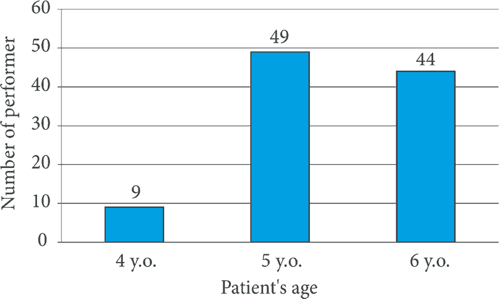
At the first step (putting a cotton ball with a therapeutic agent for seven days), formocresol was used in 78 cases, Cresophene – in 22 cases and Caustinerf – in two teeth. Nine amputations were performed in four-year old patients, forty-nine – in five-year old patients, and forty-four – in six-year old patients (fig. 1).
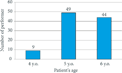
Fig. 1. Number of performer formocresol amputations in various age groups
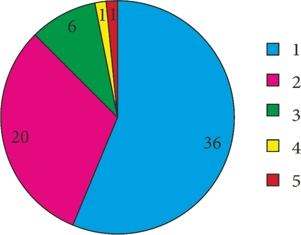
One pulpotomy procedure was performed in 37 people, 2 teeth needed to be amputated in 19 people, 3 – in 6 patients, 4 – in 1 patient and 5 baby molars in 1 patient (fig. 2). Pulpotomy was repeated as a result of a loss of glass-ionomeric filling in 19 cases.
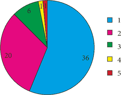
Fig. 2. Classification of patients according to the number of performer formocresol amputations from 1 to 5
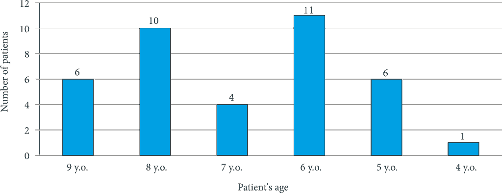
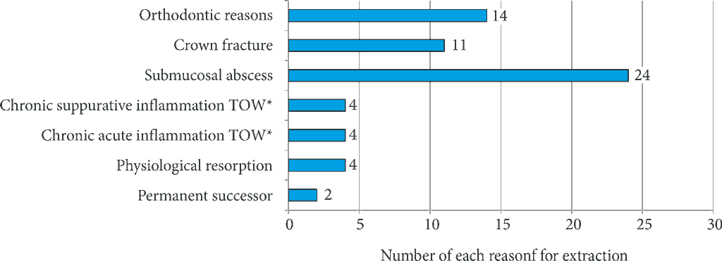
All in all, 63 teeth were pulled, including 25 on time: 14 as a result of development of a permanent tooth, 11 – due to physiological resorption. 38 teeth were extracted prematurely: 6 teeth in the 9th year of age, 10 teeth in the 8th year of age, 4 teeth in the 7th year of age, 11 teeth in the 6th year of age, 6 teeth in the 5th year of age, and 1 tooth in the 4th year of age (fig. 3). 24 among 38 teeth were referred for extraction due to symptoms characteristic to chronic acute periodontitis such as spontaneous ache or changes in periapical tissues in clinical or radiological assessment. In 4 cases, tooth crowns were fractured with no possibility of their restoration. Further 4 primary molars were removed due to a purulent exudate (found after removal of filling) or presence of fistulas (lat. periodontitis chronica purulenta). In four cases, submucosal abscess developed. Another two primary teeth were extracted due to orthodontic reasons (fig. 4). Furthermore, 8 teeth were missing without permanent successor yet there was no information about the reason for their extraction.
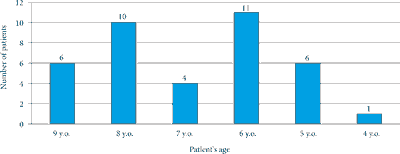
Fig. 3. Number of patients in accordance with age, in whom premature extraction was performed

Fig. 4. Reasons for the extraction if teeth in which formocresol amputation was performed
TOW – periapical tissue
Powyżej zamieściliśmy fragment artykułu, do którego możesz uzyskać pełny dostęp.
Mam kod dostępu
- Aby uzyskać płatny dostęp do pełnej treści powyższego artykułu albo wszystkich artykułów (w zależności od wybranej opcji), należy wprowadzić kod.
- Wprowadzając kod, akceptują Państwo treść Regulaminu oraz potwierdzają zapoznanie się z nim.
- Aby kupić kod proszę skorzystać z jednej z poniższych opcji.
Opcja #1
29 zł
Wybieram
- dostęp do tego artykułu
- dostęp na 7 dni
uzyskany kod musi być wprowadzony na stronie artykułu, do którego został wykupiony
Opcja #2
69 zł
Wybieram
- dostęp do tego i pozostałych ponad 7000 artykułów
- dostęp na 30 dni
- najpopularniejsza opcja
Opcja #3
129 zł
Wybieram
- dostęp do tego i pozostałych ponad 7000 artykułów
- dostęp na 90 dni
- oszczędzasz 78 zł
Piśmiennictwo
1. Godhi B, Sood PB, Sharma A: Effects of mineral trioxide aggregate and formocresol on vital pulp after pulpotomy of primary molars: an in vivo study. Contemp Clin Dent 2011; 2: 296-301.
2. Pimenta HC, Borges AH, Bandeca MC et al.: Antimicrobial activity of filling materials used in primary teeth pulpotomy. J Int Oral Health 2015; 7: 54-57.
3. Olatosi OO, Sote EO, Orenuga OO: Effect of mineral trioxide aggregate and formocresol pulpotomy on vital primary teeth: a clinical and radiographic study. Niger J Clin Pract 2015; 18: 292-296.
4. Szczepańska J, Olczak-Kowalczyk D, Postek-Stefańska L: Leczenie chorób miazgi zębów mlecznych. [W:] Olczak-Kowalczyk D, Szczepańska J, Kaczmarek U (red.): Współczesna stomatologia wieku rozwojowego. Wyd. I. Med Tour Press International, Otwock 2017: 511-523.
5. Yildirim C, Basak F, Akgun OM et al.: Clinical and radiographic evaluation of the effectiveness of formocresol, mineral trioxide aggregate, portland cement, and enamel matrix derivative in primary teeth pulpotomies: a two year follow-up. J Clin Pediatr Dent 2016; 40: 14-20.
6. Węglarz A, Olczak-Kowalczyk D: Alternatywy dla użycia formokrezolu w leczeniu chorób miazgi w uzębieniu mlecznym – przegląd piśmiennictwa. Nowa Stom 2015; 2: 65-72.
7. Walker LA, Sanders BJ, Jones JE et al.: Current trends in pulp therapy: a survey analyzing pulpotomy techniques taught in pediatric dental residency programs. J Dent Child (Chic) 2013; 80: 31-35.
8. Rolling I, Thylstrup A: A 3-year clinical follow-up study of pulpotomized primary molars treated with the formocresol technique. Scand J Dent Res 1975; 83: 47-53.
9. Winters J, Cameron AC, Widmer RP: Leczenie chorób miazgi zębów mlecznych oraz stałych niedojrzałych. [W:] Cameron AC, Widmer RP (red.): Stomatologia dziecięca. Wyd. III. Elsevier, Wrocław 2008: 95-114.
10. https://www.medme.pl/leki/formokrezol,5912.html.
11. Jose B, Ratnakumari N, Mohanty M et al.: Calcium phosphate cement as an alternative for formocresol in primary teeth pulpotomies. Indian J Dent Res 2013; 24: 522.
12. Sonmez D, Sari S, Cetinbas T: A comparison of four pulpotomy techniques in primary molars: a long-term follow-up. J Endod 2008; 34: 950-955.
13. Berger JE: Pulp tissue reaction to formocresol and zinc oxide-eugenol. J Dent Child (Chic) 1965; 32: 13-28.
14. International Agency for Research on Cancer. Press release no. 153; http://www.iarc.fr/en/media-centre/pr/2004/pr153.html.
15. Kahl J, Easton J, Johnson G et al.: Formocresol blood levels in children receiving dental treatment under general anesthesia. Pediatr Dent 2008; 30: 393-399.
16. Milnes AR: Persuasive evidence that formocresol use in pediatric dentistry is safe. J Can Dent Assoc 2006; 72: 247-248.
17. Fuks AB: Vital pulp therapy with new materials for primary teeth: new directions and treatment perspectives. Pediatr Dent 2008; 30: 211-219.
18. Zarzar PA, Rosenblatt A, Takahashi CS et al.: Formocresol mutagenicity following primary tooth pulp therapy: an in vivo study. J Dent 2003; 31: 479-485.
19. Trairatvorakul C, Koothiratrakarn A: Calcium hydroxide partial pulpotomy is an alternative to formocresol pulpotomy based on a 3-year randomized trial. Int J Paediatr Dent 2012; 22: 382-389.
20. Rolling I, Lambjerg-Hansen H: Pulp condition of successfully formocresol-treated primary molars. Scand J Dent Res 1978; 86: 267-272.
21. Ruby JD, Cox CF, Mitchell SC et al.: A randomized study of sodium hypochlorite versus formocresol pulpotomy in primary molar teeth. Int J Paediatr Dent 2013; 23: 145-152.
22. Shabzendedar M, Mazhari F, Alarni M et al.: Sodium hypochlorite vs formocresol as pulpotomy medicaments in primary molars: 1-year follow-up. Paediatr Dent 2013; 35: 329-332.
23. Peng L, Ye L, Guo X et al.: Evaluation of formocresol versus ferric sulphate primary molar pulpotomy: a systematic review and meta-analysis. Int Endod J 2007; 40: 751-757.
24. Liu J. Effects of Nd:YAG laser pulpotomy on human primary molars. J Endod 2006; 32: 404-407.
25. Rood HD, Waterhouse PJ, Fuks AB et al.: Pulp therapy for primary molars. Int J Paediatr Dent 2006; 16: 15-23.