© Borgis - New Medicine 4/2009, s. 92-94
*Anna Chmielik1, Lechosław P. Chmielik2, Magdalena Żabicka1, Mieczysław Chmielik2, Romana Bogusławska1
RADIOLOGICAL DIAGNOSTICS OF THE SPHENOID SINUS IN CHILDREN BEFORE PAEDIATRIC ENDOSCOPIC SINUS SURGERY
1Medical Radiology Unit, Military Medical Institute, Warsaw, Poland
Head of Radiology Unit: Ass. Prof. Romana Bogusławska, MD, PhD
2Department of Paediatric ENT, Warsaw Medical University, Poland
Head of Department: Prof. Mieczysław Chmielik, MD, PhD
Summary
Introduction.In the close neighbourhood of the sphenoid sinus many important structures are located, which make sphenoid sinus surgery potentially risky.
Aim. The aim of this study was to analyze the usefulness of multidimensional computed tomography reconstructions of the sphenoid sinus in children on whom endoscopic sinus surgery was performed.
Material and methods. Before the operation precise computed tomography diagnostics are needed. The authors analyzed 32 two- and three- dimensional CT images of children on whom PESS was performed. Diverse anatomy of the sphenoid sinus was found. The use of multi-dimensional computed tomography reconstructions of the sphenoid sinus improves visualization of the location and size of the pathology, as well as the anatomical variations of the sinus.
Results. There were 20 boys and 12 girls with ages varying from 6 to 17 years (mean age 10.9 years). In all analyzed cases (33) at least one septum was seen in the sphenoid sinus. Multiseptate sinus was found in 20 cases. One-side hypoplastic sphenoid sinus was seen in 3 cases, with collateral sinus well developed. One-side protrusion of the optic nerve was observed in 1 case, and in 1 case the protrusion was bilateral. In this cases pneumatization of the ipsilateral or both anterior clinoid processes was found. There were no dehiscences in the sphenoid sinus walls.
Conclusions. The sphenoid sinus anatomy varies. The use of multi-dimensional computed tomography reconstructions of the sphenoid sinus improves visualization of the location and size of the pathology, as well as the anatomical variations of the sinus, giving preoperative information facilitating surgical planning.
OBJECTIVE
The sphenoid sinus is the most posterior of the paranasal sinuses. Lateral to the sphenoid sinus lies the cavernous sinus within the internal carotid artery and cranial nerves. Over the anterolateral region of the sphenoid sinus the optic nerve passes. This close relationship to a vital anatomical structure requires the preoperative visualization of the sphenoid sinus to be extremely precise.
Paediatric endoscopic sinus surgery (PESS) is the basic method of sinus surgery in children (1). As operations in this special region are potentially risky, precise preoperative CT examination is required (2).
AIM
The aim of this study was to analyze the usefulness of multidimensional computed tomography reconstruc-tions of the sphenoid sinus in children on whom endoscopic sinus surgery was performed.
MATERIAL AND METHODS
We analyzed 32 CT examinations of paranasal sinuses, carried as a diagnostic procedure before sinus surgery in children with chronic sinusitis. The helical CT examination was performed in the Medical Radiology Unit of the Medical Military Institute using GE Light Speed 16 CT in the axial plane. The slice thickness was 0.625 mm. The secondary two- and three-dimensional reconstructions (Coronal, Sagittal, Volume Rendering, Navigation) were made (fig. 1, 2).
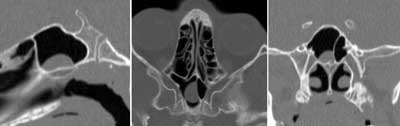
Fig. 1. Pathologic mass in the sphenoid sinus (Sagittal, Axial, Coronal views).
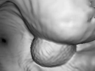
Fig. 2. 3-D reconstruction of a pathologic mass in the sphenoid sinus.
The endoscopic sinus surgery was performed in the Paediatric Otolaryngology Department of the Medical University of Warsaw. The radiological studies were correlated with intra-operative views during PESS. During operation of the sphenoid sinus two approaches were used – Wigand´s approach (3), in cases of isolated lesions of the sphenoid sinus, or modified Stammberger´s (4) or Wigand´s approach in the remaining cases (5).
RESULTS
There were 20 boys and 12 girls with ages varying from 6 to 17 years (mean age 10.9 years). In all analyzed cases (33) at least one septum was seen in the sphenoid sinus. Multiseptate sinus was found in 20 cases (fig. 3).
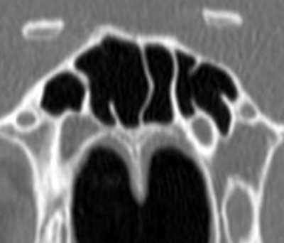
Fig. 3. The multiseptate sphenoid sinus.
One-side hypoplastic sphenoid sinus was seen in 3 cases, with collateral sinus well developed. One-side protrusion of the optic nerve was observed in 1 case, and in 1 case the protrusion was bilateral (fig. 4).
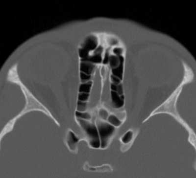
Fig. 4. Bilateral protrusion of the optic nerves.
In these cases pneumatization of the ipsilateral or both anterior clinoid processes was found. There were no dehiscences in the sphenoid sinus walls.
The course of the carotid artery in the wall of the sphenoid sinus was analyzed – pathological bulging of the carotid artery (defined as more than half of the circumference of the carotid artery) was never observed.
Hyper-developed lumen of the sphenoid sinus was seen in 3 cases; in all those cases bilateral recesses extending from the sinus laterally were observed. In 3 cases one-side lateral recess was present. In 4 cases one-side pneumatization of the anterior clinoid process was found, and in 2 cases both clinoid processes were pneumatized (fig. 5).
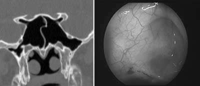
Fig. 5. Lateral recesses of the sphenoid sinus in 2-D reconstruction and view during operation.
The bulge of the optic nerve and carotid artery into the superior-lateral wall of the sphenoid sinus was visualized in the three-dimensional reconstruction of the lateral walls of the sinus (fig. 6).
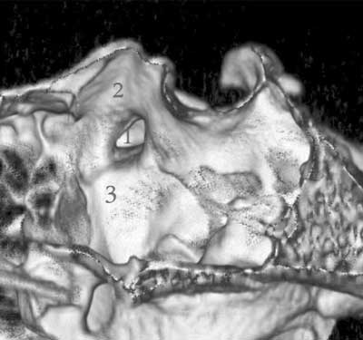
Fig. 6. 3-D reconstruction of the lateral wall of the sphenoid sinus.
1. The anterior clinoid process.
2. The bulge of the optic nerve.
3. The bulge of the carotid artery.
DISCUSSION
The sphenoid sinuses vary in size and symmetry (6, 7), which makes sphenoid sinus surgery risky. Preoperative radiological diagnostics of the sphenoid sinus and its relationship to vital anatomical structures are needed to avoid the potential risk of endoscopic sphenoid sinus surgery – threat of damage to the optic nerve or the internal carotid artery (2). Normally axial and coronal CT planes are made (7, 8). The two-dimensional reconstructions present the degree of pneumatization, additional septa and recesses, which potentially could be a source of disorientation in the operative field. The use of both two- and three-dimensional/volume rendering, navigation/computed tomography reconstructions improves visualization of the sphenoid sinus region. In case of local changes of the sphenoid sinus, precise visualization in navigation reconstruction shows the surgeon the location and volume of the lesion in the sphenoid sinus lumen (9, 10).
CONCLUSIONS
1. The sphenoid sinus anatomy varies.
2. The use of multi-dimensional computed tomography reconstructions of the sphenoid sinus improves visualization of the location and size of the pathology, as well as the anatomical variations of the sinus, providing preoperative information facilitating surgical planning.
Piśmiennictwo
1. Chmielik M, Chmielik LP: Surgery in chronic rhinosinusitis in children. New Medicine 4/2007; vol. XI: 104-105. 2. Hoseman WG, Weber RK, Keerl RE et al.: Minimally Invasive Endonasal Sinus Surgery. Thieme New York 2000; 29. 3. Wigand M: Transnasale, Endoskopischer Chirurgie der Nasennebenhohlen bie chronischer sinusitis. II Die endonasale Kieferhhohlen – Operation. HNO 1981; 29: 263-269. 4. Stammberger H, Pasawetz: Functional endoscopic sinus surgery. Concept, indications and results of the Masserklinger technique. Eur. Arch. Otorhinolaryngol. 1990; 2: 247. 5. Eryilmaz A, Dursun E: Endoscopic transnasal sphenoidotomy with or without ethmoidectomy. Kulak Burun Bogaz Ihtis Derg. 2007; 17(2): 90-95. 6. Shancar L, Evans K, Hawke M: An atlas of imaging of the paranasal sinuses. Martin Dunitz, London 1994; 50-51. 7. Bogusławska R: Badanie zatok przynosowych metodš tomografii komputerowej dla celów chirurgii endoskopowej nosa i zatok. Warszawa, 1995; 19-23. 8. Sirikci A, Bayazit Y, Bayram M et al.: Variations of sphenoid and related structures. Eur Radiol. 2000; 10(5): 844-848. 9. Chmielik LP, Chmielik A, Żabicka M et al.: Obrazowanie zatoki klinowej u dzieci w badaniu radiologicznym i klinicznym. Alergoprofil 2009; Vol. 5, Nr 1: 33-35. 10. Wankahar B, Bapuraj JR, Gupta AK et al.: Chronic sphenoid sinusitis revisited: comparison of multidetector axial sections, multiplanar reconstructions, and virtual siniscopy with endoscopic sinus surgery. Arch. Otolaryngol. Head Neck Surg. 2007; 133(7): 710-716.
