© Borgis - New Medicine 4/2009, s. 97-99
*Lidia Zawadzka-Głos1, Magdalena Fršckiewicz1, Michał Brzewski2, Agnieszka Biejat2, Mieczysław Chmielik1
TREATMENT OF LARYNGEAL CYSTS IN CHILDREN
1Department of Paediatric Otorhinolaryngology, Medical University, Warsaw, Poland
Head of Department: Prof. Mieczysław Chmielik, MD, PhD
2Department of Paediatric Radiology, Medical University, Warsaw, Poland
Head of Department: Michał Brzewski, MD
Summary
Objective.Cysts of the larynx in children are a rare cause of upper airway obstruction which may lead to serious morbidity and mortality if diagnosis and treatment are delayed.
Aim. We report the experience of the Department of Paediatric Otolaryngology of the Medical University of Warsaw, over a nine-year period from 2000 to 2009, during which twelve children with laryngeal cysts were treated.
Methods. We performed a retrospective analysis of treatment of laryngeal cysts in twelve children. Marsupialization procedures were the mainstay of treatment in this department, all carried out endoscopically by laryngeal knife and cupped forceps. We used the external approach in two children. Results: No cyst recurred more than once with endoscopic technique. We did not observe a recurrence after the external approach.
Conclusions. All children with stridor require investigation. Early endoscopic marsupialization of the laryngeal cyst is the recommended form of treatment. Nevertheless, as ventricular cysts may be deep and extensive and difficult to remove endoscopically, an external approach may still be indicated in individual cases.
INTRODUCTION
Laryngeal cysts in children are rare but the treatment is easy when the diagnosis is made. If mismanaged, the resulting respiratory obstruction can lead to high morbidity and mortality. An incidence of 1.87 per 100 000 live births was calculated by Pak et al. (8). Congenital laryngeal cysts should be differentiated from laryngomalacia, thyroglossal cysts, branchiogenic cysts, laryngocoeles, lymphangioma and neoplasms. The clinical symptoms depend on the size and location of the cyst. Location of the cyst near the orifice of the ventricle or subglottic region results in stridor. Cysts located away from the inlet of the glottis, for example in the vallecula, present initially with feeding problems, whereas stridor only develops after some time (3, 6). The diagnosis can be made by laryngoscopy. The cysts should be examined under general anaesthesia using direct laryngoscopy.
METHODS AND MATERIALS
Twelve children with laryngeal cysts, presenting to the Department of Paediatric Otolaryngology of the Medical University of Warsaw between 2000 and 2009, form the basis of this report. The symptoms were established and the diagnosis reviewed in respect of type and site of cyst. The diagnosis in all cases was confirmed by direct laryngoscopy under general anaesthesia. In two cases laryngoscopy was combined with CT scan (fig. 1).
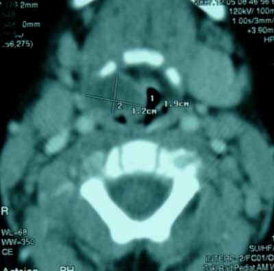
Fig. 1. CT imaging of a laryngeal cyst.
The treatment each patient received was reviewed with reference to efficacy and recurrence rate.
RESULTS
Twelve children with laryngeal cysts were treated between 2000 and 2009. Four of them were prematurely born and intubated in the first days of their lives. The most common presenting symptom was stridor. Respiratory symptoms present in nine children included stridor of different grade, inspiratory-expiratory dyspnoea and snoring. Two of them required tracheostomy. Two patients presented with feeding problems and one patient presented with hoarseness and silent weeping. In all twelve cases, the diagnosis of laryngeal cyst was confirmed by direct laryngoscopy. Four cysts were situated in the subglottic area (fig. 2), three in the aryepiglottic fold (fig. 3), three in the vallecula (fig. 5) and two in the glossal surface of the epiglottis (fig. 4).
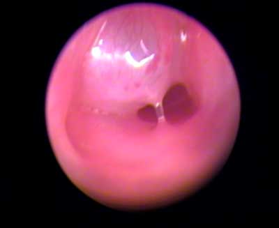
Fig. 2. Subglottic cyst.
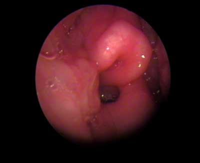
Fig. 3. Aryepiglottic fold cyst.
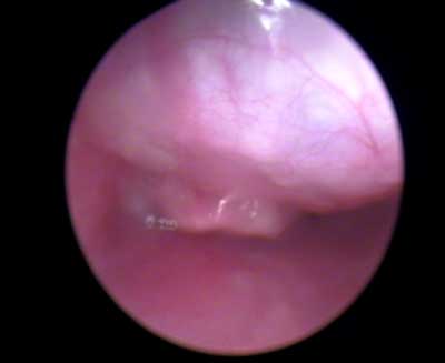
Fig. 4. Cyst of glossal surface of the epiglottis.
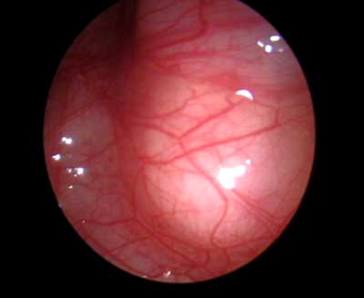
Fig. 5. Ventricular cyst closed insert of glottis.
After performing direct laryngoscopy and diagnosing the laryngeal cysts, all children were treated surgically. Endoscopic marsupialization of the cyst was carried out in eleven children and in two patients an external cervical approach was chosen. One child was operated with an external approach after failure of the endoscopic method. No recurrence was noted in any of the patients during a follow-up period lasting between eight months and eight years.
DISCUSSION
In 1970 De Santo classified laryngeal cysts as either saccular or ductal types. The saccular cyst has no communication with the laryngeal lumen. Most of them are congenital in origin. The saccular cysts are thought to arise in the saccule of the ventricle as a result of atresia of the ventricular orifice. Some of them may also appear as acquired lesions caused by various inflammatory processes and traumatic events (7). Saccular cysts are divided into anterior and lateral. They protrude into the laryngeal lumen between the true and false vocal cord. The latter are generally larger and extend towards the false vocal cord and aryepiglottic fold (1, 2, 4). In the examined group of children cysts in the ventricular area were observed in eight cases.
The ductal type is mostly situated in the vallecula. They appear to arise from an obstruction of the submucosal gland ducts. The characteristic retention of mucus in the dilated collecting ducts of the intramucosal seromucinous glands can be found anywhere in the larynx (9, 10). Ductal cysts originate from an obstruction of the glandular ducts. It is difficult to differentiate a congenital cyst from a secondary one in a newborn (10). Cysts of the subglottic area in the examined group of children occurred only in four premature infants who were intubated in the first days of their lives. This corresponds with the observation suggesting that the reason for cyst occurrence in this area is a post-intubation complication, an inflammatory state and obstruction of seromucinous gland duct causing development of a secondary cyst (5, 7).
Every infant with stridor requires investigation. Soon after the diagnosis is established, all patients should have the cysts thoroughly examined and removed under general anaesthesia. The choice of definitive surgical treatment remains a subject of controversy. In general the cysts can be removed via an endoscopic or an external approach.
All patients underwent surgical treatment; among them ten were removed endoscopically and two with the external approach. The external approach was used in two patients with ventricular cysts. One of them, who earlier underwent an endoscopic procedure, was reoperated using the external approach. In both cases the cyst obturated the lumen of the larynx and extended over the borders of the larynx. Complete recovery was achieved after surgery. The remaining ten children after endoscopic marsupialization remain healthy without any recurrence. Endoscopic treatment is a simple, effective and adequate treatment of the condition but is burdened with a risk of recurrence, which occurred in one case in the group of presented children. Ventricular cysts may be deep and extensive and difficult to remove by endoscopic procedures. In these cases an external approach may still be indicated.
CONCLUSIONS
1. Endoscopic treatment is a safe and effective primary treatment for most laryngeal cysts in children.
2. An external approach is reserved for those cases in which endoscopic treatment is hindered due to deep and extensive cysts.
3. Laryngeal cysts should be considered in the differential diagnosis of any children presenting with stridor, weak cry, or feeding difficulties.
Piśmiennictwo
1. Abramson A, Zielinski B: Congenital laryngeal saccular cyst of the newborn. Laryngoscope 1984; 94: 1580-1581. 2. Arens C, Glanz H, Kleinsasser O: Clinical and morphological aspects of laryngeal cysts. Eur Arch Otorhinolaryngol 1997; 254: 430-436. 3. Bais AS, Uppal K et al.: Congenital cysts of the larynx, The Journal of Laryngology and Otology 1989; vol 103: 966-967. 4. Forte V, Fuoco G, James A: A new classification system for congenital laryngeal cysts. Laryngoscope 2004; 114: 1123-1127. 5. Lim J, Hellier W et al.: Subglottic cysts: the Great Ormond Street experience. International Journal of Pediatric Otorhinolaryngology 2003; 67: 461-465. 6. Marien S, Jespers A et al.: Congenital laryngeal cyst: a case report. Acta Oto-rhino-laryngologica belg. 2003; 57: 119-121. 7. Mitchell DB, Irwin C, Bailey C et al.: Cysts of the Infant Larynx. The Journal of Laryngology and Otology 1987; 101: 833-837. 8. Pak MW et al.: Congenital laryngeal cysts: current approach to management. The Journal of Laryngology and Otology 1996; 110: 854-856. 9. Smith D, Callanan V et al.: Intracordal cyst in a neonate. International Journal of Pediatric Otorhinolaryngology 2000; 52: 227-281. 10. Wen-Shuan Lee et al.: Airway obstruction caused by congenital epiglottic cyst. International Journal of Pediatric Otorhinolaryngology 2000; 53: 229-233.
