© Borgis - Postępy Nauk Medycznych 12/2013, s. 896-900
Irmina Jankowska-Lech1, *Iwona Grabska-Liberek1, Piotr Chojnowski1, Agnieszka Skowyra1, Jaromir Wasyluk2
Zmiany starcze aparatu ochronnego oka
Ageing of eye adnexa
1Clinic of Ophthalmology, Medical Center of Postgraduate Education, Warszawa
Head of Clinic: Iwona Grabska-Liberek, MD, PhD, assoc. prof.
2 Clinic of Ophthalmology, Military Institute of Aviation Medicine, Warszawa
Head of Clinic: prof. Marek Prost, MD, PhD
Streszczenie
Aparat ochronny oka stanowi niezwykle ważną część narządu wzroku, narażoną na ciągły wpływ świata zewnętrznego. Należą do niego: powieki wraz z łukami brwiowymi i rzęsami, spojówki powiek i gałki ocznej oraz narząd łzowy. W warunkach fizjologicznych zapewnia on stałą ochronę gałki ocznej przed szkodliwym działaniem czynników zewnętrznych, między innymi promieniowaniem ultrafioletowym czy różnego rodzaju urazami. Wraz z upływem czasu, aparat ochronny oka ulega procesowi starzenia, a więc zmniejsza się jego zdolność do samonaprawy będącej odpowiedzią na gromadzące się w wyniku stresu środowiskowego uszkodzenia wewnątrzkomórkowe. Może to prowadzić do utraty jego funkcji ochronnych, a w konsekwencji również nieprawidłowego funkcjonowania powierzchni oka. To z kolei może wpływać ostatecznie na obniżenie funkcji widzenia. Dlatego bardzo ważna jest wczesna diagnostyka tych zmian, monitorowanie ich przebiegu i w razie konieczności wdrożenie odpowiedniego leczenia, które miałoby na celu zapobieganie ich powikłaniom.
Celem niniejszej pracy jest zestawienie i opisanie najważniejszych zmian starczych dotyczących poszczególnych elementów aparatu ochronnego oka.
Summary
The eye adnexa is one of the most important parts of the organ of vision, which is permanently influenced by environmental factors. It consists of eyelids along with eyebrows and eyelashes, conjuctivas and eyeball and also lacrimal apparatus. In physiological conditions it constantly protect eyeball from harmful influence of external factors, i.a.: ultraviolet radiation or different kind of injuries. Over time the eye adnexa is effected by an aging process, therefore, its ability to selfrepair decrease on the grounds of intracellular damages caused by environmental stress factors. It may lose its protective functions and consequently cause an abnormal functioning of the ocular surface. At least it may decrease functions of vision. So, it is very important to diagnose this type of changes in early phase, control proceedings and implement apropriate treatment to prevent complications.
The aim of this study is to compare and describe the major senile changes regarding the particular elements of the eye adnexa.

INTRODUCTION
Ageing is a natural, irreversible and intensifying process of changes in the metabolism and physicochemical properties of cells, which leads to an inhibition of autoregulation and regeneration of the body and to morphological and functional changes of its tissues and organs (according to the PWN Encyclopedia).
Eye adnexa is a group of tissues and anatomical structures which cover the eyeball and enable proper functioning of the eye. The eye adnexa consists of eyelids along with superciliary arches, palpebral and bulbar conjunctivae and lacrimal apparatus. The eyelids provide mechanical protection to the anterior part of the eyeball. Eyelashes and hair on the superciliary arch protect the eye from sweat and small particles. Apart from that, the eyelashes serve as sensors which trigger the eye-closing reflex. Blinking spreads tears across the anterior surface of the eye, moisturizing it. During blinking, a thin layer of tears is moved across the anterior surface of the cornea and the eyeball conjunctiva, washing any potential foreign bodies to the lower fold of lid conjunctiva and to the eyeball. Tear film creates a very smooth surface of the eyeball which allows the light to be properly refracted by the cornea, supplies the superficial layers of the cornea with oxygen as it does not have its own vessels. Tears have a slightly higher concentration of sodium chloride than other tissue fluids and, additionally. They contain small amounts of protein bodies with buffer capacity, as well as certain amount of lysozyme of antibacterial properties. Mechanical rinsing and the lysozyme protect the eye against infections.
SENILE CHANGES OF EYELIDS
The eyelid skin of elderly people is wrinkled. It is a result of dermis thinning, subcutaneous tissue atrophy, lowered elasticity and excessive skin dryness. The amount of lipids on the surface of skin is lowered parallel to the atrophy of lipids in the skin itself, which is caused by a significant drop in the number of fibroblasts synthesising and accumulating skin cholesterol. A lowered activity of sebaceous glands, especially in women, also takes place. In some people, a gland overgrowth occurs, which can lead to a formation of senile sebaceous adenomas.
A decrease in the skin’s elasticity and a lowered immunity of capillaries leads to their dilation (telangiectasia). A tendency to focal, brown discolourations (senile lentigines) occurs. In the case of both dry skin and seborrhoea, an increased tendency to inflammatory reactions occurs as a result of chemical, physical, thermal and bacterial factors.
The orbital fatty tissue atrophies in elderly people. The eyeball collapses and the palpebral fissure appears to be narrowed. A slow atrophy of the orbital septum can take place and the orbital fat can move forward (baggy eyelids). The eyeball sheath and palpebral ligaments become less elastic. The tarsus and eyelid muscles slowly atrophy which leads to a senile ptosis of the upper eyelid and to lower eyelid ectropion (fig. 1) (1). The treatment of the upper eyelid ptosis is based on a surgical correction under local anaesthesia and consists of cutting the palpebral sulcus, dissecting free the aponeurosis of the levator palpebrae superioris, suspending it to the tarsus and layer suturing.
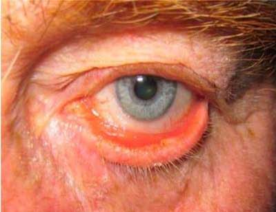
Fig. 1. Eyelid ectropion.
Eyelid ectropion causes the palpebral conjunctiva to become exposed. This leads to its hyperaemia, inflammatory overgrowth and epiphora. The latter is a consequence of lacrimation being increased by inflammation as well as by an impairment of the outflow of tears, which is a result of the lacrimal point (the first step of draining the tears) protruding out of the eyeball (2). The senile eyelid ectropion requires surgical correction which can be performed in 4 ways:
– tarsal-conjunctival resection: rhombus of approx. 3-4 mm vertically and 6-8 mm horizontally below the lacrimal point,
– “T” plastics: additional resection of a full-thickness pentagonal fragment of the eyelid, lateral from the lacrimal point,
– horizontal shortening of the eyelid: by resecting a proper, full-thickness fragment of the eyelid with an optional blepharoplasty of the lower eyelid,
Powyżej zamieściliśmy fragment artykułu, do którego możesz uzyskać pełny dostęp.
Mam kod dostępu
- Aby uzyskać płatny dostęp do pełnej treści powyższego artykułu albo wszystkich artykułów (w zależności od wybranej opcji), należy wprowadzić kod.
- Wprowadzając kod, akceptują Państwo treść Regulaminu oraz potwierdzają zapoznanie się z nim.
- Aby kupić kod proszę skorzystać z jednej z poniższych opcji.
Opcja #1
29 zł
Wybieram
- dostęp do tego artykułu
- dostęp na 7 dni
uzyskany kod musi być wprowadzony na stronie artykułu, do którego został wykupiony
Opcja #2
69 zł
Wybieram
- dostęp do tego i pozostałych ponad 7000 artykułów
- dostęp na 30 dni
- najpopularniejsza opcja
Opcja #3
129 zł
Wybieram
- dostęp do tego i pozostałych ponad 7000 artykułów
- dostęp na 90 dni
- oszczędzasz 78 zł
Piśmiennictwo
1. Orłowski W: Okulistyka współczesna. Tom 2, Wydawnictwo lekarskie PZWL, Warszawa 1977: 643-687.
2. Niżankowska MH: Okulistyka. Podstawy kliniczne. Wydawnictwo lekarskie PZWL, Warszawa 2007: 128-137.
3. Basic and Clinical Science Course: Oczodół, powieki i układ łzowy (BCSC 7). I wyd. pol., Urban & Partner, Wrocław 2005: 239-371.
4. Lam H, Bleiden L, de Paiva CS et al.: Tear Cytokine Profiles in Dysfunctional Tear Syndrome. American Journal Of Ophthalmology 2009; 147: 198-205.
