© Borgis - Postępy Nauk Medycznych 5/2012, s. 402-405
*Michał Przyszlak, Tomasz Jargiełło, Maciej Szajner, Małgorzata Szczebo-Trojanowska
Wewnątrznaczyniowe leczenie zewnątrzczaszkowych tętniaków tętnicy szyjnej wewnętrznej
Endovascular Treatment of Extracranial Internal Carotid Artery Aneurysms
Department of Interventional Radiology and Neuroradiology, Medical University of Lublin
Head of the department: prof. Małgorzata Szczerbo-Trojanowska
Streszczenie
Wstęp. Celem poniższego artykułu jest prezentacja wewnątrznaczyniowych metod leczenia zewnątrzoponowych tętniaków tętnicy szyjnej wewnętrznej oraz ich ocena pod kątem stopnia wyleczenia i występowania powikłań.
Materiały i metody. Od 1999 roku do dziś, w naszym zakładzie, 52 pacjentów było leczonych z powodu zewnątrzoponowych tętniaków (zarówno tętniaków prawdziwych, jak i rzekomych) tętnicy szyjnej wewnętrznej. Nasze metody obejmowały: stentowanie (stenty zwykłe, stenty pokrywane), implantacje odczepialnych spiral platynowych (czasem przy użyciu technik remodellingu z balonem lub stentem), zamykanie tętnicy macierzystej tętniaka. Każdą procedurę poprzedzała próba Matas’a.
Wyniki. Leczenie wewnątrznaczyniowe spowodowało pełne wyłączenie tętniaka z krążenia we wszystkich 52 przypadkach. Dwóch pacjentów miało epizod TIA około miesiąca po zabiegu (zamknięcie tętniaka odczepialnymi spiralami platynowymi). U jednego pacjenta wystąpił udar dokonany po terapeutycznym zamknięciu tętnicy szyjnej wewnętrznej.
Wnioski. Nasze ponad 10-letnie doświadczenie w leczeniu zewnątrzoponowych tętniaków tętnicy szyjnej wewnętrznej pokazuje, że leczenie wewnątrznaczyniowe jest bezpieczne i skuteczne. Jednakże dobre rezultaty zależą od wybrania odpowiedniej techniki wewnątrznaczyniowej.
Summary
Introduction. The purpose of this article is to present endovascular treatment options for extradural ICA aneurysms and evaluate their usefulness based on treatment success rate and complications.
Materials and methods. We have treated 52 patients with extradural aneurysms, both true and false aneurysms, of the internal carotid artery (ICA) since 1999. Our techniques involved: stenting with bare and covered stents, primary coiling, remodeling with stent or balloon, ICA trapping and parental artery occlusion. Each procedure was performed after successful Balloon Test Occlusion (BTO).
Results. Endovascular treatment was successful in all cases. Two patients had a TIA one month after primary coiling and one patient had a major stroke after parental artery occlusion.
Conclusions. Our experience with extradural carotid aneurysms management shows that endovascular treatment of those lesions is both safe and efficient and should be considered a primary treatment option. However, good results depend heavily on proper choice of endovascular equipment and technique.

INTRODUCTION
Extradural internal carotid artery aneurysms are very rare. They are significantly less common than the intracranial aneurysms. Their incidence is still not established at this point. They also differ in clinical presentation. Symptoms include distal embolism (60%), a pulsatile mass on the neck, murmur, Horner’s syndrome, headaches, tinnitus, vertigo and local mass effect. The most common location for extradural aneurysms is the ICA (Internal Carotid Artery), or less frequently, the VA (Vertebral Artery). In some very rare cases extradural aneurysms form on the ECA (External Carotid Artery) or it’s branches. There is no single cause for extradural aneurysm formation. Typically those lesions can be secondary to atherosclerosis, vessel wall defects, trauma, infection, radiation, or special flow conditions. Sometimes there are no obvious underlying conditions and the etiology is unknown. The vast majority of extradural aneurysms interventional neuroradiologists and neurosurgeons encounter, are located in the cavernous segment of the ICA (1). The anatomy of this location poses a major challenge for surgical treatment, wheras the endovascular approach in reaching aneurysms of these locations is relatively easy and safe.
According to one of the most comprehensive books for neuroradiologists, the Surgical Neuroangiography, the most common cause of extradural aneurysms is trauma. Authors also acknowledge that in the past, infectious aneurysms were much more frequent, but now are very rare. Finally, there are reports of false aneurysms after failed attempt to place a central line (1). In our department, we have been treating extradural ICA aneurysms since 1999. To this day we have treated 52 lesions of this kind. The purpose of this study is to demonstrate and evaluate the current endovascular treatment options employed in these cases.
MATERIAL AND METHODS
Since 1999 we have treated 52 extradural aneurysms in 52 patients (27 female, 25 male) with mean age of 36 years. Aneurysms varied in etiology, location, morphology and clinical presentation. Of 52 lesions, 24 where due to atherosclerosis, 19 were traumatic, 1 was infectious and the rest 8 were not connected to any underlying condition, therefore were considered to be of congenital/developmental origin.
The clinical of the aneurysm were: symptoms TIA in case 8, mass effect in 23, dysphagia in 2 and a pulsatile mass on the neck in 2 patients. Aneurysms varied in size with the mean diameter of 28 mm (range 5-56 mm).
Patients were qualified for endovascular treatment based on Doppler Ultrasound, CT-Angio or Digital Subtraction Angiography (DSA)
Before each procedure a Balloon Test Occlusion (BTO) was performed to assess efficiency of the circle of Willis. Under local anesthesia with 2% Lignocaine two 5Fr sheaths were introduced – one into each femoral artery, giving us access to the aorta with all of it’s branches. A 5Fr Headhunter catheter was introduced and navigated to the ICA on the opposite side to the lesion. Using the second arterial access via the femoral artery a balloon was navigated through aorta and cervical vessels, and positioned in the aneurysms parental artery. Patients were given 5000 iu of heparin and aneurysms parental artery was occluded with the balloon. Contrast medium was then injected through the catheter placed on the other side. This showed if the collateral circulation through anterior communicating artery (AcomA) is sufficient according to neuroradiologic criteria (based mostly on the symmetrical filling of the cortical veins of both hemispheres). At the same time, each patients neurological status was examined. Should any neurological deficits or additional symptoms appear, the BTO would be terminated and patient disqualified from endovascular treatment (2).
All patients included in this study had sufficient blood circulation during BTO therefore were the candidates for the endovascular treatment.
All patients were treated under local anesthesia with 2% lignocaine. In each case via femoral arterial access an intraarterial sheath was introduced and a cerebral angiography was performed using 5Fr Headhunter catheter. After visualizing the aneurysm on the DSA, a “working projection” was established. It had to show the aneurysmal neck, dome and surrounding vessels anatomy.
Various methods were used to exclude an aneurysm from the circulation.
In primary coiling we have used platinum Detachable Guglielmi Coils (GDC) which were introduced to the aneurysmal sac through microcatheters (fig. 1).
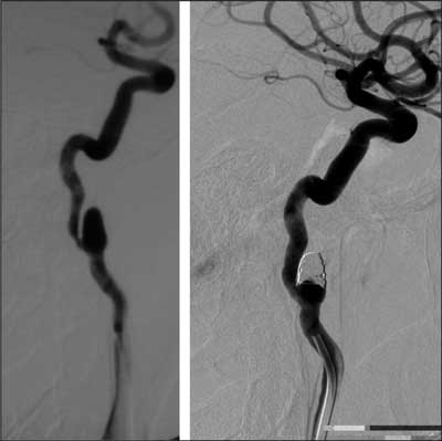
Fig. 1. Internal carotid artery angiography, lateral projection. Internal carotid artery aneurysm before and after embolization with platinum detachable coils (GDC).
Powyżej zamieściliśmy fragment artykułu, do którego możesz uzyskać pełny dostęp.
Mam kod dostępu
- Aby uzyskać płatny dostęp do pełnej treści powyższego artykułu albo wszystkich artykułów (w zależności od wybranej opcji), należy wprowadzić kod.
- Wprowadzając kod, akceptują Państwo treść Regulaminu oraz potwierdzają zapoznanie się z nim.
- Aby kupić kod proszę skorzystać z jednej z poniższych opcji.
Opcja #1
29 zł
Wybieram
- dostęp do tego artykułu
- dostęp na 7 dni
uzyskany kod musi być wprowadzony na stronie artykułu, do którego został wykupiony
Opcja #2
69 zł
Wybieram
- dostęp do tego i pozostałych ponad 7000 artykułów
- dostęp na 30 dni
- najpopularniejsza opcja
Opcja #3
129 zł
Wybieram
- dostęp do tego i pozostałych ponad 7000 artykułów
- dostęp na 90 dni
- oszczędzasz 78 zł
Piśmiennictwo
1. Berenstein A, Lasjaunias P, Ter Brugge KG et al.: Surgical Neuroangiography Vol. 2; Chapter 7.2 Extradural Aneurysms. 2nd edition. Germany: Springer-Verlag, Berlin, Heidelberg, New York 2004; 382-425.
2. Gevers S, Heijtel D, Ferns SP et al.: Cerebral Perfusion Long Term after Therapeutic Occlusion of the Internal Carotid Artery in Patients Who Tolerated Angiographic Balloon Test Occlusion; AJNR Am J Neuroradiol 2012; 33: 329-335.
3. Sluzewski M, Menovsky T, Jan van Wooij W et al.: Douwe Wijnalda; Coiling of Very Large Cerebral Aneurysms: Long-Term Clinical and Serial Angiography Results; AJNR Am J Neuroradiol 2003; 24: 257-262.
4. Nelson PK, Levy DI: Balloon-assisted Coil Embolization of Wide-necked Aneurysms of the Internal Carotid Artery: Medium-term Angiographic and Clinical Follow-up in 22 Patients; AJNR Am J Neuroradiol 2001; 22: 19-26.
5. Layton KF, Cloft HJ, Gray LA et al.: Balloon-Assisted Coiling of Intracranial Aneurysms: Evaluation of Local Thrombus Formation and Symptomatic Thromboembolic Complications; AJNR Am J Neuroradiol June 2007; 28: 1172-1175.
6. Bodily KD, Cloft HJ, Lanzino G et al.: Stent-Assisted Coiling in Acutely Ruptured Intracranial Aneurysms: A Qualitative, Systematic Review of the Literature; AJNR Am J Neuroradiol 2011; 32: 1232-1236.
7. Li MH, Zhu YQ, Fang C et al.: The Feasibility and Efficacy of Treatment with a Willis Covered Stent in Recurrent Intracranial Aneurysms after Coiling; AJNR Am J Neuroradiol August 2008; 29: 1395-1400.
8. Bodily KD, Cloft HJ, Lanzino G et al.: Stent-Assisted Coiling in Acutely Ruptured Intracranial Aneurysms: A Qualitative, Systematic Review of the Literature; AJNR Am J Neuroradiol 2011; 32: 1232-1236.
9. Hwang G, Park H, Bang JS et al.: Comparison of 2-Year Angiographic Outcomes of Stent- and Nonstent-Assisted Coil Embolization in Unruptured Aneurysms with an Unfavorable Configuration for Coiling. AJNR Am J Neuroradiol 2011; 32: 1707-1710.
10. van Rooij WJ, Sluzewski M: Unruptured Large and Giant Carotid Artery Aneurysms Presenting with Cranial Nerve Palsy: Comparison of Clinical Recovery after Selective Aneurysm Coiling and Therapeutic Carotid Artery Occlusion. AJNR Am J Neuroradiol May 2008; 29: 997-1002.
11. Field M, Jungreis CA, Chengelis N et al.: Symptomatic Cavernous Sinus Aneurysms: Management and Outcome After Carotid Occlusion and Selective Cerebral Revascularization. AJNR Am J Neuroradiol 2003; 24: 1200-1207.
12. Saatci I, Saruhan Cekirge H, Oztork MH et al: Treatment of Internal Carotid Artery Aneurysms with Covered Stent: Experience in 24 Patients with Mid-term Follow-up Results. AJNR Am J Neuroradiol 2004; 25:1742-1749.
13. Redekop G, Marotta T, Weill A: Treatment of traumatic aneurysms and arteriovenous fistulas of the skull base by using endovascular stents. J Neurosurg 2001; 95: 412-419.
14. Dai D, Ding YH, Kadirvel R et al.: Patency of Branches after Coverage with Multiple Telescoping Flow-Diverter Devices: An In Vivo Study in Rabbits. AJNR Am J Neuroradiol 2012; 33: 171-174.
