© Borgis - New Medicine 1/2001, s. 51-54
Andrzej Koczynski1, Zofia Rajtar-Leontiew2
Ultrasonographic scanning of the central nervous system in the newborn and neonates: a relative assessment
1Department of Radiology and Ultrasonography Clinical Hospital
2Department of Neonatal Pathology The Medical University of Warsaw
Summary
The quality of ultrasound examination is associated with technical equipment and diagnostic methods. Interpretation based on „overreading ” or „underreading ” images may yield false results. The development of imaging techniques has resulted in eliminating invasive diagnostic x-ray procedures.
Visualisation depends on technical conditions and a diagnostic technique. Every imaging procedure has a limited reliability which provides the grounds for proper assessment based on appropriate perception. In the assessment of a complex picture the measurement of reliability consists of a purposeful disregard of areas which might lead to „overreading ” or „underreading ”. It is equally important to eliminate artefacts. Assessment of such areas may yield false results. Each imaging techniques has a definite diagnostic power which is a vehicle for a particular amount of relevant information affecting the quality of results.
Ultrasound examination (USG) is a highly valued means of clinical diagnosis because it is an easily accessible, simple, noninvasive, informative and cost-effective procedure.
Cranial sonography is the basic imaging assessment technique in the central nervous system in the newborn and infants, and a vital part of a thorough clinical examination. The significance of the procedure can be measured by its well-established place in almost every diagnostic, monitoring, and follow-up process. The well-documented role of the technique has been demonstrated by the increasing number of procedures and growing clinical and imaging experience reported in literature (1, 2, 3, 4, 5).
The present paper focusses on the necessity of establishing an objective assessment of the USG examination by indicating the likelihood of inadequate and even false images. Another aim is to emphasise the significance of the procedural evolution in the basic imaging technique. The paper also includes results of USG examinations carried out on patients admitted to the Department of Neonatal Pathology. The radiological pictures have been submitted by the Radiological Department archives and demonstrate cases treated before the USG procedure was introduced.
The technological progress has become a valid factor in enforcing a changing approach to the diagnostic process. A good example might be to compare the value of different imaging techniques of the head: a radiological scan showing the skull (fig. 1) and cranial sonography showing the brain (fig. 2). Regardless of the different value of the techniques and time period in which they were used, each is distincly exclusive (fig. 3).
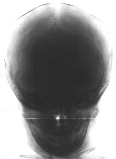
Fig. 1. A normal radiological scan of the neurocranium.
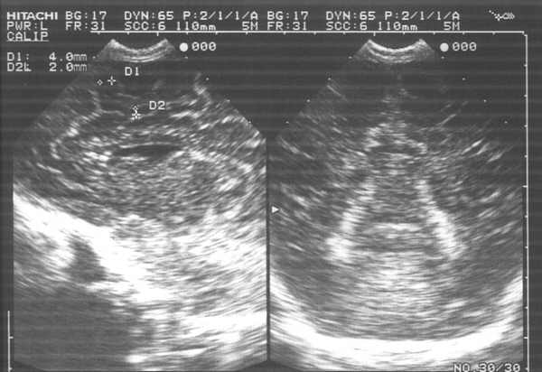
Fig. 2. A normal USG scan of the brain.
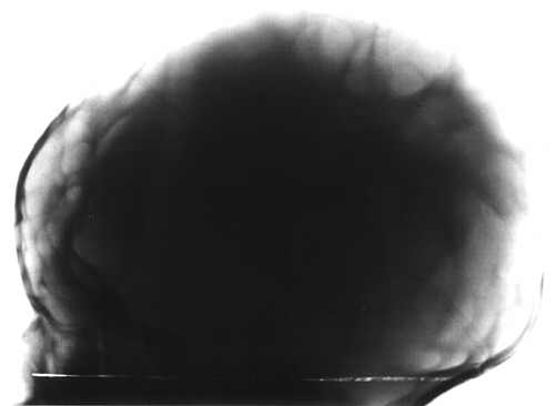
Fig. 3. Craniofenestria - `windows´ in the cranial vault.
A rational joint use of imaging techniques, strictly dependent on clinical indications, increases the evidence area in terms of both quantity and quality.
A positive development in procedural evolution was the complete replacement of pneumocephalography, ventriculography and subdurography by USG techniques (fig. 4, 5, 6, 7).
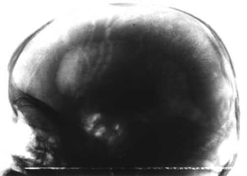
Fig. 4. Ventriculography: markedly enlarged lateral ventricles.
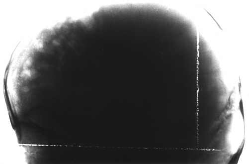
Fig. 5. Subdurography: extensive defects of the brain tissue in the frontal area.

Fig. 6. Cranial sonography: marked enlargement of the lateral ventricles.
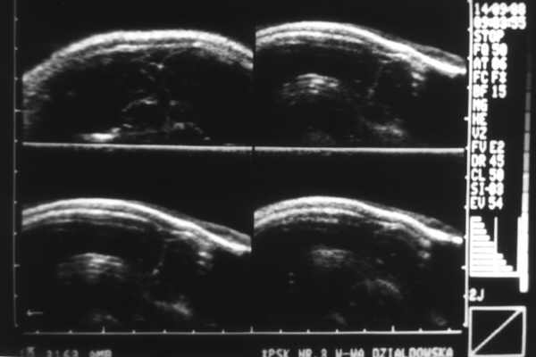
Fig. 7. Cranial sonography: marked dilation of the submeningeal spaces.
Due to a wide range of practical, technical (different apparatus) and technological standards (different properties of apparatus and equipment), normal images may have a different appearance, which, in turn, may result in extremely variable assessments (fig. 8, 9). Technical conditions may also be responsible for producing false images within normal structures. Thus, for example, boundary amplification of curvatures particularly abundant in the brain may produce images resulting in overinterpretation (fig. 10) and, owing to this, hypodensity areas may be diagnosed as fluid cisterns.
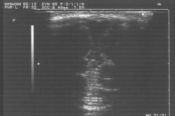
Fig. 8. Cranial sonography: appropriate examination technique.

Fig. 9. The same infant (fig. 8): inappropriate examination technique showing false fluid areas.
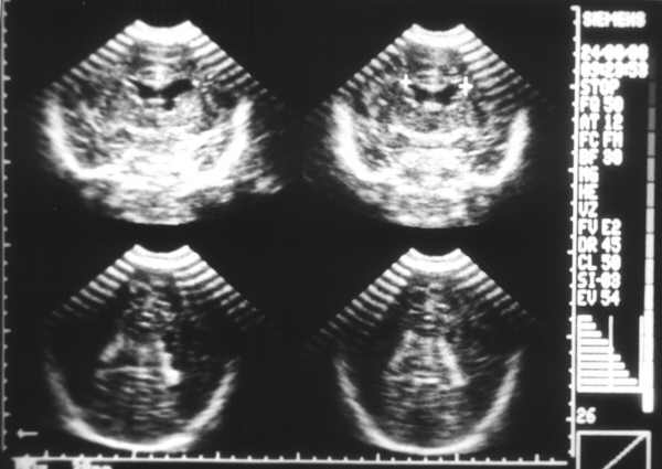
Fig. 10. An amplified curvature boundary shows false fluid areas.
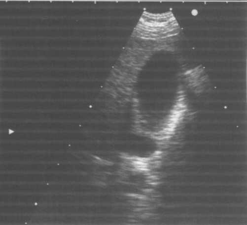
Fig. 10a. An amplified boundary is a physical phenomenon difficult to eliminate: the body of the gall bladder.
Abnormal artefact formation may also be a source of a false interpretation substrate (fig. 11).
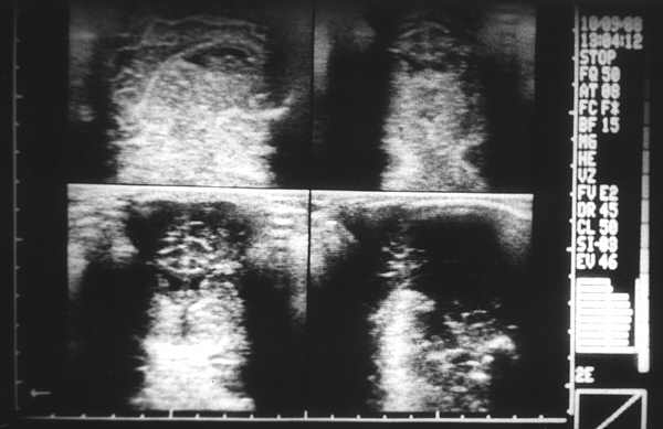
Fig. 11. A USG artefact scan: extreme differentiation of the impedance effect.
Resulting from the above facts, borderline normal and pathology may be the areas lacking in imaging and clinical cohesion, which may subsequently lead to methodological abuse in diagnostic imaging, and deficient treatment in clinical management.
An inadequately selected penetration depth and excessively differentiated intensity of ultrasound wave reflection (impedance) may result in the formation of false hyperdensity areas (fig. 12), resembling haemorrhage or abnormal hyperplastic masses. Due to the fact that spinal cord abnormalities in the newborn and infants are rare, the USG examination of the spinal canal (fig. 13) is less frequent than cranial sonography. However, in literature much attention is devoted to that part of the central nervous system (4).

Fig. 12. A false image: excessive hyperdensity areas may be indicative of haemorrhage into the brain tissue.
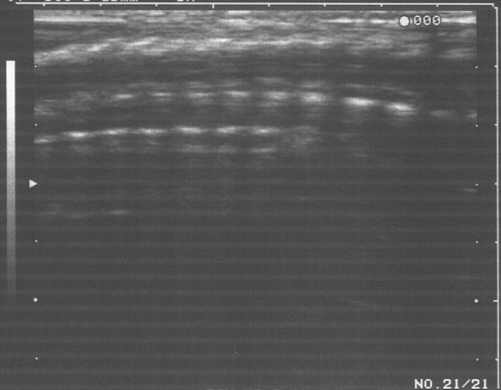
Fig. 13. A USG scan shows a normal spinal canal.
Careful and detailed assessment of both methodology and the range of CNS pathology in the newborn and neonates is included in an appropriate chapter of `Ultrasonografia pediatryczna´ (Paediatric sonography) (4). Special attention is paid to the significant variability of the photographic material with respect to technical conditions. The degree of visualised abnormalities is substantial and renders no doubt.
Everyday clinical experience shows that in many cases the abnormalities searched for are the initial ones, sometimes very discrete, but their diagnosis may affect both the introduction of adequate management and the planning of supervision and control. In such situations accuracy, caution and relative assessment become relevant.
Conclusions
1. The USG examination should not be mandatory.
2. Technical and technological conditions affect the quality of imaging.
3. In the diagnostic process `overreading´ is as harmful as `underreading´.
4. The most vital, and the most difficult, issue is to establish objective assessment.
Piśmiennictwo
1. Daneman A: Cranial Sonography, Ped Radiol, Merit Communications, London 1992:87-105. 2. Daniel B: Mózgowie, Atlas Anatomii Radiologicznej Czlowieka, PZWL, Warszawa 1996:190-191. 3. Kossoff G: Ultrasound, Excerpta Medica, Amsterdam 1989:29-32. 4. Marcinski A: Badanie ultrasonograficzne ukladu nerwowego, Ultrasonografia pediatryczna, PZWL, Warszawa 1994:17-79. 5. Rajtar-Leontiew Z, Lipska E: Krwotoki sródczaszkowe u noworodków, Nowa Ped 2000, 18, 1:3-8.
