© Borgis - Postępy Nauk Medycznych 8/2016, s. 588-590
*Katarzyna Pawelec1, Katarzyna Grzeszkiewicz1, Przemysław Bombiński2, Agnieszka Biejat2, Michał Matysiak1
Mediastinal enterogenous cyst in a child with ALL – case report
Torbiel enterogenna śródpiersia u dziecka z ALL – opis przypadku
1Department of Pediatric, Hematology and Oncology, Medical University of Warsaw
Head of Department: Professor Michał Matysiak, MD, PhD
2Department of Pediatic Radiology, Medical University of Warsaw
Head of Department: Michał Brzewski, MD, PhD
Streszczenie
Nienowotworowe torbiele śródpiersia (NNMC) stanowią rzadką grupę wrodzonych zmian, o częstości od 7 do 25% wszystkich guzów tej przestrzeni. Najczęściej rozpoznawane są u mężczyzn, w przedniej części śródpiersia, po stronie prawej. Torbiele enterogenne powstają na skutek nieprawidłowego pączkowania grzbietowej części pierwotnej cewy pokarmowej. Większość pacjentów zgłasza objawy związane z niewydolnością oddechową. Obecność objawów koreluje z lokalizacją i wielkością zmiany. Nierzadko torbiele enterogenne bywają bezobjawowe, wykrywane przypadkowo. Objawy zależą od umiejscowienia i rozmiarów guza. Mogą to być objawy związane z uciskiem guza na otaczające narządy – bóle w klatce piersiowej, kaszel, krwioplucie. Bóle mogą wystąpić także w związku ze zwiększeniem się ciśnienia wewnątrz torbieli. Objawy ostre pojawiają się przy zakażeniu torbieli lub przy owrzodzeniu bądź przedziurawieniu torbieli wysłanej błoną śluzową żołądka. W populacji pediatrycznej torbiele enterogenne stanowią 70%, a zarazem najczęstszy rodzaj torbieli wywodzących się z głowowej części jelita pierwotnego. Ze względu na bardzo często asymptomatyczny przebieg torbieli, dopiero wykonanie takich badań obrazowym jak TK czy MRI pozwala na ustalenie rozpoznania i zaplanowanie leczenia. W pracy przedstawiono przypadek bezobjawowej torbieli enterogennej u 3-letniaj dziewczynki leczonej z powodu ALL.
Summary
Mediastinal cysts are rare congenital pathological findings, comprising 7 to 25% of all the lesions in this region. They are most commonly found in men, in the frontal right part of mediastinum. Enterogenous cysts develop as a consequence of an abnormal development of the dorsal primitive intestine. Symptoms vary regarding the localization and size of the tumor. The majority of patients declare symptoms related to respiratory insufficiency. Others include chest pains, persistent cough and haemoptysis. Those are often a result of compression of the cyst on the surrounding structures. Also, the symptoms may be observed as an effect of higher tension inside the cyst. Not so infrequently however, they canbe asymptomatic. In the pediatric population, enterogenous cysts are the most common cysts of mediastinum, accounting for 70% of cases. They are often primarily diagnosed using CT or MRI scans as accidental findings. We report a case of an asymptomatic mediastinal enteropathogenic cysts in a 3 year old girl with ALL.

Introduction
The mediastinum is an anatomical cavity in the thorax, enclosed by pleurae, thoracic spine and sternum. It starts at the superior thoracic aperture and ends at the diaphragm. There is a wide range of tissues in this area, therefore mediastinal cysts and tumors presents various histopathological origin and diverse clinical symptoms. Mediastinal cysts are rare congenital pathological findings, comprising 7 to 25% of all the lesions in this region. They are most commonly found in men, in the frontal right part of mediastinum.
Enterogenous cysts develop as a consequence of an abnormal development of the dorsal primitive intestine. They are most frequently lined with stratified squamous epithelium and filled with mucus. They can be distinctly separated from surrounding tissues, connected with esophagus or develop within the esophagus. Symptoms vary regarding the localization and size of the tumor.
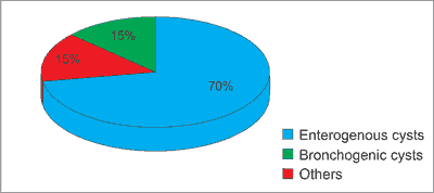
In the pediatric population, enterogenous cysts are the most common cysts of mediastinum, accounting for 70% of cases. In figure 1 show the distribution of mediastinal cystis in peadiatric population. They are often primarily diagnosed using CT or MRI scans as accidental findings. We report a case of an asymptomatic mediastinal enteropatogenic cyst in a 3 year old girl with ALL.

Fig. 1. Distribution of mediastinal cysts
Case report
A 2.5 year old girl presented to the clinic with suspicion of proliferative disease of the hematopoietic system. Based on the clinical picture and the laboratory tests she was diagnosed with acute lymphoblastic leukemia (common ALL) and qualified for protocol ALL IC BFM 2009 treatment, HR group due to poor response to steroids on day 8. There were no pathological findings in standard CT and RTG scans at the beginning of treatment.
Powyżej zamieściliśmy fragment artykułu, do którego możesz uzyskać pełny dostęp.
Mam kod dostępu
- Aby uzyskać płatny dostęp do pełnej treści powyższego artykułu albo wszystkich artykułów (w zależności od wybranej opcji), należy wprowadzić kod.
- Wprowadzając kod, akceptują Państwo treść Regulaminu oraz potwierdzają zapoznanie się z nim.
- Aby kupić kod proszę skorzystać z jednej z poniższych opcji.
Opcja #1
29 zł
Wybieram
- dostęp do tego artykułu
- dostęp na 7 dni
uzyskany kod musi być wprowadzony na stronie artykułu, do którego został wykupiony
Opcja #2
69 zł
Wybieram
- dostęp do tego i pozostałych ponad 7000 artykułów
- dostęp na 30 dni
- najpopularniejsza opcja
Opcja #3
129 zł
Wybieram
- dostęp do tego i pozostałych ponad 7000 artykułów
- dostęp na 90 dni
- oszczędzasz 78 zł
Piśmiennictwo
Shields TW: Overview of primary mediastinal tumors and cysts. [In:] Shields TW, LoCicero J III, Ponn RB (eds.): General Thoracic Surgery. 5th ed. Williams & Wilkins, Philadelphia 2000; vol. 2, 5th ed. 2105-2109.
Le Pimpec-Barthes F, Cazes A, Bagan P et al.: Mediastinal cysts: clinical approach and treatment. Rev Pneumol Clin 2010; 66: 52-62.
Gawrychowski J, Kluczewska E, Gabriel A: Guzy śródpiersia. Wyd. l. PZWL, Warszawa 2011: 97-100.
Wright CD: Mediastinal tumors and cysts in the pediatric population. Thorac Surg Clin 2009; 19(1): 47-61.
Ribet ME, Copin MC, Gosselin B: Bronchogenic cysts of the mediastinum. J Thorac Cardiovasc Surg 1995; 109(5): 1003-1010.
Zhang KR, Jia HM, Pan EY et al.: Diagnosis and treatment of mediastinal enterogenous cysts in children. Chin Med Sci J 2006; 21: 201-203.
Jain P, Sanghvi B, Shah H et al.: Thoracoscopic excision of mediastinal cysts in children. J Minim Access Surg 2007; 3(4): 123-126.
Michel JL, Revillon Y, Montupet P et al.: Thoracoscopic treatment of mediastinal cysts in children. J Pediatr Surg 1998; 33(12): 1745-1748.

